Introduction
Back pain is one of the most common diseases in
modern society, with 60–80% of people experiencing it at least once
in their lifetime (1). Lumbar disc
herniation (LDH) is the most common back pain disorder in clinical
practice, with a morbidity of 20–30% according to previous reports
(2–4). In recent years, with the continuous
development of society and the change of working and living habits,
LDH is affecting an increasing number of young patients (5). The highest prevalence of LDH is among
adults aged 30–50 years, with a male to female ratio of 2:1
(6). LDH is defined as a recurrent
symptom of lower back pain and sciatica whose pathophysiology
includes mechanical compression, autoimmunity and chemical
inflammation of the nerve roots (7,8).
However, the exact causes of lower back pain and sciatica are not
fully understood and as such, there is no effective treatment for
primary symptoms.
The treatment of symptomatic LDH includes
non-invasive therapy, minimally invasive procedures and surgery
(9). It has been demonstrated that
surgical and non-surgical treatment effectively treats symptomatic
LDH (10). However, the incidence of
operation-associated complications is 15–30% and the recurrence
rate after operation is 2–25% (11,12).
Thus, non-surgical treatment remains the first choice for most
patients. Physical therapy serves an important role in the
management of LDH and is often recommended for the treatment of
pain and for the restoration of functional and neurological
deficits (13). Active exercise
therapy is a form of physical therapy that is more popular than
passive therapy and includes activities such as walking, cycling or
yoga (14). However, to the best of
our knowledge, no studies have explored the possible role of
swimming in LDH.
Combined with the pathophysiological characteristics
of LDH, the present study constructed an animal model of LDH to
determine the expression of phospholipase A2 (PLA2), interleukin
(IL)-6, tumor necrosis factor-α (TNF-α) and NF-κBp65, and the
apoptosis of nucleus pulposus cells following swimming therapy. The
present study may provide a theoretical basis for LDH exercise
treatment and may also determine the optimal forms of exercise that
can be applied for therapy.
Materials and methods
Animals
A total of 72 male Sprague Dawley rats (weight,
215±15 g; age, 8 weeks) were purchased from the Animal Center of
the West China Medical College of Sichuan University (Chengdu,
China). All animals were housed in the animal laboratory under
controlled, conventional conditions (temperature, 24±1°C; humidity,
60±10%; 12-h light-dark cycle), and were given free access to food
and water during the experimental session. Following a week of
adaptation, rats were randomly divided into three groups
(n=24/group): The sham operation, model and exercise intervention
groups. The experimental protocol for the care and use of
laboratory animals was approved by the Experimental Animal Ethics
Committee of West China Hospital of Sichuan University (Chengdu,
China). The associated permit number is 2018/66A.
Establishment of rat LDH model
The lumbar disc herniation model was established in
the rats as previously reported (15,16). All
rats were anesthetized by an intraperitoneal injection of 10%
chloral hydrate (350 mg chloral hydrate/kg rat body weight; Animal
Center of the West China Medical College of Sichuan University).
The hair on the rats' abdomen and lower back was clipped. The left
side of the body was incised and routine disinfection was performed
to avoid wound infection. The incision was 3–3.5 cm long. The
nucleus pulposus (NP) was harvested between the 2nd and 3rd
coccygeal intervertebral disc of rat tails. In the model and
exercise intervention groups, the L5 and L6 nerve roots were cut
after exposing the ventral posterior wall. Harvested NP was then
placed on the top of the left L5 and L6 nerve roots. Next, the
wound was washed with saline and bandaged with sterile gauze. In
the sham group, the intervertebral disc was snipped without NP
transplantation. Rats were separately fed in a single cage and
fasted for solids and liquids following surgery. Animals were
administered with penicillin for 3 successive days. The rats had
normal appetite without obvious infection signs, peritonitis or
death following administration of 10% chloral hydrate.
Exercise intervention
Rats in the exercise intervention group were treated
on day 5 post-surgery. The rats were placed in a pool sized
100×70×60 cm with a water depth of 50 cm. The water temperature was
set to 30±2°C. Initially, the animals were subjected to exercise
twice for 10 min. Next, animals trained for 20, 30 and 45 min on
days 1, 2 and 3, respectively. The training occupied 60 min a day,
6 days a week for 4 weeks.
Detection of the hind paw withdrawal
threshold in LDH rats
The hind paw withdrawal threshold was detected as
previously described (17,18). Rats were placed in the test box of a
heat pain checker (PL-200) with thermal light exposure. The thermal
light was focused on the back half of one side of the rat foot
through the bottom glass plate, and the spot diameter was 5 mm. The
time from exposure to withdrawal was recorded. The interval between
tests lasted for a minimum of 5 min, and the mean value was
measured three times.
Material and specimen handling
Rats were sacrificed via cervical dislocation
following anesthesia by intraperitoneal injection of 10% chloral
hydrate (350 mg chloral hydrate/kg rat body weight) at 7, 14 and 28
days following exercise intervention. After being soaked in 70%
ethanol for 5 min, the skin of rats was cut along the posterior
median line, paravertebral muscle was separated and the lumbar
vertebral segment was removed. Then, intervertebral disc fiber ring
was cut open with a sharp knife under microscopy, and the NP tissue
was removed, fixed with 4% paraformaldehyde at 4°C for 48–72 h, and
the remaining NP tissues were preserved at −80°C.
Reverse transcription quantitative
polymerase chain reaction (RT-qPCR)
The relative expression of PLA2, TNF-α, IL-6 and
β-actin were evaluated by RT-qPCR. Total RNA was extracted from NP
tissue using TRIzol reagent (Thermo Fisher Scientific, Inc.) and
cDNA was synthesized using PrimeScript™ RT reagent kit (Takara
Biotechnology Co., Ltd.) at 37°C for 15 min and 85°C for 5 sec, in
accordance with the manufacturer's protocol. The expression levels
of PLA2, TNF-α, IL-6 and β-actin were then detected with SYBR
Premix Ex Taq II (Takara Biotechnology Co., Ltd). The thermocycling
conditions were as follows: 3 min at 95°C; 40 cycles of 95°C for 5
sec and 60°C for 30 sec; followed by 72°C for 30 sec. The data was
analyzed using Bio-Rad CFX Manager software (version 3.0; Bio-Rad
Laboratories, Inc.). The 2−∆∆Cq method was used for
comparative quantitation (19).
β-actin was used as endogenous controls. Primer sequences are
presented in Table I.
 | Table I.Primer sequences for reverse
transcription-quantitative polymerase chain reaction. |
Table I.
Primer sequences for reverse
transcription-quantitative polymerase chain reaction.
| Gene | Primer sequence
(5′→3′) |
|---|
| PLA2 |
F-CATGAAGGTCCTCCTGTTGCT |
|
|
R-AGCAACTGGGCGTCTTCCC |
| TNF-α |
F-CGGTGCCTATGTCTCAGCCTCTTCTC |
|
|
R-TGGTGGTTTGTGAGTGTGAGGGTCTG |
| IL-6 |
F-TGGAGTCACAGAAGGAGTGGCTAAGG |
|
| R-
GCATAACGCACTAGGTTTGCCGAGTA |
| β-actin |
F-GAAGATCAAGATCATTGCTCCT |
|
|
R-TACTCCTGCTTGCTGATCCA |
Terminal
deoxynucleotidyl-transferase-mediated dUTP nick end labeling
(TUNEL) staining
NP tissues were fixed with 4% paraformaldehyde for
48–72 h at room temperature. NP was then embedded in paraffin and
5-µm-thick paraffin sections were prepared. Apoptotic cells were
detected using an in situ cell death detection kit (Roche
Applied Science) at 37°C for 1 h in the dark, in accordance with
the manufacturers protocol. Samples were then rinsed in 0.1 M PBS
three times for 5 min, incubated in Converter peroxidase (POD) for
30 min at 37°C, rinsed in 0.1 M PBS three times for 5 min and
color-developed with 3,3′-diaminobenzidine for 5 min at room
temperature. Samples were subsequently counterstained with
hematoxylin at room temperature for 1–3 sec. Following complete
washing with distilled water, sections were mounted with Rhamsan
gum (Beijing Solarbio Science & Technology Co., Ltd.) and
observed under a fluorescence microscope (Olympus Corporation;
magnification, ×400). Apoptotic cells exhibited brown staining
within the nucleus.
Annexin-V/propidium iodide (PI)
double-staining assay
The collected NP cells were resuspended in 100 µl
Annexin V binding buffer (1×10⁵ cells) with 5 µl Annexin
V-fluorescein isothiocyanate and 5 µl PI (cat. no. 556547; BD
Biosciences). Subsequently, cell apoptosis was detected using a
FACSCalibur™ flow cytometer (BD Biosciences) within 1 h. Data were
analyzed using FlowJo 10.07 software (Tree star, Inc.).
Detection of secretory (s)PLA2
activity and TNF-α and IL-6 contents
NP tissue (100 mg) was rinsed with 1×PBS,
homogenized in 1 ml of 1×PBS and stored overnight at −20°C. After
two freeze-thaw cycles at −20°C overnight were performed to break
cell membranes, homogenates were centrifuged at 4°C for 5 min at
5,000 × g. The supernatant was removed and assayed immediately.
sPLA2 (cat. no. CSB-E13206r; Cusabio Technology LLC) activity,
TNF-α content (cat. no. PT516; Beyotime Institute of Biotechnology)
and IL-6 content (cat. no. PI328; Beyotime Institute of
Biotechnology) was detected via the colorimetry method using a
microplate reader (Thermo Fisher Scientific, Inc.) at 450 nm,
according to kit instructions.
Western blotting
Protein samples were prepared from NP tissues using
radioimmunoprecipitation assay lysis buffer (Wuhan Boster
Biological Technology, Ltd.). Protein concentration was measured
using a bicinchoninic acid Protein assay kit (Wuhan Boster
Biological Technology, Ltd.). Protein samples (20 µg) were then
separated using 10% SDS-PAGE gel and transferred onto a
polyvinylidene difluoride membrane (EMD Millipore). The membrane
was subsequently blocked with 5% skimmed milk powder for 1 h at
room temperature and incubated with primary anti-nuclear factor
(NF)-κBp65 (cat. no. 8242) and β-actin (cat. no. 4970) antibodies
(Cell Signaling Technology, Inc.) at a dilution of 1:1,000
overnight at 4°C. Samples were then incubated with horseradish
peroxidase-conjugated goat anti-rabbit immunoglobulin G secondary
antibodies (cat. no. 7074; 1:5,000; Cell Signaling Technology,
Inc.) for 1 h at room temperature. Protein bands were visualized
using an ECL chemiluminescence kit (EMD Millipore). Protein levels
were calculated relative to β-actin and Image-ProPlus software
(version 6.0; Media Cybernetics, Inc.) was used for densitometry
analysis.
Statistical analysis
SPSS 20.0 software (IBM Corp., Armonk, NY, USA) was
used for the statistical analysis. Quantitative data are presented
as the mean ± standard deviation. One-way analysis of variance and
Dunnett's test were used to determine significance among groups,
and P<0.05 was considered to indicate a statistically
significant difference.
Results
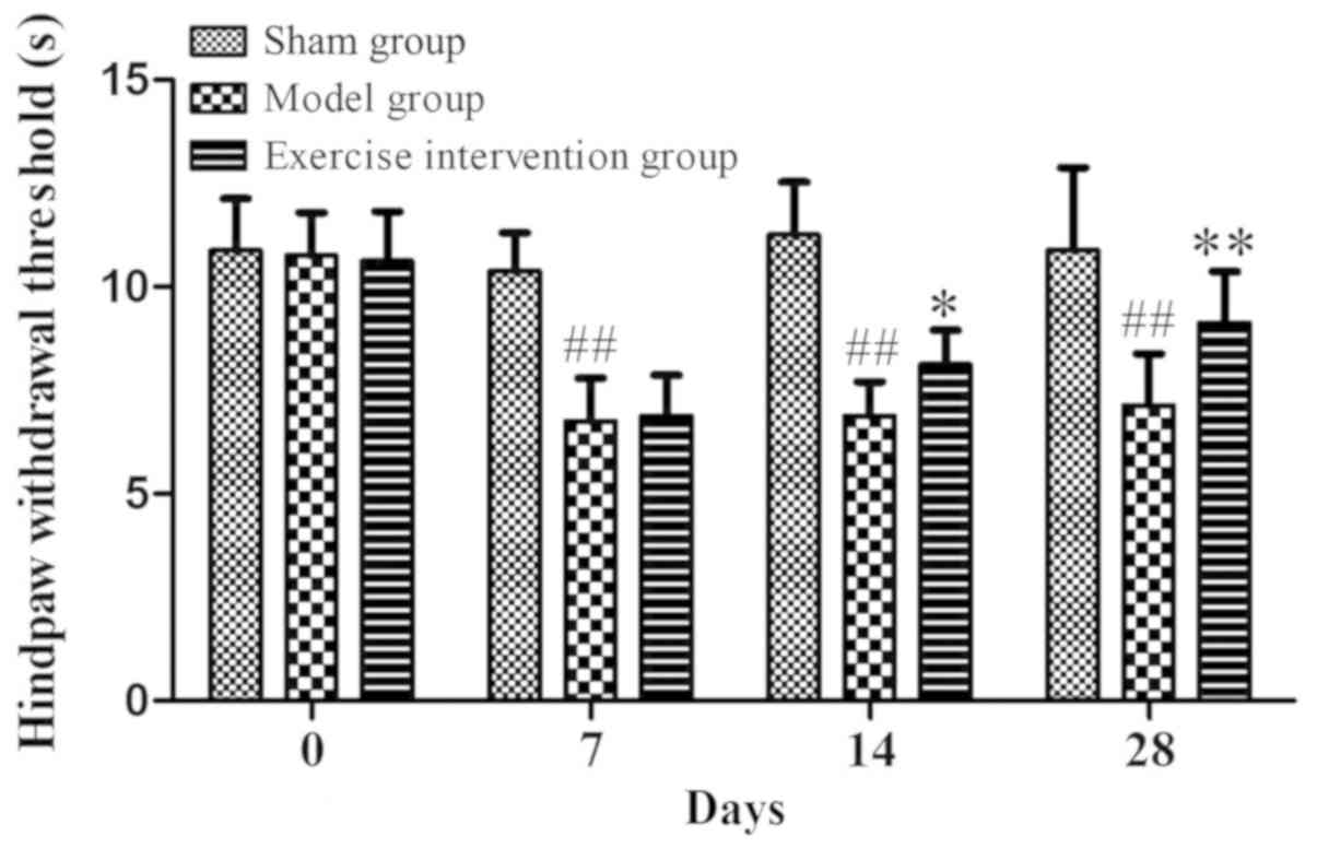
Hind paw withdrawal threshold is
increased following swimming in LDH rats
Prior to surgery, the hind paw withdrawal threshold
was ~10 sec in all three groups. Compared with the model group, the
hind paw withdrawal threshold was significantly increased in the
exercise intervention group on days 14 and 28 following surgery. As
compared with the sham operation group, the hind paw withdrawal
threshold of the model group was significantly reduced (Fig. 1). These data indicated that swimming
could alleviate nerve root pain in LDH rats as the training time
increased.
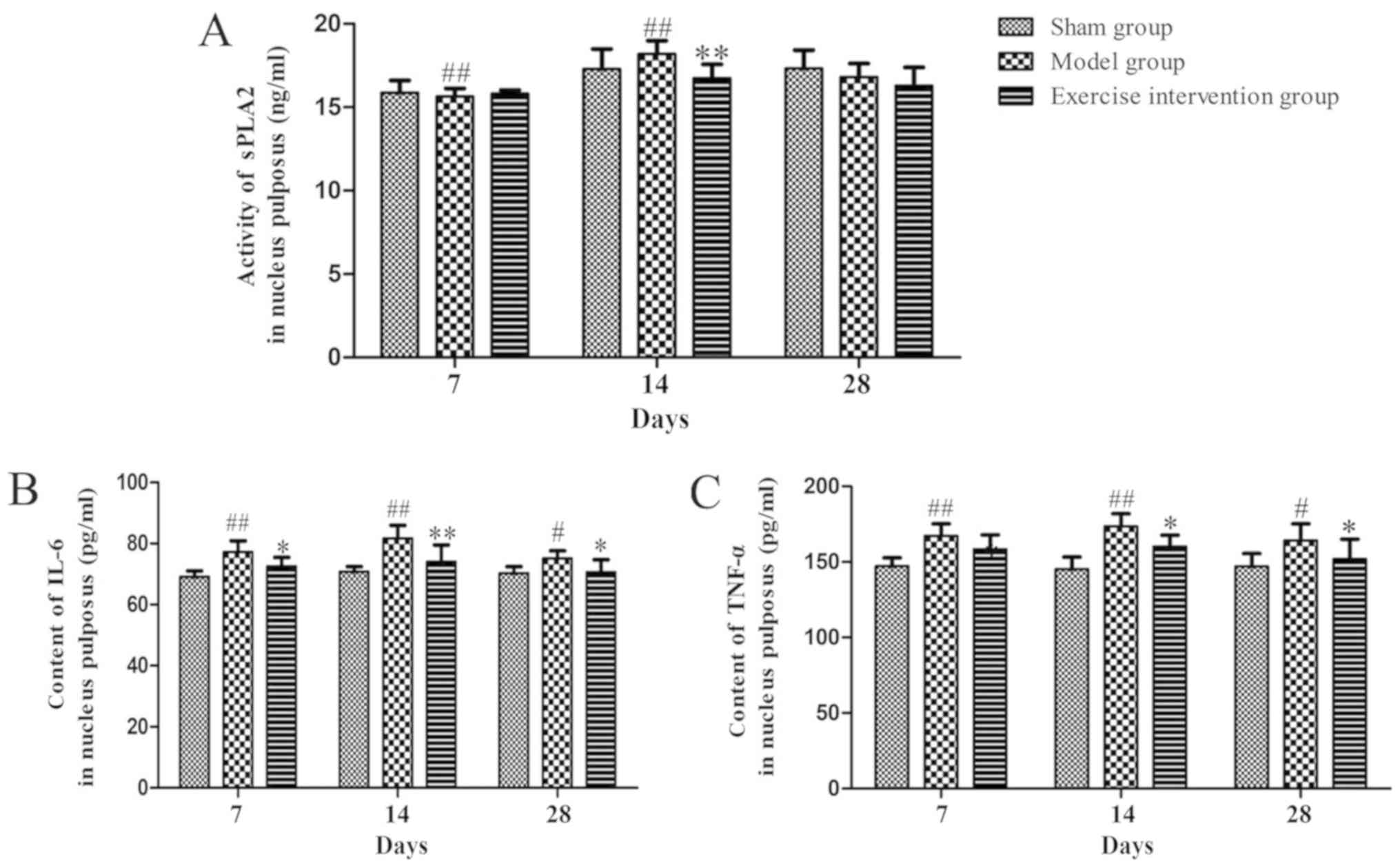
sPLA2 expression and IL-6 and TNF-α
content decreased in LDH rats following swimming
The sPLA2 expression in the exercise intervention
group was decreased on day 14, as compared with that in the model
group. sPLA2 expression in the model group was significantly
increased on days 7 and 14, as compared with that in the sham group
(Fig. 2A). As compared with the
model group, the IL-6 and TNF-α content was reduced on days 14 and
28 following surgery. As compared with the sham group, the IL-6 and
TNF-α content clearly increased on days 7, 14 and 28 in the model
group (Fig. 2B and C).
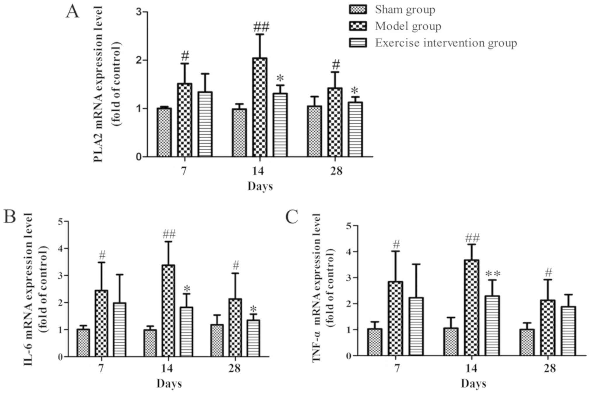
PLA2, IL-6 and TNF-α mRNA expression
decreases in LDH rats following swimming
The mRNA expression level of PLA2 in the exercise
intervention group significantly decreased on days 14 and 28
following surgery, when compared with the sham group, and the same
expression in the model group was higher than that in the sham
group from day 7 following surgery (Fig.
3A). The mRNA expression level of IL-6 in the exercise
intervention group was significantly decreased on days 14 and 28,
when compared with that in the model group, and that of the model
group was increased from day 7 following surgery, when compared
with that in the sham group (Fig.
3B). The mRNA expression level of TNF-α in the exercise
intervention group was significantly lower than that in the model
group on day 14, while that in the model group was clearly
increased from day 7 following surgery, compared with the sham
group (Fig. 3C).
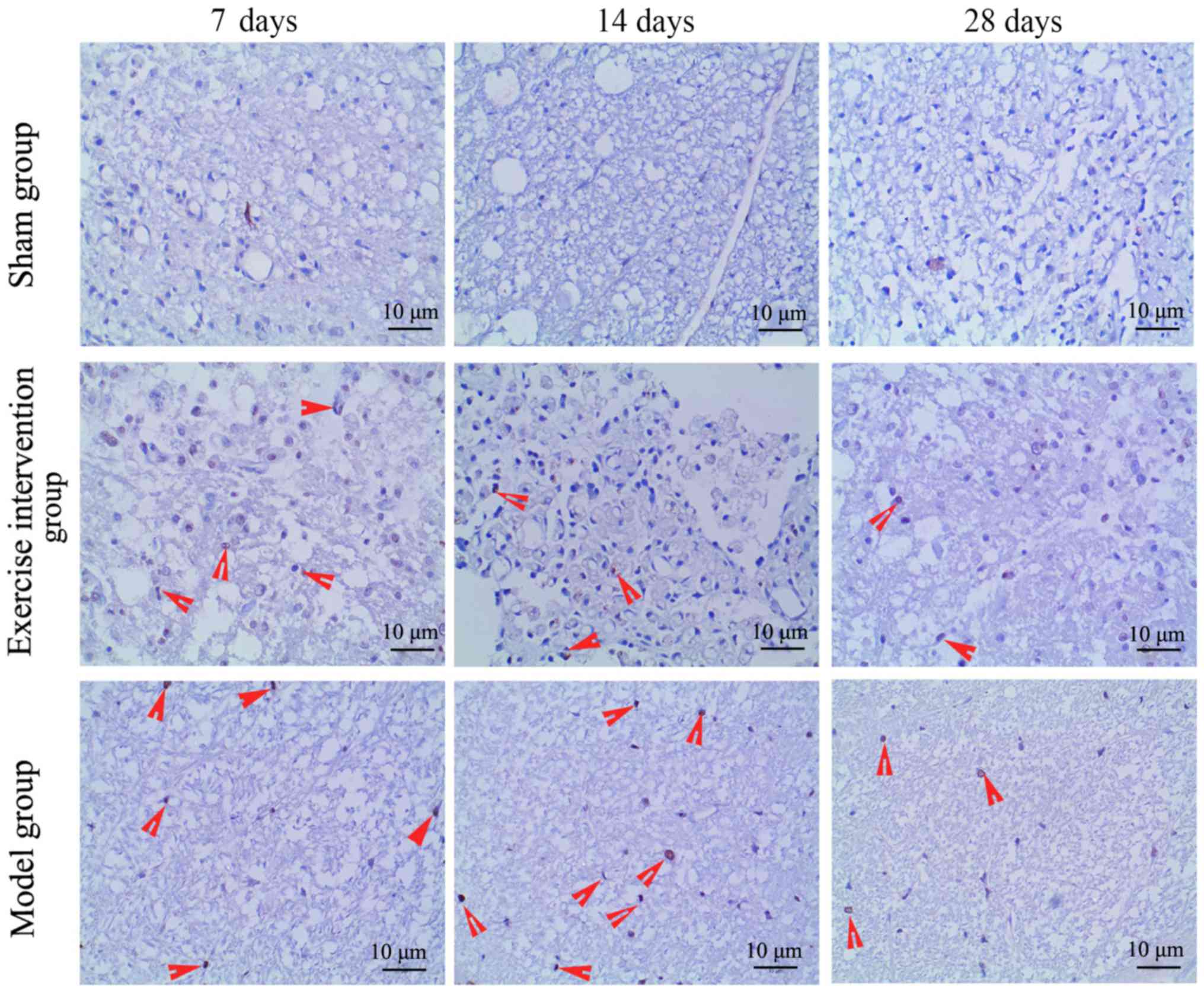
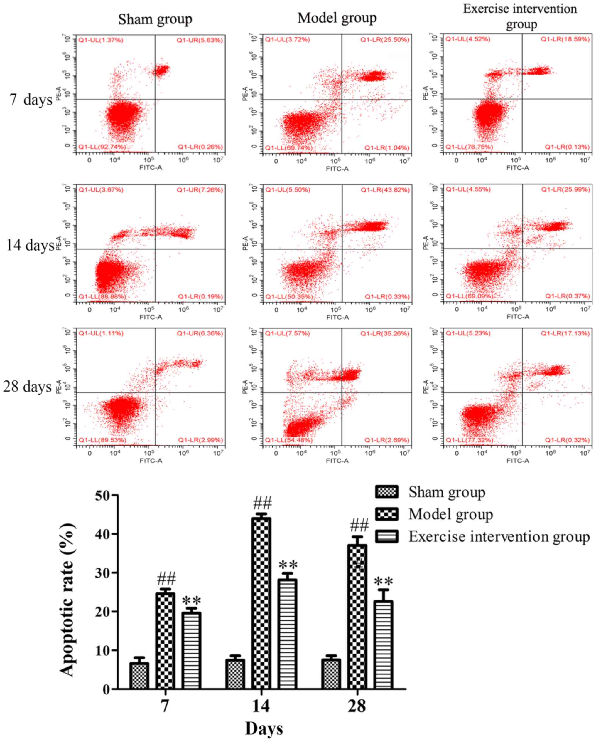
Swimming inhibits cell apoptosis in
LDH rats
Apoptotic cells were detected by TUNEL and Annexin
V/PI. The TUNEL staining results demonstrated that TUNEL-positive
cells were observed in the model and exercise intervention groups,
but those in the exercise intervention group were markedly
decreased, as compared to those in the model group (Fig. 4). The flow cytometry results
demonstrated that the apoptotic rate in the exercise intervention
group was significantly decreased from day 7 following surgery, as
compared with that in the model group. The rate in the model group
was significantly increased from day 7 following surgery, as
compared with that in the sham group (Fig. 5).
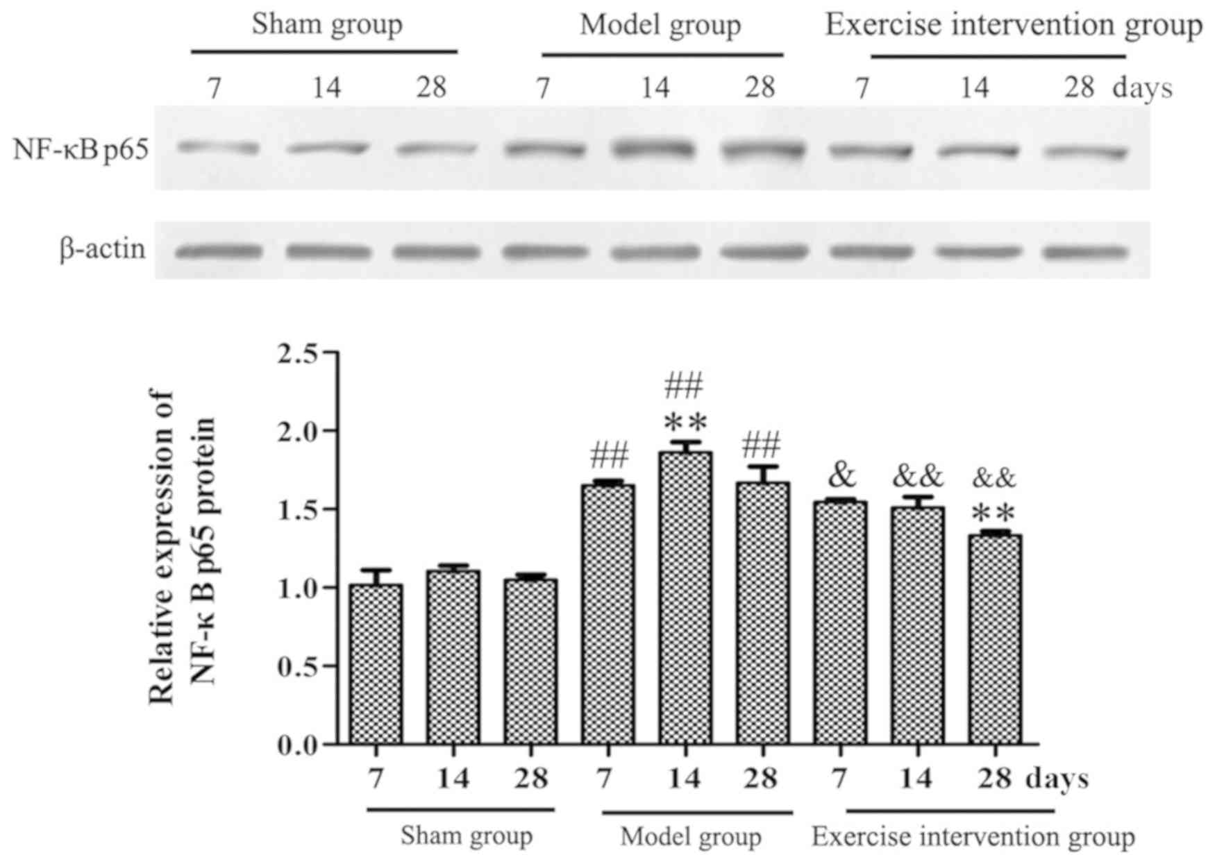
Swimming inhibits the activation of
NF-κBp65 in LDH rats
Western blotting results demonstrated that the
NF-κBp65 protein expression level in the model group was
significantly increased, when compared with that in the sham group
on days 7, 14 and 28 following surgery, and was significantly
higher on day 14, as compared with day 7. In the exercise
intervention group, the NF-κBp65 protein expression was
significantly decreased, as compared with that in model group, and
was significantly lower on day 28, as compared to day 7 (Fig. 6).
Discussion
Modern medical studies have demonstrated that
LDH-induced nerve root pain is mainly caused by mechanical
compression, autoimmunity and chemical stimulation of inflammatory
factors (7,20). Among them, mechanical oppression is
the main pathogenic factor. Early modern medicine's understanding
of LDH mainly focused on mechanical oppression, which was
considered the only pain-causing factor (21). With the progress and development of
modern medicine, the theory of molecular biology was introduced in
LDH. A previous study demonstrated that the inflammatory factors in
the body fluids and tissues of the lumbar process have toxic
effects on the nervous system, and can stimulate nerves and produce
the feeling of pain, causing nerve root pain in LDH (22). This finding demonstrated that
mechanical compression was not the only cause of lumbar and leg
pain in LDH, and that relief from compression injury was no longer
the only treatment.
The generation and maintenance of chronic
neuropathic pain is the common result of many inflammatory factors.
PLA2 is a key enzyme that produces other inflammatory substances,
which can hydrolyze glycosphospholipids specifically, and produce
free fatty acids and hemolytic phospholipids, mainly arachidonic
acid (23), causing inflammation in
the lumbar region and stimulating local sensors that ultimately
cause pain. TNF-α is released by macrophages and is a cytokine with
a strong pain-causing effect, it can cause root pain while
continuing to damage cells, promoting the production of other
inflammatory factors, such as cyclooxygenase 2, IL-6 and IL-1
(24). TNF-α can gather and regulate
neutrophil and eosinophilic cells directly, disrupt the metabolism
and cellular immune response, and affect the normal process of cell
division and differentiation, and produce cytotoxicity and
neurotoxicity (25,26). IL-6 is mainly secreted by
monocytes/macrophages, as well as B and T lymphocytes, it is an
acute reactive protein and can induce acute inflammatory reaction,
promoting and regulating the inflammatory response in different
types of cell division and differentiation (27). The present study demonstrated that
the sPLA2 expression, TNF-α and IL-6 content, and the PLA2, TNF-α
and IL-6 mRNA expression levels in the exercise intervention group
decreased significantly, as compared with those in the model group,
which was consistent with the trend of hind paw withdrawal
threshold in rats. These results indicated that the increased PLA2,
TNF-α, and IL-6 activity in the NP has an important role in
neuropathic pain of LDH, also suggested that swimming can inhibit
inflammatory reaction and improve the nerve root pain of LDH
rats.
A number of studies have indicated that apoptosis is
involved in intervertebral disc tissue degeneration of
pathophysiological changes (28,29).
Excessive cell apoptosis results in a reduction in the activity of
intervertebral disc cells and a subsequent decrease in
extracellular matrix change in synthesis and composition,
contributing to the pathology of intervertebral disc degeneration
(30). In the present study, it was
observed that swimming markedly reduced the number of apoptotic
cells in NP of LDH rats. And the apoptosis trend was consistent
with the change of inflammatory factors. Wang et al
(31) previously reported that the
IL-1, IL-4, and TNF-α expression levels contribute to the apoptosis
of disc cells. Therefore, it was hypothesized that excessive
activation of inflammatory factors may promote apoptosis in LDH
rats.
NF-κBp65 is the main effect factor of NF-κB, which
can promote the release of various inflammatory factors by
activating the downstream inflammatory signal, and increase the
chronic inflammatory injury of intestinal mucosa tissues (32). The activation of NF-κBp65 promoted
inflammation and hyperalgesia in rat adjuvant-induced arthritis
(33). The present findings
demonstrated that the NF-κBp65 protein expression level was
significantly decreased following swimming, which indicated that
the reduction of neuronal pain and inflammation in LDH rats by
swimming may be associated with the inhibition of NF-κBp65
activity.
In conclusion, the present findings indicate that
swimming can effectively reduce the nerve root pain in LDH rats,
which may be associated with the downregulation of inflammatory
factors, inhibition of the activity of NF-κBp65 pathway in NP, and
inhibition of apoptosis of NP cells. These findings are encouraging
and suggest that swimming might be employed as a novel and
effective therapy for LDH patients in the future.
Acknowledgements
Not applicable.
Funding
The present study was supported by a grant from the
Sichuan Health and Family Planning Commission Research Project
(grant no. 130198).
Availability of data and materials
The datasets used and/or analyzed during the current
study are available from the corresponding author on reasonable
request.
Authors' contributions
YH, ZZ and DY conceived and designed the
experiments. YH, ZZ, DY, LH, FH and DL performed the experiments.
LY, RW, LZ, XH and JH analyzed the data and provided assistance
with the experiments. YH and ZZ wrote the manuscript. All authors
have read and approved the final version of this manuscript.
Ethics approval and consent to
participate
The experimental protocol for the care and use of
laboratory animals was approved by the Experimental Animal Ethics
Committee of West China Hospital of Sichuan University (Chengdu,
China).
Patient consent for publication
Not applicable.
Competing interests
The authors declare that they have no competing
interests.
References
|
1
|
Krekoukias G, Gelalis ID, Xenakis T,
Gioftsos G, Dimitriadis Z and Sakellari V: Spinal mobilization vs
conventional physiotherapy in the management of chronic low back
pain due to spinal disk degeneration: A randomized controlled
trial. J Man Manip Ther. 25:66–73. 2017. View Article : Google Scholar : PubMed/NCBI
|
|
2
|
Haines SJ, Jordan N, Boen JR, Nyman JA,
Oldridge NB and Lindgren BR; LAPDOG/LEAPDOG Investigators, :
Discectomy strategies for lumbar disc herniation: Results of the
LAPDOG trial. J Clin Neurosci. 9:411–417. 2002. View Article : Google Scholar : PubMed/NCBI
|
|
3
|
Ahn SH, Cho YW, Ahn MW, Jang SH, Sohn YK
and Kim HS: mRNA expression of cytokines and chemokines in
herniated lumbar intervertebral discs. Spine (Phila Pa 1976).
27:911–917. 2002. View Article : Google Scholar : PubMed/NCBI
|
|
4
|
Orief T, Orz Y, Attia W and Almusrea K:
Spontaneous resorption of sequestrated intervertebral disc
herniation. World Neurosurg. 77:146–152. 2012. View Article : Google Scholar : PubMed/NCBI
|
|
5
|
Karademir M, Eser O and Karavelioglu E:
Adolescent lumbar disc herniation: Impact, diagnosis, and
treatment. J Back Musculoskelet Rehabil. 30:347–352. 2017.
View Article : Google Scholar : PubMed/NCBI
|
|
6
|
Karaaslan B, Aslan A, Borcek AO and Kaymaz
M: Clinical and surgical outcomes of upper lumbar disc herniations:
A retrospective study. Turk J Med Sci. 47:1157–1160. 2017.
View Article : Google Scholar : PubMed/NCBI
|
|
7
|
Cho HK, Kim SY, Choi MJ, Baek SO, Kwak SG
and Ahn SH: The effect of GCSB-5 a new herbal medicine on changes
in pain behavior and neuroglial activation in a rat model of lumbar
disc herniation. J Korean Neurosurg Soc. 59:98–105. 2016.
View Article : Google Scholar : PubMed/NCBI
|
|
8
|
Schroeder GD, Guyre CA and Vaccaro AR: The
epidemiology and pathophysiology of lumbar disc herniations.
Seminars Spine Surg. 28:2–7. 2016. View Article : Google Scholar
|
|
9
|
Gugliotta M, da Costa BR, Dabis E, Theiler
R, Jüni P, Reichenbach S, Landolt H and Hasler P: Surgical versus
conservative treatment for lumbar disc herniation: A prospective
cohort study. BMJ Open. 6:e0129382016. View Article : Google Scholar : PubMed/NCBI
|
|
10
|
Pearson AM, Blood EA, Frymoyer JW,
Herkowitz H, Abdu WA, Woodward R, Longley M, Emery SE, Lurie JD,
Tosteson TD and Weinstein JN: SPORT lumbar intervertebral disk
herniation and back pain. Spine (Phila Pa 1976). 33:428–435. 2008.
View Article : Google Scholar : PubMed/NCBI
|
|
11
|
Shepard N and Cho W: Recurrent lumbar disc
herniation: A review. Global Spine J. 9:202–209. 2019. View Article : Google Scholar : PubMed/NCBI
|
|
12
|
Righesso O, Falavigna A and Avanzi O:
Correlation between persistent neurological impairment and clinical
outcome after microdiscectomy for treatment of lumbar disc
herniation. Neurosurgery. 70:396–397. 2012. View Article : Google Scholar
|
|
13
|
Brotz D, Kuker W, Maschke E, Wick W,
Dichgans J and Weller M: A prospective trial of mechanical
physiotherapy for lumbar disk prolapse. J Neurol. 250:746–749.
2003. View Article : Google Scholar : PubMed/NCBI
|
|
14
|
Pourahmadi MR, Taghipour M, Ebrahimi
Takamjani I, Sanjari MA, Mohseni-Bandpei MA and Keshtkar AA: Motor
control exercise for symptomatic lumbar disc herniation: Protocol
for a systematic review and meta-analysis. BMJ Open. 6:e0124262016.
View Article : Google Scholar : PubMed/NCBI
|
|
15
|
Yan J, Hu S, Zou K, Xu M, Wang Q, Miao X,
Yu SP and Xu GY: Inhibition of cystathionine β-synthetase
suppresses sodium channel activities of dorsal root ganglion
neurons of rats with lumbar disc herniation. Sci Rep. 6:381882016.
View Article : Google Scholar : PubMed/NCBI
|
|
16
|
Zhu X, Cao S, Zhu MD, Liu JQ, Chen JJ and
Gao YJ: Contribution of chemokine CCL2/CCR2 signaling in the dorsal
root ganglion and spinal cord to the maintenance of neuropathic
pain in a rat model of lumbar disc herniation. J Pain. 15:516–526.
2014. View Article : Google Scholar : PubMed/NCBI
|
|
17
|
Zhao LR, Xing RL, Zhang L, Xu B, Li XC and
Wang PM: Transient receptor potential signaling pathways influence
the hyperalgesia in a rat model of mechanical lumbar disc
herniation. Myopain. 23:118–124. 2017. View Article : Google Scholar
|
|
18
|
Kameda T, Kaneuchi Y, Sekiguchi M and
Konno SI: Measurement of mechanical withdrawal thresholds and gait
analysis using the CatWalk method in a nucleus pulposus-applied
rodent model. J Exp Orthop. 4:312017. View Article : Google Scholar : PubMed/NCBI
|
|
19
|
Livak KJ and Schmittgen TD: Analysis of
relative gene expression data using real-time quantitative PCR and
the 2(-Delta Delta C(T)) method. Methods. 25:402–408. 2001.
View Article : Google Scholar : PubMed/NCBI
|
|
20
|
de Souza Grava AL, Ferrari LF and Defino
HL: Cytokine inhibition and time-related influence of inflammatory
stimuli on the hyperalgesia induced by the nucleus pulposus. Eur
Spine J. 21:537–545. 2012. View Article : Google Scholar : PubMed/NCBI
|
|
21
|
diZerega GS, Traylor MM, Alphonso LS and
Falcone SJ: Use of temporary implantable biomaterials to reduce leg
pain and back pain in patients with sciatica and lumbar disc
herniation. Materials (Basel). 3:3331–3368. 2010. View Article : Google Scholar
|
|
22
|
Yan J, Zou K, Liu X, Hu S, Wang Q, Miao X,
Zhu HY, Zhou Y and Xu GY: Hyperexcitability and sensitization of
sodium channels of dorsal root ganglion neurons in a rat model of
lumber disc herniation. Eur Spine J. 25:177–185. 2016. View Article : Google Scholar : PubMed/NCBI
|
|
23
|
Russell RL, Levine JM, Jeffery ND, Young
C, Mondragon A, Lee B, Boudreau CE, Welsh CJ and Levine GJ:
Arachidonic acid pathway alterations in cerebrospinal fluid of dogs
with naturally occurring spinal cord injury. BMC Neurosci.
17:312016. View Article : Google Scholar : PubMed/NCBI
|
|
24
|
Gui HQ, Zhi-Sheng MO and Yang J: Value of
TNF-α inhibitor in the treatment of lumbar disc herniation. Med
Innovation China. 2017.
|
|
25
|
Shang QS, Huang B and Sheng WH: Detection
of tumor necrosis factor-α and interleukin-18 in herniated
intervertebral disc and their significance. Orthopedic J China.
17:385–387. 2009.
|
|
26
|
Barichello T, dos Santos ID, Savi GD,
Florentino AF, Silvestre C, Comim CM, Feier G, Sachs D, Teixeira
MM, Teixeira AL and Quevedo J: Tumor necrosis factor alpha
(TNF-αlpha) levels in the brain and cerebrospinal fluid after
meningitis induced by Streptococcus pneumoniae. Neurosci Lett.
467:217–219. 2009. View Article : Google Scholar : PubMed/NCBI
|
|
27
|
Hunter CA and Jones SA: IL-6 as a keystone
cytokine in health and disease. Nat Immunol. 16:448–457. 2015.
View Article : Google Scholar : PubMed/NCBI
|
|
28
|
Alini M, Eisenstein SM, Ito K, Little C,
Kettler AA, Masuda K, Melrose J, Ralphs J, Stokes I and Wilke HJ:
Are animal models useful for studying human disc
disorders/degeneration? Eur Spine J. 17:2–19. 2008. View Article : Google Scholar : PubMed/NCBI
|
|
29
|
Chen J, Hou C, Chen X, Wang D, Yang P, He
X, Zhou J and Li H: Protective effect of cannabidiol on hydrogen
peroxide-induced apoptosis, inflammation and oxidative stress in
nucleus pulposus cells. Mol Med Rep. 14:2321–2327. 2016. View Article : Google Scholar : PubMed/NCBI
|
|
30
|
Kermani HR, Hoboubati H, Esmaeili-Mahani S
and Asadi-Shekaari M: Induction of intervertebral disc cell
apoptosis and degeneration by chronic unpredictable stress. J
Neurosurg Spine. 20:578–584. 2014. View Article : Google Scholar : PubMed/NCBI
|
|
31
|
Wang SL, Yu YL, Tang CL and Lv FZ: Effects
of TGF-β1 and IL-1β on expression of ADAMTS enzymes and TIMP-3 in
human intervertebral disc degeneration. Exp Ther Med. 6:1522–1526.
2013. View Article : Google Scholar : PubMed/NCBI
|
|
32
|
Gan H, Ouyang Q, Chen Y and Xia Q:
Activation of nuclear factor-kappaB and effects of
anti-inflammatory treatment thereon in intestinal mucosa of
patients with ulcerative colitis. Zhonghua Yi Xue Za Zhi.
82:384–388. 2002.(In Chinese). PubMed/NCBI
|
|
33
|
Luo JG, Zhao XL, Xu WC, Zhao XJ, Wang JN,
Lin XW, Sun T and Fu ZJ: Activation of spinal NF-κB/p65 contributes
to peripheral inflammation and hyperalgesia in rat adjuvant-induced
arthritis. Arthritis Rheumatol. 66:896–906. 2014. View Article : Google Scholar : PubMed/NCBI
|