Introduction
Lead acetate is a chemical compound. It is a white
crystalline substance with a sweetish taste. Like other lead
compounds, it is very poisonous. Lead acetate is used as a mordant
in textile printing and dyeing, as a drier in paints and varnishes,
and in preparing other lead compounds. It is an environmental and
occupational toxicant which has been known to damage vital organs
and suppress cellular processes. It is hazardous when swallowed,
inhaled or absorbed through the skin (1). There is a risk of cancer depending on
the level and duration of exposure (2). It causes irritation to skin, eyes and
the respiratory tract. It also has effects on the gum tissue,
central nervous system, kidneys, blood and reproductive system
(2).
Specific population groups are disproportionately at
a high risk for lead exposure, especially the lower age group, such
as fetuses, infants and young children, and the working class with
direct contact with lead in industries (3). Manser et al in 1990 showed
elevated lead levels and provided firm evidence of the
contamination of the water supply, fish and other food sources by
industrial effluent in big cities (4). Due to health concerns, lead from
paints and ceramic products, caulking and pipe solder has been
reduced dramatically in recent years. However, water pipes in some
older homes may still contain lead solder. Lead can leach out into
the water and enter the human body via oral ingestion or skin
contact (1).
Historically, lead is a known abortifacient and
spermicidal agent in the case of high exposure (5). Studies have shown that lead exposure
affects the endocrine function as well as the spermatogenesis
capability of the testis (5).
Landrigan in 1990 also showed that occupational exposure of lead to
men decreases their fertility (6).
Rodamilans et al suggested that prolonged lead exposure
initially produces a direct testicular toxicity followed by
hypothalamic or pituitary disturbances at long exposures (7). Saxena et al concluded that
lead exposure during the growing period when spermatogenesis is
proceeding may result in reproductive impairment during adulthood
(8).
Etlingera elatior (torch ginger) is a coarse
herb widely distributed in Malaysia and Indonesia. In Malaysia, it
is called kantan. Local communities ingest it by chopping the
flower and adding them into laksa (various curries or soups made
with rice noodles). Extracts from Etlingera species are
proven to have high anti-oxidant and anti-bacterial activity
(9). A study by Mohamad et
al reported that 1,7-bis(4-hydroxyphenyl)-2,4,6-heptatrienone
and 16-hydroxylabda-8(17),11,13-trien-15,16-olide are the two
important bioactive compounds from Etlingera elatior, which
are believed to have anti-oxidant properties (10). However, there are no reports
available on the use of this extract in reducing lead
acetate-induced testicular toxicity.
A previous study confirmed that Etlingera
elatior extract successfully protects the bone marrow against
lead toxicity (11). In the
present study, we examined the effects of lead acetate toxicity on
the spermatogenesis capabilities and functional damage in rat
testis and the protective effect of Etlingera elatior
extract on lead acetate-induced damage in rat testis.
Materials and methods
Etlingera elatior (4 kg) powder was extracted
using a soxhlet extractor, concentrated using a rotary evaporator
(Buchi, R-200, S/N 10328562, Germany) and then freeze-dried using a
freeze dryer (Freezone 4.5, Labconco, S/N 040622016S, USA) for 2
days.
All experimental procedures were in accordance with
the ethical guidelines for animal experimentation, and the study
protocol was approved by the Institute Research and Ethics
Committee.
A total of 36 male adult Sprague-Dawley rats were
purchased from UKM Animal Holdings Facility (University Kebangsaan
Malaysia). They were 2 months old, with their body weight ranging
from 230 to 270 g.
Experimental design
The rats obtained were divided into six groups
(Table I). Lead acetate was
administered via drinking water and was recorded as per drinking
water. Etlingera elatior extract was diluted with ultra-pure
water to the described concentration, and forced-fed via feeding
tube daily between 4 and 6 pm. At the end of the treatment period,
the rats were sacrificed. Serum was collected, the testes were
excised and one was preserved in 10% neutral buffered formalin. The
other testis was cut and homogenized in 5 ml PBS using a
homogenizer, and the supernatant was collected and stored at
−80°C.
 | Table I.Grouping criteria for the rats based
on dosage. |
Table I.
Grouping criteria for the rats based
on dosage.
| Group | Substance | Dose | Duration |
|---|
| 1 | Ultra-pure filter
water (control) | - | 14 days |
| 2 | Lead acetate | 500 ppm | 14 days |
| 3 | Etlingera
elatior extract | 100 mg/kg body
weight/day | 14 days |
| 4 | Lead acetate +
Etlingera elatior extract | 500 ppm + 100 mg/kg
body weight/day | 14 days |
| 5 | Lead acetate +
Etlingera elatior extract | 500 ppm + 100 mg/kg
body weight/day | 14 days+14days |
| 6 | Etlingera
elatior extract + Lead acetate | 100 mg/kg body
weight/day + 50 ppm | 14 days+14days |
The serum collected was used for the testosterone
assay, while the homogenized testis was used for superoxide
dismutase (SOD), glutathione peroxidase (GPx) and protein carbonyl
content (PCC) ELISA kits. Both the serum sample and the homogenized
testis were sent to the National Toxicological Centre in University
Sains Malaysia (USM) for lead analysis.
For histopathological analysis, the testis
maintained in the formalin solution was cut into half and then
soaked in formalin for 1 week. The tissue was then processed and
cut into 5-μm sections using a microtome. The tissue that
was made into slides was stained with H&E and observed under a
microscope.
Statistical analysis
All analytical data were expressed as the means with
standard deviation, with a 95% confidence interval. The level of
significance α was set at 0.05. p-value obtained from statistical
tests was compared against α for significance. Data regarding the
area of the seminiferous tubules, as well as the total cell count,
were analyzed using Kolmogorov-Smirnov test and histograms to
confirm a normal distribution. Comparison of data between various
groups for the area of seminiferous tubules and total cell count
was carried out using one-way ANOVA, with the post hoc test
as Bonferroni test.
For the data of biochemical tests, non-parametric
tests were used for statistical analysis. The level of significance
was determined through the p-value which was set at 0.05. Global
comparison was carried out using the Kruskal-Wallis non-parametric
test, while pair-wise comparison between individual groups was
carried out using the Mann-Whitney U test.
Results
Blood lead level (BLL)
The lead acetate alone group showed a significant
increase in the BLL in comparison to the control (p<0.05). The
BLL in the Etlingera elatior alone group was comparable to
the control, and it was significantly lower than that of the lead
acetate alone group (p<0.05). The lead acetate + Etlingera
elatior (concurrent treatment), lead acetate + Etlingera
elatior (post-treatment) and Etlingera elatior + lead
acetate groups all had a BLL significantly higher when compared to
that of the control (p<0.05), due to administration of lead in
these groups. However, the BLL of these three groups was also
significantly lower than that in the lead acetate alone group
(p<0.05), suggesting that Etlingera elatior successfully
reduced the amount of lead in the blood circulation. Comparing
among the three groups, the post-treatment group had the most
significant reduction compared to the other two.
Lead level in the testis
Using pair-wise comparison, the lead acetate alone
group had a testis lead level significantly higher than that of the
control (p<0.05), suggesting that lead does accumulate in the
testis. The Etlingera elatior alone group showed a testis
lead level comparable to that of the control, and was significantly
lower than the level in the lead acetate alone group (p<0.05).
Similar to the BLL result, the testis lead level in the lead
acetate + Etlingera elatior (concurrent treatment), lead
acetate + Etlingera elatior (post-treatment) and
Etlingera elatior + lead acetate was significantly higher
compared to that of the control (p<0.05). It was also
significantly lower when compared to the lead acetate alone group
(p<0.05), suggesting that Etlingera elatior successfully
reduced the amount of lead in the testis.
Testosterone
The serum level of testosterone in the lead acetate
alone group was significantly lower than that in the control
(p<0.05), showing that lead administration indeed affected
testosterone production. The serum level of testosterone in the
Etlingera elatior alone group and the lead acetate +
Etlingera elatior group (concurrent treatment), when
compared to the lead acetate alone group, were both significantly
higher (p<0.05), but they were of similar level to the control.
The lead acetate followed by Etlingera elatior
(post-treatment) group also had a serum level of testosterone
significantly higher than the lead acetate alone group (p<0.05),
and it showed a slight increase when compared to the control
(p<0.05), suggesting a reversal of damage by Etlingera
elatior. In the last group, Etlingera elatior followed
by lead acetate, the serum testosterone level was slightly higher
when compared to the lead acetate alone group, and was
significantly lower compared to the control (p<0.05).
Superoxide dismutase
The level of SOD in the lead acetate alone group was
significantly lower than that in the control (p<0.05), showing
that lead acetate reduced the production of SOD. In the
Etlingera elatior alone group, the level of SOD was the
highest among all groups. It was significantly higher than both the
control (p<0.05) and lead acetate alone group (p<0.05),
suggesting that the production of SOD was boosted by Etlingera
elatior. The level of SOD in the lead acetate + Etlingera
elatior (concurrent treatment) group was significantly higher
than that in the lead acetate alone group (p<0.05), and was of
comparable level to the control. The post-treatment group (lead
acetate followed by Etlingera elatior) had the second
highest level of SOD. It was also significantly higher than both
the control (p<0.05) and the lead acetate alone group
(p<0.05), suggesting a reversal of damage in terms of SOD. The
Etlingera elatior followed by lead acetate group had a SOD
level slightly lower than that of the control, but was
significantly higher compared to the lead acetate alone group
(p<0.05).
Glutathione peroxidase
Using pair-wise comparison, the lead acetate alone
group had a significantly lower level of GPx compared to the
control (p<0.05), proving that lead acetate reduced GPx
production. GPx in the Etlingera elatior alone group was
significantly higher than in the lead acetate alone group
(p<0.05), but also significantly lower than the control
(p<0.05). In the lead acetate + Etlingera elatior
(concurrent treatment) group, the GPx level was almost similar to
the Etlingera elatior alone group. It was significantly
higher than the lead acetate alone group (p<0.05), but also
significantly lower than the control (p<0.05). In the lead
acetate followed by Etlingera elatior (post-treatment)
group, the GPx level was significantly higher when compared to the
lead acetate alone group (p<0.05). It was lower than the
control, but not significantly. The last group, Etlingera
elatior + lead acetate, had a GPx level significantly higher
compared to the lead acetate alone group (p<0.05), but also
significantly lower when compared to the control (p<0.05).
Protein carbonyl content
Using pair-wise comparison, the lead acetate alone
group had a significantly higher level of PC than that of the
control (p<0.05), showing that the testicular tissue was greatly
damaged by lead acetate. In the Etlingera elatior alone
group, the PCC was of comparable level to the control group. When
compared to the lead acetate alone group, PC in the Etlingera
elatior alone group was significantly lower (p<0.05). PC for
the lead acetate + Etlingera elatior (concurrent treatment)
group was significantly higher compared to that of the control
(p<0.05), but also significantly lower than the lead acetate
alone group (p<0.05). The lead acetate followed by Etlingera
elatior (post-treatment) group and the Etlingera elatior
+ lead acetate group were both also significantly higher than the
control (p<0.05), but significantly lower compared to the lead
acetate alone group (p<0.05), suggesting a reversal of
damage.
Histopathological analysis (Table II)
In the control group, seminiferous tubules showed
normal architecture with adequate cellularity. All cells of
spermatogenic lineage were present. The basement membrane appeared
normal. There was no thickening of the basement membrane. Tubules
appeared healthy and no areas of fibrosis were found. Healthy
quantities of Sertoli and Leydig cells were present.
By contrast, the lead acetate alone group showed
shrunken seminiferous tubules and reduced cellularity. There was
more reduction in matured forms when compared to immature forms,
suggesting a maturation arrest. There was thickening of the
basement membrane and areas of fibrosis involving both the basement
membrane and the interstitium. Patches of interstitial oedema were
also present. Few tubules showed inflammatory activities with
residual macrophages. The blood vessels were also noted to be
dilated and congested. All these suggested that the testis was
heavily damaged by the administration of lead acetate.
The Etlingera elatior group showed healthy
testicular tissue similar to the control, with a slight increase in
the interstitial tissue. The concurrent treatment and
post-treatment groups showed mild thickening of the basement
membrane noted in certain seminiferous tubules, but otherwise
appeared generally healthy. This, when compared to the lead acetate
group, indicates that administration of Etlingera elatior
successfully reversed the damage caused by lead acetate in the
testicular tissue. The Etlingera elatior followed by the
lead acetate group appeared to be less healthy, with mild atrophy
and a paucity of cells inside the tubules suggesting growth arrest.
Moderate thickening of the basement membrane was also noted with
some interstitial oedema and focal areas of fibrosis. This shows
that Etlingera elatior administered as a treatment option
may produce a beneficial outcome.
The lead acetate group showed a reduction in total
cells when compared to the control. The Etlingera elatior
group showed an increase in cellularity, which was nearly similar
to the control and marginally lower. The post-treatment group
showed a good increase in cellularity, which was higher compared to
the control. The concurrent treatment group showed moderate
cellularity, which was almost similar to the control. The
Etlingera elatior followed by the lead acetate group showed
a reduction in cellularity and was the lowest of all groups.
Discussion
Observing the BLL results, the administration of
lead in drinking water induced a high BLL in the lead acetate alone
group, confirming that lead is highly absorbed into the body
through the gastrointestinal tract. The BLL of the lead acetate
alone group was much higher than the safe level of 10 μg/dl
(11). From the result of the
obtained levels of lead in the testis, it was observed that a high
concentration of lead was deposited in the testis in the lead
acetate alone group, which was approximately half the concentration
of lead in the blood circulation. This proves that the testis is a
site at risk from lead intoxication. It was observed that
administration of Etlingera elatior either concurrently with
lead acetate, post-administration of lead acetate, or prior to
administration of lead acetate, does have an effect in lowering the
level of lead in both blood and testis. However, the BLL in all
three groups did not drop back to the acceptable safe level of 10
μg/dl, although this could probably be achieved by
prolonging the treatment period.
Comparing among the three groups, the post-treatment
group had the most significant reduction in lead level in the blood
and testis, suggesting that Etlingera elatior treatment is
most effective in removing lead in the body post-exposure to lead.
While administering it concurrently, the active contents of
Etlingera elatior may be inhibited or used up. Administered
before lead acetate, Etlingera elatior may be utilized
against other oxidative challenge and thus the effect against lead
is reduced.
Lead is known to bring about damage in tissues by
inducing oxidative stress. It creates a high content of reactive
oxygen species in our body and also depletes anti-oxidant enzymes
(12). This was proven once again
based on the biochemical result obtained, where SOD and GPx were
reduced in the lead acetate alone group, while the protein carbonyl
content was increased. SOD and GPx are the two main enzymes
involved in the anti-oxidant defense mechanism in our body. SOD
works against superoxide anions, while GPx protects tissues and
cells against lipid hydroperoxide and hydrogen peroxide (13). There are different types of SODs,
depending on the metals that co-factor with the protein, including
Cu, Zn, Fe, Mn and Ni (14). Lead
is believed to displace metals in the functional group of SOD. This
is based on the fact that lead being itself bivalently charged in
ionic form is able to displace bivalent ions, such as
Zn2+, Cu2+ and Fe2+ (19), rendering the resultant enzyme
useless in catalyzing the redox reaction. The same principle holds
true for the reduction in GPx level. Lead displaces the
selenocysteine group from the active site of GPx, which is
responsible for its catalytic functions (15,16).
PC is formed when amino acid residues on protein molecules, such as
lysine, arginine, proline and histidine, are oxidized (17). The oxidizing agent for PC is ROS.
Hence, in the result obtained, an increase in PC is directly
attributed to an increased ROS level induced by lead acetate.
In another study by Sharma et al (18), rats were administered lead nitrate
for 7 days. Biochemical tests afterwards showed a significant
increase in PC and a significant decrease in SOD level. There was
also a significant increase in tissue lead level. The results of
this study are in accordance with our study, and our results are
also in accordance with other studies, in which a significantly
higher level of lead was detected by chemiluminescence in groups
treated with lead for 6 months. The elevated lead level was found
in blood, testicular tissue and epididymis (19–21).
In our study, Etlingera elatior was also used
as a treatment, both concurrently to lead acetate administration
and also post-exposure to lead acetate. The SOD level was
significantly increased when compared to both the control and lead
acetate alone group. This suggests that Etlingera elatior
enhances the production of SOD. In the concurrent treatment group,
SOD was brought back to the control level, while in the
post-treatment group, SOD level was increased almost to the level
of the Etlingera elatior alone group. In the Etlingera
elatior followed by the lead acetate group, however, the SOD
level was increased, but still slightly lower compared to the
control. This suggests that Etlingera elatior is most
effective when administered post-exposure to lead, and that the GPx
has a very similar result as SOD. In the concurrent treatment
group, GPx was increased to a significantly higher level compared
to the lead acetate alone, although it was still significantly
lower than the control. This result was similar for the
Etlingera elatior followed by the lead acetate group. Again,
the post-treatment group appeared to be the most effective group in
terms of the efficacy of Etlingera elatior against lead
toxicity, with the GPx level reverted back to almost the level of
the control. Comparing the PC, which indicates tissue or cell
injury from oxidative stress, all of the treatment and prevention
groups showed a significant decrease compared to the lead acetate
alone group. Among the three groups, the post-treatment group again
confirmed that Etlingera elatior is best administered
post-exposure to lead. The results obtained confirm that
post-treatment is the most optimal option compared to concurrent
treatment, with administration of Etlingera elatior prior to
lead acetate administration conferring a lesser protective
effect.
The serum testosterone level was significantly
reduced in the lead acetate alone group. In the concurrent
treatment group, Etlingera elatior successfully brought back
the testosterone level to the control level. In the Etlingera
elatior followed by the lead acetate group, the testosterone
level increased, but still remained significantly lower than the
control. The post-treatment group exhibited the highest level of
testosterone, even slightly higher than the control, suggesting
that Etlingera elatior is also most effective when
administered post-exposure to lead in recovering the testosterone
level.
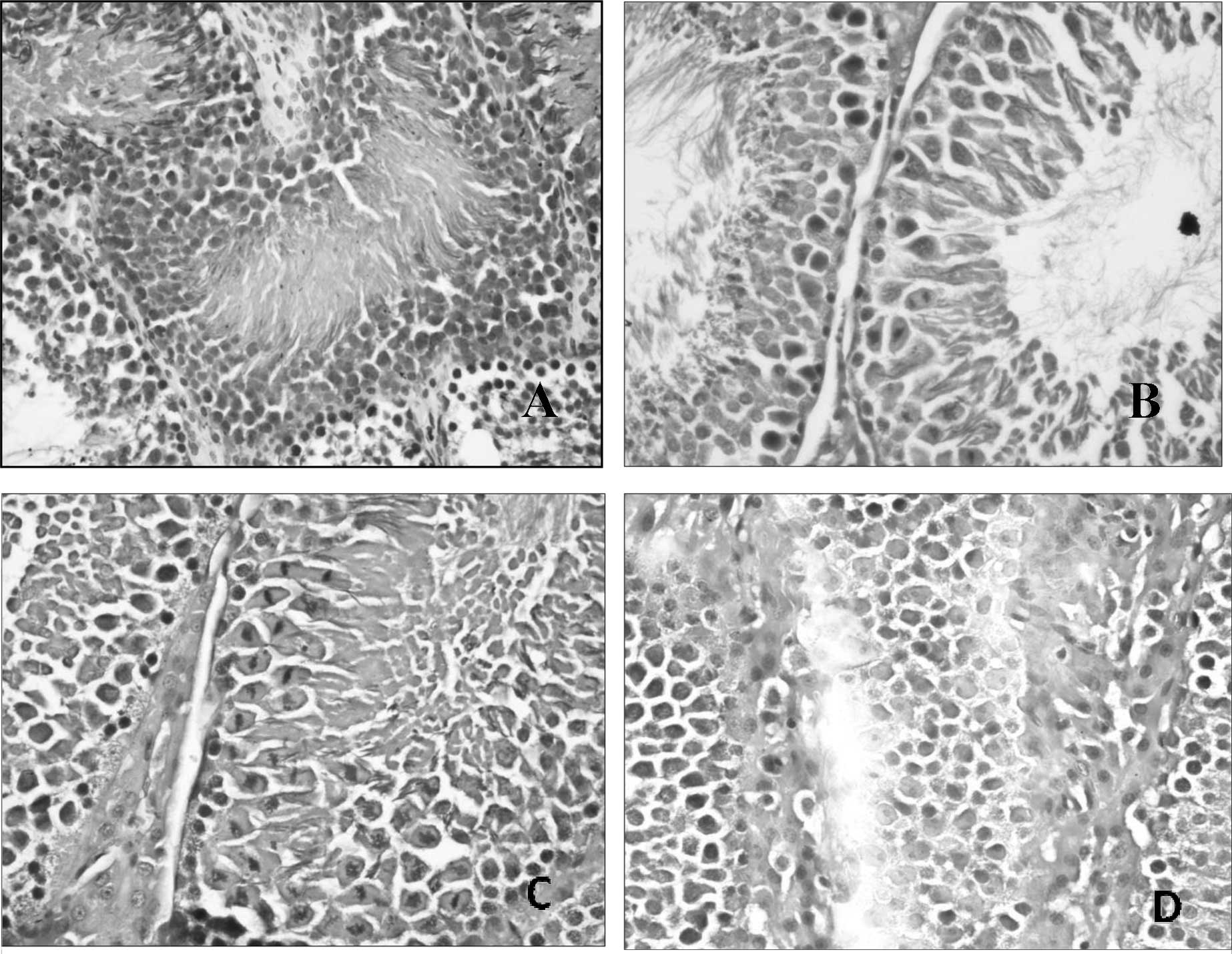
Histopathological alterations noted under the
microscope included shrunken seminiferous tubules, decreased
cellularity with reduction in matured forms when compared to
immature forms, suggesting a maturation arrest, thickening of
basement membrane, fibrosis affecting both the basement membrane
and the interstitium (Fig. 1). Our
findings are consistent with other studies (22,23),
in which many changes were observed after lead administration for
30 days, including shrunken seminiferous tubules with a wavy
outline, thickened basement membrane with hyalinization, maturation
arrest with abnormal ratio of spermatogenic cells and also
abnormalities in interstitial Leydig cells.
In the Etlingera elatior alone group, normal
spermatogenesis was noted to be actively ongoing (Fig. 1B) with abundant mature spermatozoa
formed. However, it was also noted that the interstitial tissue was
increased with certain areas of interstitial oedema, suggesting
that Etlingera elatior promotes the growth of interstitial
tissue. In the concurrent treatment group, the histology of the
testicular tissue was similar to the Etlingera elatior alone
group, with normal spermatogenesis (Fig. 1C) noted and abundant mature
spermatozoa formed. There was also an increased interstitial tissue
observed, but in addition to that, congestion of blood vessels was
also noted and a moderate thickening of the basement membrane. This
suggests that Etlingera elatior protects the testicular
tissue with only minor changes occurring. In the post-treatment
group, normal architecture of seminiferous tubules was noted, with
actively ongoing spermatogenesis. However, it was observed that
there was mild thickening of the basement membrane (Fig. 1D), increased interstitial tissue
with abundant Leydig cells, congestion of blood vessels and the
presence of few macrophages in some of the seminiferous tubules.
The testicular tissue in this group, although showing all the
above-mentioned minor changes, generally appeared healthy, showing
that the damage that lead had caused was repaired by the
Etlingera elatior post-exposure to lead acetate. The
presence of macrophages again was deduced to aid in removing
excessive lead present in the testicular tissue. The Etlingera
elatior followed by the lead acetate group, on the other hand,
appeared to sustain more damage than the two treatment groups.
Moderate thickening of the basement membrane was noted with some
areas of interstitial oedema (Fig.
1D), and a paucity of cells also suggest that there was a
maturation arrest of spermatogenic cells. There was also some
congestion of blood vessels noted. Collectively, the tissues
appeared to be mild to moderately atrophic, confirming with the
biochemical tests that Etlingera elatior administered prior
to lead acetate administration is not as beneficial as a treatment.
Again, comparison to other studies was not possible due to the lack
of studies on the effect of Etlingera elatior against lead
toxicity in the testis. However, our result is still supported by
the fact that Etlingera elatior contains a high level of
anti-oxidant activity (24),
bringing about the promising result obtained.
In conclusion, Etlingera elatior is indeed a
beneficial treatment option against lead-induced oxidative stress
in testicular tissue. Among the treatment method options,
administration of Etlingera elatior post-exposure to lead
yields the most beneficial result. Further investigations are
warranted as Etlingera elatior can be a potential
complimentary agent in treating chronic lead intoxication.
Acknowledgements
This study was done under the research
grant no. BMS-I-02/2010(06) provided by the International Medical
University, Malaysia
References
|
1.
|
H FuP BoffettaCancer and occupational
exposure to inorganic lead compounds: a meta-analysis of published
dataOccup Environ Med527381199510.1136/oem.52.2.737757170
|
|
2.
|
T DamstraToxicological properties of
leadEnviron Health Perspect19297307197710.1289/ehp.7719297
|
|
3.
|
JI RaderJT PeelerKR MahaffeyComparative
toxicity and tissue distribution of lead acetate in weanling and
adult ratsEnviron Health
Perspect42187195198110.1289/ehp.81421877333253
|
|
4.
|
WW ManserR LalaniS HaiderMA KhanTrace
element studies on Karachi populations. Part V: Blood lead levels
in normal healthy adults and grammar school childrenJ Pak Med
Assoc4015015419901703243
|
|
5.
|
F GorbelM BoujelbeneF Makni-AyadiF
GuermaziF CrouteJP SoleilhavoupA el FekiCytotoxic effects of lead
on the endocrine and exocrine sexual function of pubescent male and
female rats. Demonstration of apoptotic activityC R
Biol325927940200212481686
|
|
6.
|
PJ LandriganCurrent issues in the
epidemiology and toxicology of occupational exposure to leadEnviron
Health Perspect896166199010.1289/ehp.9089612088757
|
|
7.
|
M RodamilansMJ OsabaJ To-FiguerasF Rivera
FillatJM MarquesP PerezJ CorbellaLead toxicity on endocrine
testicular function in an occupationally exposed populationHum
Toxicol7125128198810.1177/0960327188007002033132417
|
|
8.
|
DK SaxenaRS SrivastavaB LalSV ChandraThe
effect of lead exposure on the testis of growing ratsExp
Pathol31249252198710.1016/S0232-1513(87)80010-53622725
|
|
9.
|
EWC ChanYY LimM OmarAntioxidant and
antibacterial activity of leaves of Etlingera species
(Zingiberaceae) in Peninsular MalaysiaFood
Chem10415861593200710.1016/j.foodchem.2007.03.023
|
|
10.
|
H MohamadNH LajisF AbasAM AliMA SukariH
KikuzakiN NakataniAntioxidant constituents of Etlingera
elatiorJ Nat Prod68285288200510.1021/np040098l
|
|
11.
|
A WuTietz Clinical Guide to Laboratory
Tests4th editionSaunders ElsevierSt. Louis, MO6586592006
|
|
12.
|
M MarchlewiczB WiszniewskaB GonetI
Baranowska-BasiockaK SafranowA KolasaW GlabowskiR KurzawaK
JakubowskaME RacIncreased lipid peroxidation and ascorbic acid
utilization in testis and epididymis of rats chronically exposed to
leadBiometals201319200710.1007/s10534-006-9009-z16699871
|
|
13.
|
Q RanH LiangY IkenoW QiTA ProllaLJ Roberts
IIN WolfH van RemmenA RichardsonReduction in glutathione peroxidase
4 increases life span through increased sensitivity to apoptosisJ
Gerontol A Biol Sci Med
Sci62932942200710.1093/gerona/62.9.93217895430
|
|
14.
|
S MarklundDistribution of Cu Zn superoxide
dismutase and Mn superoxide dismutase in human tissues and
extracellular fluidsActa Physiol Scand492S19S2319806939305
|
|
15.
|
F UrsiniM MaiorinoC GregolinThe
selenoenzyme phospholipid hydroperoxide glutathione
peroxidaseBiochim Biophys
Acta8396270198510.1016/0304-4165(85)90182-5
|
|
16.
|
JW ForstromJJ ZakowskiAL
TappelIdentification of the catalytic site of rat liver glutathione
peroxidase as
selenocysteineBiochemistry1726392644197810.1021/bi00606a028678534
|
|
17.
|
ER StadtmanCN OliverMetal-catalyzed
oxidation of proteins: physiological consequencesJ Biol
Chem2662005200819911989966
|
|
18.
|
V SharmaL KansalA SharmaProphylactic
efficacy of Coriandrumsativum (Coriander) on testis of
lead-exposed miceBiol Trace Elem Res1363373542010
|
|
19.
|
M MarchlewiczT MichalskaB
WiszniewskaDetection of lead-induced oxidative stress in the rat
epididymis by
chemiluminescenceChemosphere5715531562200410.1016/j.chemosphere.2004.08.10215519400
|
|
20.
|
N HaleagraharaT JackieS ChakravarthiM RaoT
PasupathiProtective effects of Etlingera elatior extract on
lead acetate-induced changes in oxidative biomarkers in bone marrow
of ratsFood Chem Toxicol48268826942010
|
|
21.
|
N HaleagraharaT JackieS ChakravarthiM RaoA
KulurProtective effect of Etlingera elatior (torch ginger)
extract on lead acetate – induced hepatotoxicity in ratsJ Toxicol
Sci356636712011
|
|
22.
|
I AhmadM SabirKF YasinStudy of the effects
of lead poisoning on the testes in albino ratsPak J Med
Res4222562622003
|
|
23.
|
NW GhelbergE BordasLead-induced
experimental lesions of the testis and their treatmentJ Appl
Toxicol1284286198110.1002/jat.25500105097185889
|
|
24.
|
EWC ChanYY LimLF WongFS LiantoSK WongKK
LimCE JoeTY LimAntioxidant and tyrosinase inhibition properties of
leaves and rhizomes of ginger speciesFood
Chem109477483200810.1016/j.foodchem.2008.02.016
|