Introduction
Skin aging is a degenerative process including
intrinsic aging which is characterized by dryness, generalized
wrinkling and thin appearance. Solar ultraviolet (UV) radiation is
the most significant extrinsic factor which causes photoaging of
the skin, manifesting as deep wrinkling, severe roughness and
dryness. The photoaging of the skin partly overlaps and
superimposes the intrinsic dryness (1). Solar UV reaching earth is comprised
of UVA (320–400 nm in wavelength) and UVB (280–320 nm). UVB is
mostly absorbed by the epidermis and predominantly affects
keratinocytes (1,2).
Water movement across the plasma membrane occurs via
two pathways: diffusion through the lipid bilayer and via
aquaporins (AQPs) (1,3–5). The AQPs act primarily as
water-selective pores and facilitate water transport across cell
plasma membranes (6). There are
at least 13 mammalian AQPs (AQP0-AQP12), which have been divided
into two groups on the basis of their permeability. AQP 1, 2, 4, 5
and 8 are primarily water-selective transporters; AQP 3, 7, 9 and
10 transport water, glycerol and other small solutes (7,8).
It has been demonstrated that AQP3 expressed in the
basal layer of the epidermis and the deficiency reduce the stratum
corneum hydration and glycerol content (4,8,9).
After exposure to UV radiation, AQP3 down-regulation reduces the
stratum corneum hydration; the deficient water conditions damage
the function of the skin, leading to dryness and wrinkle formation
(1,10). However, the functions of AQP3 in
human skin keratinocytes remain to be further elucidated.
Adequate photoprotection is essential to prevent
UV-related damage. Photoprotective agents, such as polyphenols and
baicalin, have been demonstrated to be effective in photoprotection
via influencing pertinent cell signaling pathways (11). Kanglaite is a mixture consisting
of extractions from Coix seed, a Chinese herb, which has been
demonstrated to be effective in anticancer treatment via inhibition
of COX-2, MMP9, protein kinase C and NF-κB (12,13). We carried out this study to
investigate whether Kanglaite has any protective effects against
UVB-induced AQP3 down-regulation in cultured human skin
keratinocytes.
Materials and methods
UV light apparatus
The UV radiation apparatus used in the study was the
Waldmann UV801KL (Waldmann GmbH Co., Germany). The UVB wavelength
was 285–350 nm (peak 312 nm). As previously described, (5,15)
before UVB radiation, cultured human skin keratinocytes were washed
with 1 ml PBS buffer and then changed to 0.5 ml PBS in each well.
The cells were radiated at the desired intensity without a plastic
dish lid. After UVB radiation, the cells were returned to
incubation in basal medium with treatments for various times prior
to harvest.
Chemicals and reagents
Rabbit anti-AQP3 was obtained from Chemicon
(Temecula, CA). Monoclonal mouse anti-β-actin was obtained from
Sigma (St. Louis, MO). Kanglaite was obtained from the Zhejiang
Kanglaite Pharmaceutical Co. (Hangzhou, China).
Cell culture
As previously described (5,14),
spontaneously immortalized human keratinocytes (HaCaT) were
maintained in DMEM medium (Sigma) supplemented with 10% fetal
bovine serum (Invitrogen, Carlsbad, CA), penicillin/streptomycin
(1:10; Sigma) and 4 mM L-glutamine (Sigma), in a CO2
incubator at 37°C. For Western blotting, cells were reseeded in
6-well plates at a density of 0.5×10⁶ cells/ml with
fresh complete culture medium.
MTT assay
The cell proliferation effect of Kanglaite was
determined by the MTT assay. The cells (4×10³ cells/ml)
were cultured on a 96-well plate in a DMEM medium with different
concentrations of Kanglaite (0.5×10⁻⁴,
1×10⁻³, 5×10⁻³, 1×10⁻²,
5×10⁻², 1×10⁻¹ ml/ml) for 24 h. The cells
were next washed with PBS and 200 μl of MTT (0.05 mg/ml) was added
to each well, followed by incubation for 4 h at 37°C. The
supernatant was removed, and 200 μl of dimethylsulfoxide was added
to each well to dissolve the formazan product. Wells without cells
were used as blank controls. Absorbance was determined at 570 nm,
spectrophotometrically, using an ELISA reader (Tecan, Salzburg,
Austria). The results are expressed as the percentage of control
cells obtained from six experiments conducted under the same
culture conditions.
RNA extraction, reverse transcription and
PCR
Total-RNA from cells was extracted using the SV
total-RNA Isolation System (Zhongshan, China) following the
manufacturer’s instructions. The concentration of total-RNA was
determined by measuring the optical density at 260 nm. Total-RNA (1
μg) was converted into first-strand cDNA using the ImProm-II
Reverse Transcription system with random primers following the
manufacturer’s instructions (Zhongshan). Parallel reactions for
each RNA sample were run in the absence of reverse transcriptase to
assess any genomic DNA contamination of the RNA.
For the semi-quantitative reverse transcription PCR
experiment, the product was amplified using specific primers
designed as described before (15,16): AQP3 forward, 5′-GCT GTC ACT CTG
GGC ATC CTG-3′ and reverse primers, 5′-GCG TCT GTG CCA GGG TGT
AG-3′, amplifying a 131-bp product and the GAPDH forward, 5′-TCC
TGT GGC ATC CAC GAA ACT-3′ and reverse primers, 5′-GAA GCA TTT GCG
GTG GAC GAT-3′, amplifying a 313-bp product.
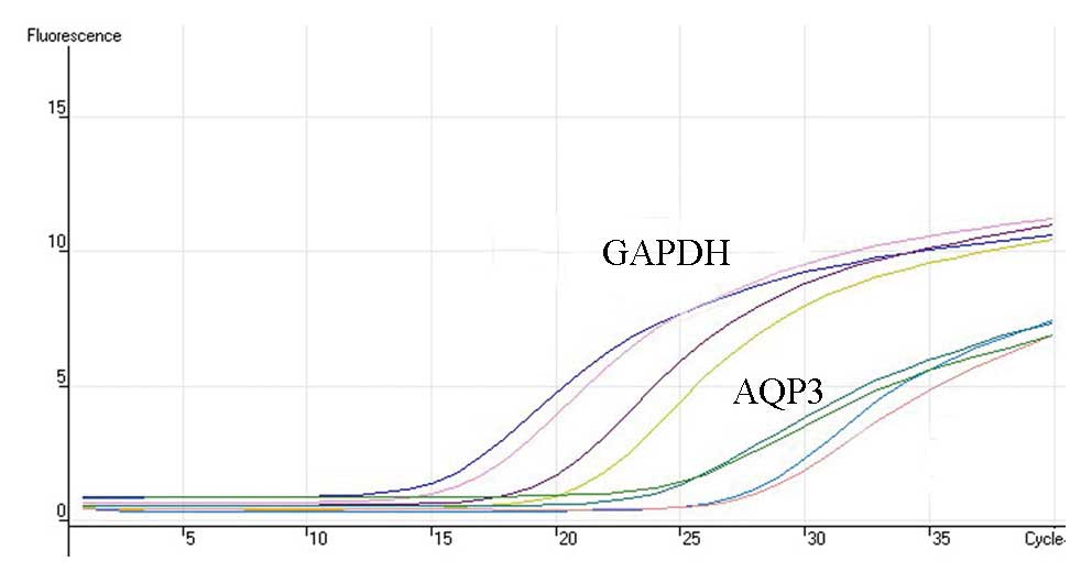
The real-time quantitative PCR experiments were
carried out in an Rotor-gene 3000 (Australia), using a SYBR-Green
PCR Mastermix (Zhongshan). Each sample was analyzed iduplicate
along with standard and no-template controls. The reaction
contained 30 ng cDNA in 1 μl Mastermix, including pre-set
concentrations of deoxyribonucleotide triphosphates,
MgCl2, and buffers, along with 300 nM forward and
reverse primers and the SYBR-Green reporter dye. The PCR parameters
were 95°C for 2 min, 40 cycles at 95°C for 15 sec, 60°C for 1 min
and 72°C for 30 sec (Fig. 1). RNA
concentrations were determined by comparing cDNA-generated signals
in samples with those generated from known amounts of cDNA. RNA
levels were corrected with the GAPDH cDNA signal for variations in
the amounts of input RNA. The product purity was confirmed using a
dissociation standard curve.
Western blot analysis
As reported previously (5,14),
cultured skin keratinocytes with or without treatment were washed
with cold PBS and harvested by scraping into 100 μl of RIPA buffer.
Cell lysates were incubated at 4°C for 30 min. Proteins (20 μg)
were denatured in 5X SDS-PAGE sample buffer for 5 min at 95°C.
Proteins were separated by 10 or 12% SDS-PAGE gels and transferred
onto PVDF membranes (Millipore, Bedford, MA). Nonspecific binding
was blocked with 10% dry milk in TBST for 1 h at room temperature.
After blocking, membranes were incubated with specific antibodies
in dilution buffer (2% BSA in TBS) overnight at 4°C. Blots were
incubated with horseradish peroxidase conjugated anti-rabbit or
anti-mouse IgG at appropriate dilutions and room temperature for 1
h. Antibody binding was detected using the enhanced
chemiluminescence (ECL) detection system (Amersham Biosciences)
following the manufacturer’s instructions and visualized by
autoradiography with Hyperfilm.
Statistical analysis
The values in the figures were expressed as the
means ± standard error (SE). The data in this study are
representative of more than three different experiments. Repeated
measures of one factor ANOVA was used to analyze the data. The
SNK-q assay was performed between the treated groups. The Student’s
t-test was performed to detect differences between the Kanglaite
and vehicle groups. P<0.05 was considered significant.
Results
Effect of Kanglaite on proliferation in
cultured human skin keratinocytes
Cultured skin keratinocytes were treated with 0,
5×10⁻⁴, 1×10⁻³, 5×10⁻³,
1×10⁻², 5×10⁻², 1×10⁻¹ ml/ml
Kanglaite. The results of the MTT assay showed proliferation rates
of 0.093±0.008, 0.963±0.280, 1.140±0.201, 1.073±0.132, 1.055±0.233,
1.068±0.208 and 0.857±0.218, respectively. Kanglaite in all the
examined concentrations exhibited no inhibitory effect on the
proliferation of cultured skin keratinocytes (P>0.05).
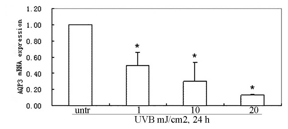
UVB radiation down-regulates AQP3 mRNA in
cultured human skin keratinocytes
Cultured skin keratinocytes were radiated with 1,
10, 20 mJ/cm2 UVB. Cells were collected after 24 h of
culture. The effect of UVB radiation on gene expression was
determined by means of real-time quantitative PCR. The results of a
relative quantification analysis revealed that the UVB-induced AQP3
mRNA down-regulation was dose-dependent. The AQP3 mRNA expression
was down-regulated after radiation with 1 mJ/cm2 UVB and
the down-regulation was most obvious after 20 mJ/cm2 of
UVB irradiation (F=19.88, P<0.0005) (Fig. 2A). The UVB-induced down-regulation
of AQP3 mRNA was also time-dependent. The AQP3 mRNA expression was
first found to be down-regulated at 6 h and was most obvious at 24
h after radiation with 10 mJ/cm2 UVB (F=25.30,
P<0.0002) (Fig. 2B).
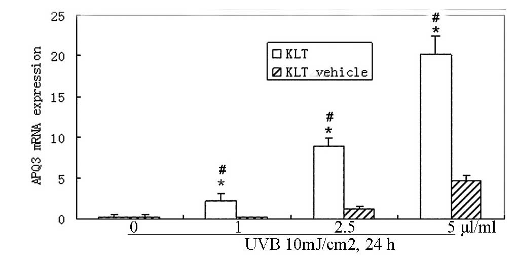
Kanglaite inhibits UVB-induced
down-regulation of AQP3 mRNA in cultured human skin
keratinocytes
Cultured skin keratinocytes were radiated with 10
mJ/cm2 UVB. Cells were collected after 24 h of
incubation with 1, 2.5 or 5 μl/ml Kanglaite and the same
concentrations of Kanglaite vehicles were used as controls.
Real-time quantitative PCR was performed. Application of 1 μl/ml
Kanglaite significantly up-regulated the AQP3 mRNA expression after
24 h incubation (F=−3.84 P=0.0184); 2.5, 5 μl/ml Kanglaite
incubation significantly up-regulated the AQP3 transcripts by
8.92±1.04 and 20.20±2.25-fold, respectively. Significant
up-regulation was observed in all 1, 2.5 and 5 μl/ml
Kanglaite-treated groups vs. the Kanglaite vehicle groups (Fig. 3A). After 6, 12 or 24 h of
incubation with 2.5 μl/ml Kanglaite and the vehicles, significant
up-regulation of the AQP3 transcripts was only observed in the 24 h
samples (F=−4.80, P=0.0086) (Fig.
3B). There were no significant changes in the 6 and 12 h
samples between the Kanglaite and vehicle groups.
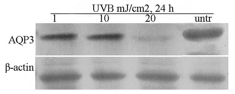
UVB irradiation down-regulates AQP3
protein in cultured skin keratinocytes
Cultured skin keratinocytes were radiated with 1, 10
or 20 mJ/cm2 UVB. Cells were collected after 24 h of
culture. Western blot analysis showed that UVB irradiation
down-regulated the AQP3 protein expression in a dose-dependent
manner. AQP3 protein expression began to decrease after 1
mJ/cm2 UVB radiation, and more significantly when
radiated with 20 mJ/cm2 UVB compared to untreated
samples (Fig. 4A). After being
radiated with 10 mJ/cm2 UVB and cultured for 6, 12 or 24
h, AQP3 protein expression was again measured by Western blotting.
The results showed that AQP3 began to decrease at 6 h and further
decreased at 12 h. After 24 h culture, AQP3 expression began to
return towards basal levels compared to that at 12 h (Fig. 4B).
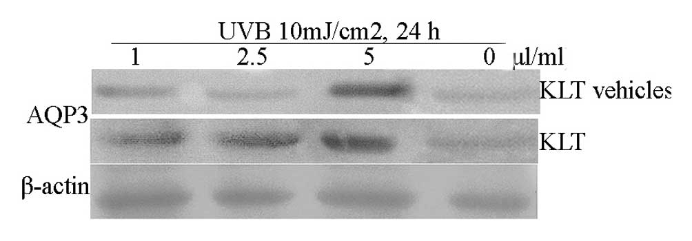
Kanglaite inhibits UVB-induced
down-regulation of AQP3 protein expression in cultured skin
keratinocytes
To analyze the effect of Kanglaite on AQP3 protein
expression after UVB irradiation, cultured skin keratinocytes were
radiated with 10 mJ/cm2 UVB and incubated with 1, 2.5, 5
μl/ml Kanglaite and Kanglaite vehicles, respectively. Application
of 1 μl/ml Kanglaite significantly up-regulated AQP3 protein
expression after 24 h incubation; 2.5, 5 μl/ml Kanglaite also
significantly up-regulated the AQP3 protein expression (Fig. 5A). After 6, 12 or 24 h of
incubation with 2.5 μl/ml Kanglaite and the vehicles, significantly
up-regulated AQP3 protein expression was observed in the
Kanglaite-treated groups. No significant changes in AQP3 protein
expression were observed in the vehicle-treated groups (Fig. 5B).
Discussion
Some intracellular and extracellular proteins are
involved in skin photoaging through regulation of cell signaling
pathways such as JNK, ERK, p38 MAP kinase and PI3K/AKT kinases
(14,17). Antioxidants and botanical agents
such as polyphenols, epigallocathechin-3-gallate, flavonoids,
isoflavonoids and all-trans retinoic acid have been demonstrated to
be able to block certain pathways to exert their protective effect
on skin photoaging (14,18–21).
Dehydration is one of the major events of skin
photoaging. Previous studies have shown that AQP3 plays an
important role in the regulation of water permeability (22). UV radiation down-regulates AQP3
expression in cultured skin keratinocytes, then decreases water
permeability of keratinocytes. Meanwhile the reduction of water
permeability is also due to the reduced glycerol transport through
AQP3 (14).
UV-induced H2O2, oxidized
lipid hydroperoxides and ROS also induce AQP3 down-regulation via
activating the MEK/ERK pathway in cultured skin keratinocytes
(23). The MEK/ERK pathway is one
of the most important pathways in mediating the UV-induced skin
photoaging and skin cancer (5,14,24).
In the present study, the results showed that UVB
radiation down-regulated AQP3 mRNA and protein expression in a dose
and time-dependent manner in cultured skin keratinocytes, which was
in accordance with previous reports (5,14).
trans-Zeatin (tZ), retinoids and nicotinamide can
up-regulate skin AQP3 expression (17,19,25). In a previous study it was found
that tZ inhibits UV-induced MEK-ERK activation. tZ up-regulates
AQP3 expression in a dose and time-dependent manner (5). In another study, UV radiation was
shown to down-regulate AQP3 expression in cultured skin
keratinocytes via reactive oxygen species mediated MEK/ERK
pathways. All-trans retinoic acid inhibits the UV-induced AQP3
down-regulation and increases the water permeability of cultured
keratinocytes (14). This may
provide us the new agents with protective effects against
UV-induced photoaging.
The Coix seed has long been used in traditional
Chinese medicine for treatment of various diseases, particularly
cancer and skin HPV infection. Kanglaite injection is an acetone
extract of herbal medicine Coix seed using high performance liquid
chromatography pharmaceutical technology. The injection has been
approved for the treatment of lung, hepatic, colon, prostate, and
esophageal cancer via inhibiting tumor cell mitosis at the boundary
of the G2/M phase and inducing apoptosis through activation of the
Fas/FasL pathway. Kanglaite treatment results in a significant
down-regulation of PTGS2 mRNA, the gene which encodes COX-2
(12). Kanglaite significantly
inhibits the growth of human MDA-MB-231 breast cancer cell via
inhibiting NF-κB signaling and protein kinase C activity (13).
In this study, we found that Kanglaite inhibits
UVB-induced down-regulation of AQP3 expression. The mode of action
of Kanglaite is unlike certain ingredients of some cosmetics
products which claim to increase epidermal AQP3 expression, and in
fact, high AQP3 level may be associated with high risk of skin
tumors (7,26). Our findings may provide a new
agent with protective effects against UV-induced photoaging and may
contribute to potential therapeutic strategies for the treatment
and prevention of skin photoaging. The mechanism of the inhibitory
effect of Kanglaite needs to be further investigated.
References
|
1
|
M BerneburgH PlettenbergJ
KrutmannPhotoaging of human skinPhotodermatol Photoimmunol
Photomed16239244200010.1034/j.1600-0781.2000.160601.x
|
|
2
|
AR YoungCumulative effects of ultraviolet
radiation on the skin: cancer and photoagingSemin
Dermatol9253119902203440
|
|
3
|
M ZeleninaH BrismarOsmotic water
permeability measurements using confocal laser scanning
microscopyEur Biophys J29165171200010.1007/PL00006645
|
|
4
|
S Verdier-SévrainF BontéSkin hydration: a
review on its molecular mechanismsJ Cosmet
Dermatol67582200717524122
|
|
5
|
C JiY YangB YangTrans-Zeatin attenuates
ultraviolet induced down-regulation of aquaporin-3 in cultured
human skin keratinocytesInt J Mol Med26257263201020596606
|
|
6
|
M Hara-ChikumaAS VerkmanAquaporin-3
functions as a glycerol transporter in mammalian skinBiol
Cell97479486200510.1042/BC2004010415966863
|
|
7
|
M NakakoshiY MorishitaK UsuiM OhtsukiK
IshibashiIdentification of a keratinocarcinoma cell line expressing
AQP3Biol Cell9895100200610.1042/BC2004012715898954
|
|
8
|
M Hara-ChikumaAS VerkmanRoles of
aquaporin-3 in the epidermisJ Invest
Dermatol12821452151200810.1038/jid.2008.70
|
|
9
|
C CaoY SunS HealeyEGFR-mediated expression
of aquaporin-3 is involved in human skin fibroblast
migrationBiochem J400225234200610.1042/BJ2006081616848764
|
|
10
|
G RoupeSkin of the aging human
beingLakartidningen7109110952001
|
|
11
|
L VerschootenS ClaerhoutA Van LaethemP
AgostinisM GarmynNew strategies of photoprotectionPhotochem
Photobiol8210161023200610.1562/2006-04-27-IR-884.116709145
|
|
12
|
F QiA LiY InagakiChinese herbal medicines
as adjuvant treatment during chemo-or radio-therapy for
cancerBiosci Trends4297307201021248427
|
|
13
|
JH WooD LiK WilsbachCoix seed extract, a
commonly used treatment for cancer in China, inhibits NF-kappa B
and protein kinase C signalingCancer Biol
Ther620052011200710.4161/cbt.6.12.516818087221
|
|
14
|
C CaoS WanQ JiangAll-trans retinoic acid
attenuates ultraviolet radiation-induced down-regulation of
aquaporin-3 and water permeability in human keratinocytesJ Cell
Physiol215506516200810.1002/jcp.2133618064629
|
|
15
|
X WangZ BiW ChuY WanIL-1 receptor
antagonist attenuates MAP kinase/AP-1 activation and MMP1
expression in UVA-irradiated human fibroblasts induced by culture
medium from UVB-irradiated human skin keratinocytesInt J Mol
Med1611171124200516273295
|
|
16
|
C MoonR RousseauJC SoriaAquaporin
expression in human lymphocytes and dendritic cellsAm J
Hematol75128133200410.1002/ajh.1047614978691
|
|
17
|
D PeusRA VasaA BeyerleA MevesC
KrautmacherMR PittelkowUVB activates ERK1/2 and p38 signaling
pathways via reactive oxygen species in cultured keratinocytesJ
Invest
Dermatol112751756199910.1046/j.1523-1747.1999.00584.x10233767
|
|
18
|
GJ FisherSC DattaHS TalwarMolecular basis
of sun-induced premature skin ageing and retinoid
antagonismNature379335339199610.1038/379335a08552187
|
|
19
|
HI MoonJ LeeJH KwakOP ZeeJH
ChungIsoflavonoid from Viola hondoensis, regulates the expression
of matrix metalloproteinase-1 in human skin fibroblastsBiol Pharm
Bull28925928200510.1248/bpb.28.92515863909
|
|
20
|
GJ FisherHS TalwarJ LinRetinoic acid
inhibits induction of c-Jun protein by ultraviolet radiation that
occurs subsequent to activation of mitogen-activated protein kinase
pathways in human skin in vivoJ Clin
Invest10114321440199810.1172/JCI21539502786
|
|
21
|
GJ FisherS DattaZ Wangc-Jun-dependent
inhibition of cutaneous procollagen transcription following
ultraviolet irradiation is reversed by all-trans retinoic acidJ
Clin Invest106663670200010.1172/JCI9362
|
|
22
|
AS VerkmanPhysiological importance of
aquaporin water channelsAnn
Med34192200200210.1080/71378213812173689
|
|
23
|
S KangJH ChungJH LeeTopical
N-acetylcysteine and genistein prevent ultraviolet-light-induced
signaling that leads to photoaging in human skin in vivoJ Invest
Dermatol120835841200310.1046/j.1523-1747.2003.12122.x12713590
|
|
24
|
MS KimYK KimHC EunKH ChoJH ChungAll-trans
retinoic acid antagonizes UV-induced vegf production and
angiogenesis via the inhibition of ERK activation in human skin
keratinocytesJ Invest
Dermatol12626972706200610.1038/sj.jid.570046316810296
|
|
25
|
X SongA XuW PanNicotinamide attenuates
aquaporin 3 overexpression induced by retinoic acid through
inhibition of EGFR/ERK in cultured human skin keratinocytesInt J
Mol Med22229236200818636178
|
|
26
|
AS VerkmanA cautionary note on cosmetics
containing ingredients that increase aquaporin-3 expressionExp
Dermatol17871872200810.1111/j.1600-0625.2008.00698.x18312385
|