Introduction
Asian countries consume a traditional diet high in
seaweed (1). Brown seaweeds are a
potential source of bioactive ingredients. They also contain large
amounts (∼40% of dry matter) of polysaccharides, which are
considered dietary fibers (2,3).
These polysaccharides include laminarin, fucoidan, and alginates.
Of these, laminarin is a storage glucan found in brown algae
(4), and is composed of β-glucan
(β1–3, β1–6-glucan) (5). Due to
these characteristics, laminarin is assumed to have biological
activities similar to those of other glucans. Glucans are highly
functional materials that are FDA-approved for lowering
cholesterol. They have been shown to stimulate immunity, and to
have antitumor effects and antibacterial activity (6–8).
Moreover, they have been studied extensively for their
immunological and pharmacological effects. However, the biological
activities of laminarin have yet to be investigated. To evaluate
its potential inhibition of colon cancer, we evaluated the effects
of laminarin in vitro.
Apoptosis is important in the normal development and
differentiation of a wide variety of tissues. Apoptosis is
characterized by several unique features, including cell shrinkage,
chromatin condensation, DNA fragmentation, the cell surface
expression of phosphatidylserine, and membrane blebbing (9,10).
Predominantly, apoptosis may be initiated in two ways: by an
intrinsic (mitochondrial-mediated) or by an extrinsic (death
receptor-mediated) pathway (11–13). Each pathway results from the
activation of caspases and ultimately leads to apoptosis. In the
latter case, transmembrane death receptors are involved and the
apoptotic signal occurs by the interaction between the ligands and
the death receptor. A wide range of physical and chemical changes
of mitochondrial integrity may be triggered by stimulating the
intrinsic pathway of apoptosis (11–13). However, most cancer cells block
apoptosis, allowing the survival of malignant cells, despite
genetical and morphological changes. Fas and FasL are
apoptosis-inducing members of the TNF-cytokine family. Fas
activation by FasL and its receptor FADD activate caspases-3, -8
and -9, leading to apoptosis (14–16). Thus, we aimed to determine whether
laminarin inhibits cell growth and induces apoptosis in colon
cancer cells.
Insulin-like growth factor-I receptor (IGF-IR) is
significant in cell growth, differentiation, and survival (17). Overexpression of IGF-IR and
related proteins results in cancer cell proliferation and survival.
Thus, IGF-IR is involved in malignant transformation (18–20). Therefore, IGF-IR and related
proteins are attractive anticancer targets.
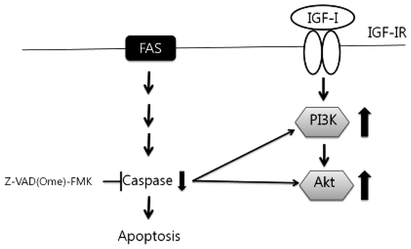
In the present study, we aimed to determine whether
laminarin induced apoptosis by molecular mechanisms involving
IGF-IR and cell death pathways. We examined the manner in which
laminarin regulates HT-29 cells, and assessed its effect on the Fas
and IGF-IR signaling pathways. The results showed that activation
of Fas-induced apoptosis blocks the IGF-IR pathway.
Materials and methods
Cell culture
Human colon adenocarcinoma cells (ATCC HTB-38) and
rat small intestine epithelial cells (IEC-6, ATCC CRL-1592) were
obtained from the American Type Culture Collection (Rockville, MD,
USA). Cells were maintained in a humidified 5% CO2, 95%
air, 37°C environment in RPMI-1640. DMEM was supplemented with
penicillin/streptomycin (P/S), and HT-29 and IEC-6 cell cultures
were supplemented with 10% fetal bovine serum (HyClone, Inc., South
Logan, UT, USA). Cells in the exponential phase were used.
Cell viability
Laminarin (L-9634) was purchased from Sigma-Aldrich
(St. Louis, MO, USA). The effects of various laminarin
concentrations on the cell proliferation of HT-29 and IEC-6 cells
were determined colorimetrically after 24 h using the
3-(4,5-dimethylthiazol-2-yl)-5-(3-carboxymethoxyphenyl)-2-(4-sulfophenyl)-2H-tetrazolium
(MTS) assay with Cell Titer 96® AQueous One Solution
Reagent (Promega, Madison, WI, USA). Cells were seeded onto 96-well
plates at 2×10⁴ cells/well in 100 μl medium and
incubated for 24 h. Attached cells were maintained in serum-free
medium (SFM) for 12 h, followed by laminarin treatment (0-5 mg/ml)
for 24 h. Subsequently, cells were incubated with MTS solution at
37°C for 30–60 min and the absorbance of each well was measured at
490 nm using a microplate reader. The OD490 values of the control
cells were designated as 100%.
Caspase activity
Caspase activities were measured using caspase-3
substrate I (Ac-DEVD-pNA; 235400), caspase-8 substrate I
(Ac-IETD-pNA) and caspase-3 inhibitor
[Z-D(Ome)-E-(Ome)-V-D(OMe)-FMK; 368057; Calbiochem, San Diego, CA,
USA]. Cells were seeded in culture dishes and grown to 60%
confluence. These cells were treated with 50 μM caspase
inhibitor for 1 h and laminarin for 24 h, after which caspase lysis
buffer (2.5 mM HEPES, pH 7.5, 5 mM EDTA, 2 mM DTT, 0.1% CHAPS) was
added. A total of 100 μg protein/100 μl was
collected, and 2 μl of the substrate was added to the wells.
Cells were incubated with a caspase substrate in a shaking
incubator at 37°C for 4 h. The absorbance at 405 nm was then
determined using an ELISA plate reader.
Western blotting
To prepare whole-cell extracts, cells were washed
with PBS and suspended in extraction buffer (50 mM Tris-HCl, pH
7.4, 150 mM NaCl, 0.25% Na-deoxycholate, 1% NP-40, and 1 mM EGTA)
containing protease inhibitors (1 mM Na3VO4,
1 μg/ml aprotinin, 1 μg/ml pepstatin, 1 μg/ml
leupeptin, 1 mM NaF, and 1 mM PMSF) on ice. The extracts were
centrifuged at 12,000 rpm for 10 min and the supernatant was used
in western blotting. Boiling sample buffer (50 μg/ml) was
added to the total cell lysate and the samples were boiled for 10
min at 100°C. Proteins were separated in 7.5–15% SDS-PAGE gels and
transferred to PVDF membranes (Millipore, Billerica, MA, USA).
Membranes were blocked for 1 h at room temperature in blocking
buffer [1% bovine serum albumin (BSA) in TBS-T]. Blots were probed
with primary antibodies (1:1,000 in 1% BSA/TBS-T) for 18 h at 4°C.
The membranes were then washed twice for 15 min in TBS-T. The
secondary antibody was a horseradish peroxidase (HRP)-conjugated
goat anti-mouse or rabbit antibody (1:10,000 in 1% BSA/TBS-T).
Signal bands were detected using an enhanced chemiluminescence
western blotting kit (Amersham Biosciences, Piscataway, NJ,
USA).
Statistical analysis
Multiple mean values were compared by analysis of
variance using the SPSS software (SPSS, Inc., Chicago, IL, USA).
Values were presented as the means ± standard deviation. P<0.05
was considered statistically significant. Values in Fig. 3, indicated with the letters a-d
were significantly different according to the Duncan’s multiple
range test.
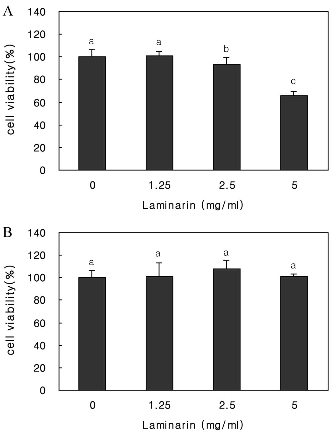
Results
Laminarin reduces the proliferation of
HT-29 cells
We determined the effect of 24-h laminarin treatment
(0, 1.25, 2.5 and 5 mg/ml) on the viability of HT-29 and IEC-6
cells by MTS assay (Fig. 1).
Laminarin treatment decreased the proliferation of HT-29 cells in a
dose-dependent manner. Exposure to 5 mg/ml laminarin inhibited cell
growth by 60%. By contrast, IEC-6 cells were unaffected. Moreover,
no toxicity to either cell type was detected.
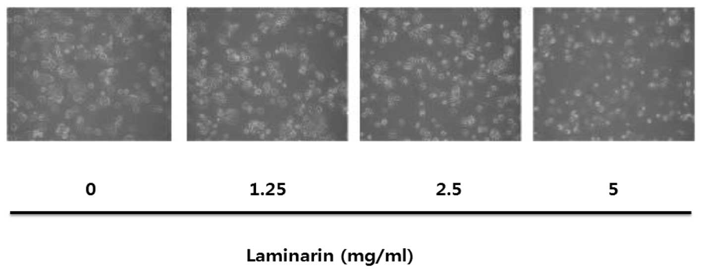
Laminarin induces morphological changes
of cells
The effect of laminarin on cell and nuclear
morphology was determined using an MTS assay and light microscopy
(Fig. 2). The survival of HT-29
cells was reduced in a laminarin concentration-dependent manner.
Cells also decreased in size in a laminarin concentration-dependent
manner.
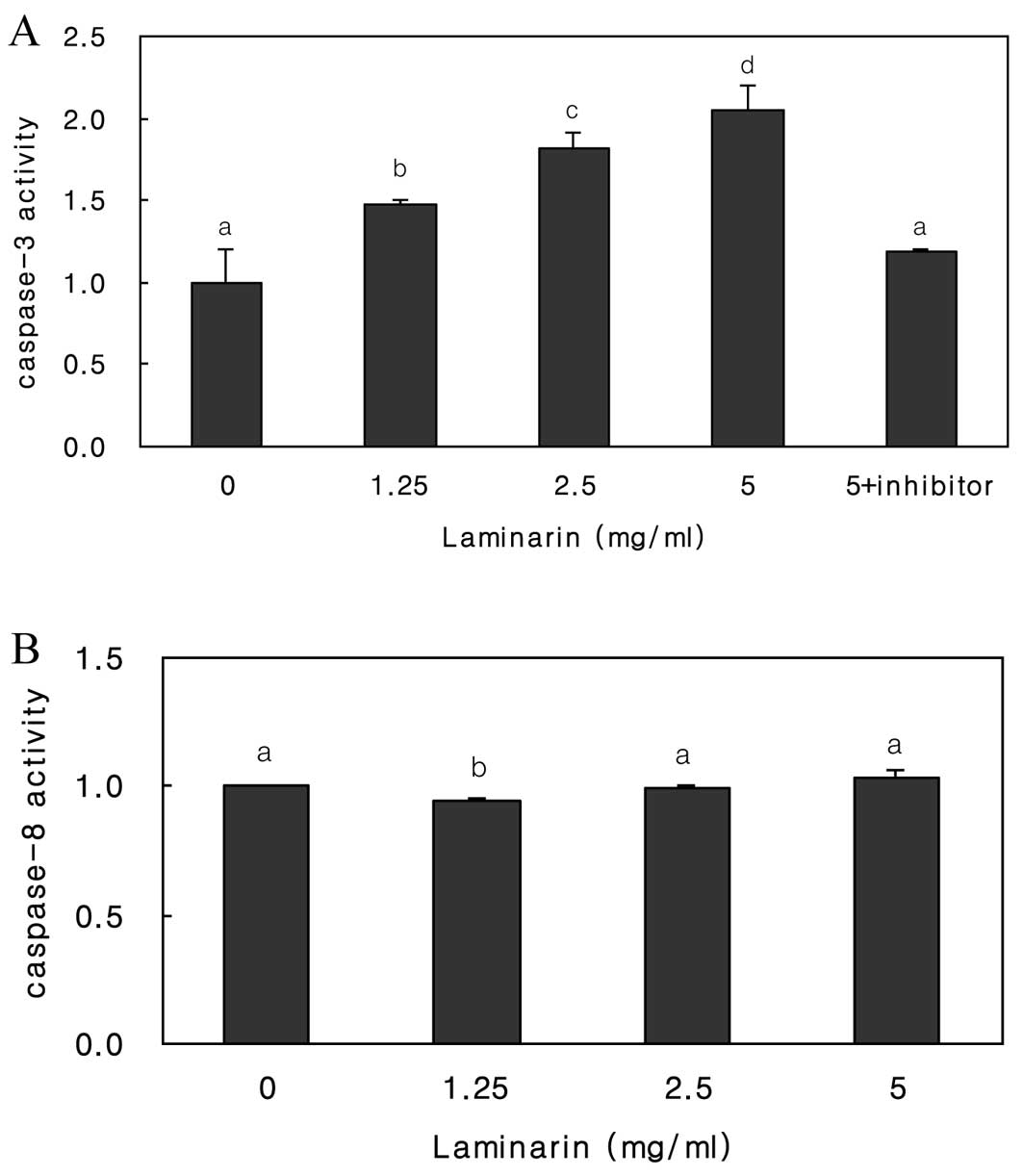
Laminarin-induced apoptosis is mediated
by caspase-3
To determine which caspases are activated by
laminarin, we identified laminarin-induced enzyme activities
(Fig. 3). A significant increase
was found in the level of caspase-3, but not caspase-8. We examined
caspase-3 activation after laminarin treatment in the presence of a
caspase-3 inhibitor. The caspase-3 inhibitor completely blocked
caspase-3 activity, suggesting that laminarin activates caspase-3,
but not caspase-8.
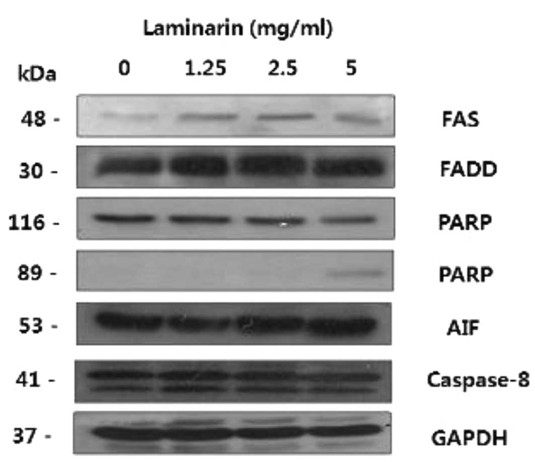
Laminarin induces the expression of
apoptosis-related proteins
A wide variety of signaling molecules are combined
with cell-surface receptors. Fas (CD95, APO-1), a member of the
tumor necrosis factor family, is a cell death receptor that plays a
key role in the regulation of homeostasis (21).
Fas and the Fas receptor induce the activation of
members of the caspase family, and subsequently the cleavage of
markers of apoptosis such as poly (ADP-ribose) polymerase (PARP)
(22). This signaling cascade is
known as the Fas signaling pathway. Following laminarin treatment,
an increase was observed in the expression of FAS and FADD
(Fig. 4). We previously reported
that laminarin treatment caused caspase-3 activation and PARP
cleavage. These results suggest that laminarin induced apoptosis
via the Fas signaling pathway.
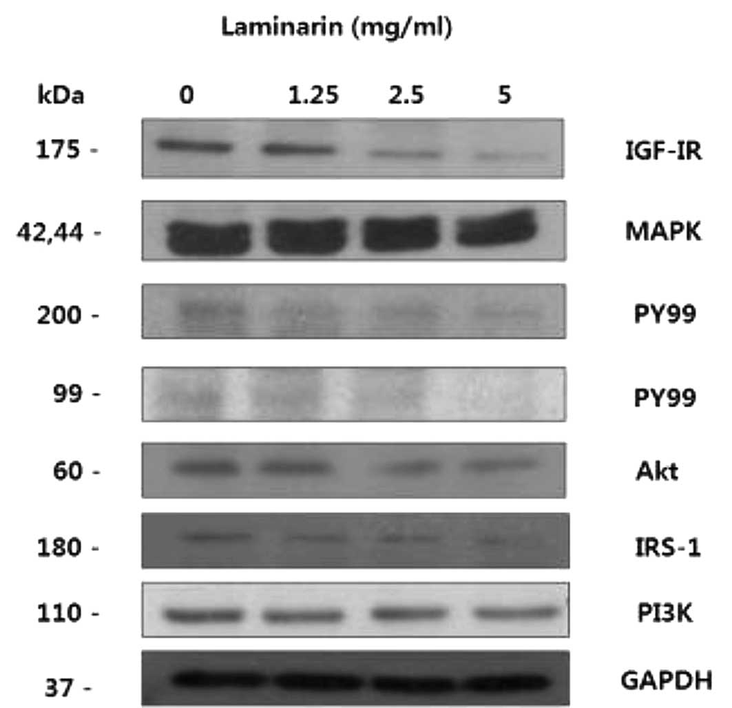
Laminarin induces the expression of
IGF-IR signaling pathway-related proteins
Laminarin induced apoptosis via the Fas signaling
pathway. Cell death signaling mechanisms and cell growth were also
affected by laminarin. The growth-inhibitory effect of laminarin
was associated with changes in the expression of proteins involved
in the IGF-IR pathway in HT-29 cells (Fig. 5). Signaling pathways activated by
IGF-IR include the mitogen-activated protein kinases (MAPK) and
phosphatidylinositol 3-kinase (PI3K) pathways (23). This signaling is controlled by the
IGF-binding protein (IGFBP). A decreased expression of the IGF-IR
and downstream signaling proteins such as PI3K, PY99, Akt and MAPK
IRS-1 inhibits events in cancer. These results suggest that
laminarin may inhibit cancer development by regulating the IGF-IR
pathway.
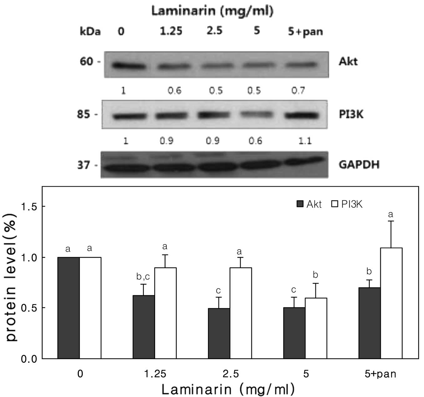
IGF-IR proteins are inhibited by
Fas-mediated caspase activation
We demonstrated that laminarin induces apoptosis in
a Fas-mediated manner. In addition, we found that laminarin
downregulates IGF-IR-related proteins.
Therefore, we determined whether Fas-induced
apoptosis blocks the IGF-IR pathway. In HT-29 cells, pancaspase
inhibitor treatment suppressed caspase activation. Results of the
western blot analysis showed that inhibitor treatment resulted in
decreased caspase-3 levels (data not shown) and the recruitment of
PI3K and Akt (Fig. 6). Thus, the
IGF-I pathway is involved in laminarin-induced apoptosis.
Discussion
The anticancer effect of seaweed has been the focus
of many recent studies. Seaweed contains large amounts (∼40% of the
dry matter) of polysaccharides, primarily laminarin, fucoidan, and
alginates. In the present study, we found that laminarin inhibits
HT-29 cell growth by decreasing cell proliferation and inducing
apoptosis.
To the best of our knowledge, we have provided the
first evidence that laminarin regulates apoptosis and the
IGF-IR-related protein expression. When HT-29 cells were incubated
with laminarin, cell viability was decreased. HT-29 cells treated
with laminarin exhibited morphological changes; cells decreased in
size in a laminarin concentration-dependent manner.
FasL and its receptor FADD are adapter molecules
required for Fas-mediated apoptosis (24,25). Laminarin regulated Fas and FADD
protein levels, suggesting that it induces Fas-mediated apoptosis.
It also increased the expression of Fas and FADD, which in turn
induced the activation of members of the caspase family (26,27). Caspases play a key role in cell
death-related apoptosis. We analyzed caspase activation during
laminarin-induced apoptosis using caspase substrates. In HT-29
cells, we detected a significant increase in the level of
caspase-3, but not caspase-8. Caspase-8 is an initial caspase in
apoptosis and is essential to the Fas-mediated apoptosis pathway
(28,29). Previous reports have shown that
caspase-8 may induce apoptosis independent of Fas. In their study,
Feng et al (30) reported
that Fas-FADD oligomerization is able to trigger a novel
caspase-8-independent pathway.
IGF-I signaling plays a role in cancer development
and progression (31,32). Remacle-Bonnet et al
(33) showed that IGF-I protected
cancer cells against apoptosis. The mechanisms by which IGF-I and
IGF-I receptors interact with cell death pathways remain unclear.
Therefore, it is important to elucidate the relationships between
IGF-I and IGF-I receptors and apoptotic pathways.
We examined the effect of laminarin on the IGF-IR
pathway. A decreased expression of IGF-IR and downstream signaling
proteins inhibits events in cancer. These results suggest that
laminarin inhibits cancer by regulating the IGF-IR pathway. As
shown in Figs. 6 and 7, a pancaspase inhibitor suppressed
caspase activation in HT-29 cells. This inhibition affected the
expression of IGF-I receptor pathway-related proteins. Therefore,
we demonstrated that the activation of Fas-induced apoptosis blocks
the IGF-IR pathway.
These data suggest that laminarin has the potential
to be used as an anticancer agent. Recently, studies have reported
anticancer effects of seaweeds (34,35). However, to the best of our
knowledge, this is the first report of laminarin activity against
human colon cancer cells. Therefore, the regulation of these two
pathways may be important for the treatment of human colon cancer
and serve as a novel target of anticancer supplements and
drugs.
Acknowledgements
This research was supported by iPET
(Korea Institute of Planning and Evaluation for Technology in Food,
Agriculture, Forestry and Fisheries), Ministry for Food,
Agriculture, Forestry and Fisheries, Republic of Korea.
References
|
1.
|
R FullerProbiotics in man and animalsJ
Appl
Bacteriol66365378198910.1111/j.1365-2672.1989.tb05105.x2666378
|
|
2.
|
GR GibsonR FullerAspects of in
vitro and in vivo research approaches directed toward
identifying probiotics and prebiotics for human useJ Nutr130Suppl
2391S395S200010721913
|
|
3.
|
K NisizawaT YamaguchiN HandaM MaedaH
YamazakiChemical nature of a uronic acid-containing polysaccharide
in the peritrophic membrane of the silkwormJ
Biochem54419426196314089735
|
|
4.
|
C MichelM LahayeC BonnetS MabeauJL BarryIn
vitro fermentation by human faecal bacteria of total and purified
dietary fibres from brown seaweedsBr J
Nutr75263280199610.1079/BJN199601298785203
|
|
5.
|
DL WilliamsOverview of (1→3)-beta-D-glucan
immunobiologyMediators Inflamm62472501997
|
|
6.
|
NK CheungS ModakA VickersB KnucklesOrally
administered beta-glucans enhance anti-tumor effects of monoclonal
antibodiesCancer Immunol Immunother51557564200212384807
|
|
7.
|
DL WilliamsA MuellerW BrowderGlucan-based
macrophage stimulators: a review of their anti-infective
potentialClin Immunother5392399199810.1007/BF03259335
|
|
8.
|
EJ KimYJ LeeHK ShinJHY ParkA study on the
mechanism by which the aqueous extract of Inonotus obliquus
induces apoptosis and inhibits proliferation in HT-29 human colon
cancer cellsJ Korean Soc Food Sci Nutr355165232006
|
|
9.
|
N KhanVM AdhamiH MukhtarApoptosis by
dietary agents for prevention and treatment of cancerBiochem
Pharmacol7613331339200810.1016/j.bcp.2008.07.01518692026
|
|
10.
|
C BurzI Berindan-NeagoeO BalacescuA
IrimieApoptosis in cancer: key molecular signaling pathways and
therapy targetsActa
Oncol48811821200910.1080/0284186090297417519513886
|
|
11.
|
D BrennerTW MakMitochondrial cell death
effectorsCurr Opin Cell
Biol21871877200910.1016/j.ceb.2009.09.004
|
|
12.
|
SY JeongDW SeolThe role of mitochondria in
apoptosisBMB Rep411122200810.5483/BMBRep.2008.41.1.011
|
|
13.
|
G MellierS HuangK ShenoyS PervaizTRAILing
death in cancerMol Aspects
Med3193112201010.1016/j.mam.2009.12.002
|
|
14.
|
S HodgeFJ NovembreL WhetterHA GelbardS
DewhurstInduction of fas ligand expression by an acutely lethal
simian immunodeficiency virus,
SIVsmmPBj14Virology252354363199810.1006/viro.1998.94779878614
|
|
15.
|
PR WalkerP SaasPY DietrichTumor expression
of Fas ligand (CD95L) and the consequencesCurr Opin
Immunol10564572199810.1016/S0952-7915(98)80225-29794830
|
|
16.
|
RA FreibergDM SpencerKA ChoatePD PengSL
SchreiberGR CrabtreePA KhavariSpecific triggering of the Fas signal
transduction pathway in normal human keratinocytesJ Biol
Chem2713166631669199610.1074/jbc.271.49.316668940187
|
|
17.
|
AA ButlerS YakarIH GewolbM KarasY OkuboD
LeRoithInsulin-like growth factor-I receptor signal transduction:
at the interface between physiology and cell biologyComp Biochem
Physiol B Biochem Mol
Biol1211926199810.1016/S0305-0491(98)10106-29972281
|
|
18.
|
M RubiniA HongoC D’AmbrosioR BasergaThe
IGF-1 receptor in mitogenesis and transformation of mouse embryo
cells: role of receptor numberExp Cell
Res230284292199710.1006/excr.1996.34309024787
|
|
19.
|
K ReissB ValentinisX TuSQ XuR
BasergaMolecular markers of IGF-I-mediated mitogenesisExp Cell
Res242361372199810.1006/excr.1998.41139665833
|
|
20.
|
AA ButlerVA BlakesleyV PoulakiM TsokosTL
WoodD LeRoithStimulation of tumor growth by recombinant human
insulin-like growth factor I (IGF-I) is dependent on the dose and
the level of IGF-I receptor expressionCancer
Res583021302719989679966
|
|
21.
|
DL VauxSJ KorsmeyerCell death in
developmentCell96245254199910.1016/S0092-8674(00)80564-4
|
|
22.
|
M EnariRV TalanianWW WongS
NagataSequential activation of ICE-like and CPP32-like proteases
during Fas-mediated
apoptosisNature380723726199610.1038/380723a08614469
|
|
23.
|
J DupontD LeRoithInsulin and insulin-like
growth factor I receptors: similarities and differences in signal
transductionHorm Res55Suppl
2S22S26200110.1159/00006346911684871
|
|
24.
|
AM ChinnaiyanK O’RourkeM TewariVM
DixitFADD, a novel death domain-containing protein, interacts with
the death domain of Fas and initiates
apoptosisCell81505512199510.1016/0092-8674(95)90071-37538907
|
|
25.
|
S NagataP GolsteinThe Fas death
factorScience26714491456199510.1126/science.7533326
|
|
26.
|
FC KischkelS HellbardtI BehrmannM GermerM
PawlitaPH KrammerME PeterCytotoxicity-dependent APO-1
(Fas/CD95)-associated proteins form a death-inducing signaling
complex (DISC) with the receptorEMBO J145579558819958521815
|
|
27.
|
GS SalvesenVM DixitCaspases: intracellular
signaling by
proteolysisCell91443446199710.1016/S0092-8674(00)80430-49390553
|
|
28.
|
EE VarfolomeevM SchuchmannV LuriaN
ChiannilkulchaiJS BeckmannIL MettD RebrikovVM BrodianskiOC KemperO
KolletTargeted disruption of the mouse caspase 8 gene ablates cell
death induction by the TNF receptors, Fas/Apo1, and DR3 and is
lethal
prenatallyImmunity9267276199810.1016/S1074-7613(00)80609-39729047
|
|
29.
|
P JuoCJ KuoJ YuanJ BlenisEssential
requirement for caspase-8/FLICE in the initiation of the
Fas-induced apoptotic cascadeCurr
Biol810011008199810.1016/S0960-9822(07)00420-49740801
|
|
30.
|
H FengY ZengMW GranerL WhitesellE
KatsanisEvidence for a novel, caspase-8-independent, Fas death
domain-mediated apoptotic pathwayJ Biomed
Biotechnol20044151200410.1155/S111072430430804115123887
|
|
31.
|
R BasergaF PeruzziK ReissThe IGF-1
receptor in cancer biologyInt J
Cancer107873877200310.1002/ijc.1148714601044
|
|
32.
|
H WernerD LeRoithNew concepts in
regulation and function of the insulin-like growth factors:
implications for understanding normal growth and neoplasiaCell Mol
Life Sci57932942200010.1007/PL0000073510950308
|
|
33.
|
MM Remacle-BonnetFL GarrousteS HellerF
AndreJL MarvaldiGJ PommierInsulin-like growth factor-I protects
colon cancer cells from death factor-induced apoptosis by
potentiating tumor necrosis factor alpha-induced mitogen-activated
protein kinase and nuclear factor kappaB signaling pathwaysCancer
Res60200720172000
|
|
34.
|
S ErmakovaR SokolovaSM KimBH UmV IsakovT
ZvyagintsevaFucoidans from brown seaweeds Sargassum hornery,
Eclonia cava, Costaria costata: structural characteristics and
anticancer activityAppl Biochem Biotechnol1648418502011
|
|
35.
|
F NamvarS MohamedSG FardJ BehravanNM
MustaphaNB AlitheenF OthmanPolyphenol-rich seaweed (Eucheuma
cottonii) extract suppresses breast tumour via hormone
modulation and apoptosis inductionFood Chemistry1303763822012
|