Introduction
Foamy viruses (FVs), which comprise the
Spumaretrovirinae in the retrovirus family, are also known
as spumaretroviruses. FVs are found in primates, including humans,
as well as in non-primates, including cows, cats and horses
(1–5).
The prototype foamy virus (PFV) Tas protein, also
known as Bel1, is a 300-amino-acid nuclear protein that is
essential for virus replication (6), and can highly transactivate the PFV
promoters, LTR and IP (7–9). Similar to most typical
transcriptional activators, nuclear localization is required for
the transactivation activity of Bel1 (10). Bel1 bears a putative nuclear
localization signal (NLS) in the central highly basic region
(11,12). Earlier studies have indicated that
peptide 211-225 and/or 209-226 are necessary and sufficient for
Bel1 nuclear localization (13–15). Later studies demonstrated that
another two basic amino acids, R199H200, also
regulate Bel1 nuclear localization, which suggests that Bel1
carries a bipartite NLS consisting of residues 199-200 and residues
211-223 (10,16). However, Ma et al further
found that residues R221R222R223,
but not R199H200, are essential for the
nuclear distribution of Bel1 (17).
Importin is a type of karyopherin (18) that transports protein molecules
into the nucleus by binding to nuclear localization sequences.
Importin has two subunits, karyopherin alpha (KPNA; also known as
importin alpha) and karyopherin beta KPNB (also known as importin
beta). Members of the KPNB family can bind and transport cargoes by
themselves (19–21), or can form heterodimers with KPNA
(22,23). As part of a heterodimer, KPNB
mediates the interaction with nuclear pore complex (NPC), while
KPNA acts as an adaptor protein to bind KPNB and the NLS on the
cargo (24). The NLS-KPNA-KPNB
trimer dissociates after binding to RanGTP inside the nucleus
(25), with the two importin
proteins being recycled to the cytoplasm for further use. Although
KPNA and KPNB are used to describe importin as a whole, they
actually represent larger families of proteins that share a similar
structure and function. A variety of genes have been identified for
both KPNA and KPNB, such as KPNA1-KPNA7 and
KPNB1 (26). Different
KPNA members show preferences for particular types of NLS cargo,
although there is no absolute boundary (26,27).
In this study, we aimed to determine which adaptor
importins are required for Bel1 nuclear translocation. We found
that the 215PRQKRPR221 fragment, which
accords with the consensus sequence K(K/R)X(K/R) of monopartite
NLS, directs the nuclear localization of Bel1. Point mutation
experiments revealed that residues K218, R219
and R221 were essential for the nuclear accumulation of
Bel1. The results of GST pull-down assay revealed that the Bel1 NLS
fragment 215-221 interacted with KPNA1, KPNA6 and KPNA7. Finally,
in vitro nuclear import assays demonstrated that KPNA1,
KPNA6 and KPNA7 caused Bel1 to localize to the nucleus. Our
findings thus indicate that KPNA1, KPNA6 and KPNA7 are involved in
Bel1 nuclear translocation.
Materials and methods
Plasmids
The Bel1 gene was amplified from the PFV
full-length infectious clone, pCHFV, kindly provided by Maxine L.
Linial (28). The mammalian cell
expression plasmids, pC3-EGFP-X-GST, pC3-EGFP-NLS-GST, pC3-EGFP-
BiNLS-GST, pC3-EGFP-Bel1-GST, pC3-EGFP-215-221)-GST and other
truncated Bel1 plasmids were generated as previously described
(17). The Bel1 mutants K218R,
K218A, R219A and R221A were generated using a QuikChange™
site-directed mutagenesis kit (Stratagene, Palo Alto, CA, USA)
using the primers listed in Table
I. The coding sequences of KPNA1-KPNA7 and KPNB1 were amplified
from the HeLa cDNA library by RT-PCR with the primers listed in
Table I and inserted into the
pCMV-Tag 2B vector (Stratagene) or the pFLAG-CMV-4 vector
(Sigma-Aldrich, St. Louis, MO, USA) to express the corresponding
proteins. All the new constructs were confirmed by DNA
sequencing.
 | Table IPrimers used for PCR or site-directed
mutagenesis PCR or RT-PCR. |
Table I
Primers used for PCR or site-directed
mutagenesis PCR or RT-PCR.
| Gene | Sense primer
sequences (5′→3′) | Antisense primer
sequences (5′→3′) |
|---|
| Bel1 |
TTAGAGCTCATGGATTCCTACGAAAAAG |
CCGAAGCTTTAAAACTGAATGTTCACCT |
| K218R |
GCCTCGGCAGAGACGACCCAGGAGA |
TCTCCTGGGTCGTCTCTGCCGAGGC |
| K218A |
CCTCGGCAGGCACGACCCAGGAGA |
TCTCCTGGGTCGTGCCTGCCGAGG |
| R219A |
CCTCGGCAGAAAGCACCCAGGAGACG |
CGTCTCCTGGGTGCTTTCTGCCGAGG |
| R221A |
GAAACGACCCGCGAGACGATCCATC |
GATGGATCGTCTCGCGGGTCGTTTC |
| KPNA1 |
TTAGGATCCATGACCACCCCAGGAAAAG |
TAGCTCGAGTCAAAGCTGGAAACCT |
| KPNA2 |
CTCGAATTCATGTCCACCAACGAGAAT |
TCACTCGAGCTAAAAGTTAAAGGTCCC |
| KPNA3 |
ATAGAATTCATGGCCGAGAACCCCAGC |
GCGCTCGAGTTTTGTTTGAAGGTTGGC |
| KPNA4 |
TTAGGATCCATGGCGGACAACGAGAAAC |
GCTCTCGAGCTAAAACTGGAACCCTTCT |
| KPNA5 |
GCGGAATTCATGGATGCCATGGCTAGT |
GCGCTCGAGTTGAAATCCATCCATTGG |
| KPNA6 |
ATCGAATTCATGGAGACCATGGCGAGC |
TATCTCGAGTAGCTGGAAGCCCTCCAT |
| KPNA7 |
GTCGAATTCATGCCGACCTTAGATGCT |
CGCCTCGAGTGCTAAGCATTCATAATC |
| KPNB1 |
ATAGCGGCCGCAATGGAGCTGATCACCAT |
CCTGGATCCTCAAGCTTGGTTCTTCAG |
Cell culture and transfection, antibodies
and reagents
HeLa and 293T cells (both from the Cell Bank of the
Chinese Academy of Sciences, Shanghai, China) were grown in
Dulbecco's modified Eagle's medium (high glucose; HyClone, Logan,
UT, USA) supplemented with 10% fetal bovine serum (Gibco-BRL, Grand
Island, NY, USA) and Pen Strep Glutamine (PSG) (Gibco). The cells
were maintained in a humidified atmosphere containing 5%
CO2 at 37°C and transfected with polyethylenimines (PEI)
(Polysciences, Inc., Warrington, PA, USA) in accordance with the
manufacturer's instructions.
Anti-EGFP (sc-9996), anti-GAPDH (sc-32233), anti-GST
(sc-138) and HRP-conjugated goat anti-mouse secondary antibodies
(sc-2005) were purchased from Santa Cruz Biotechnology, Inc. (Santa
Cruz, CA, USA). Anti-Flag (F3165) antibody and
4′,6-diamidino-2-phenylindole (DAPI) were purchased from
Sigma-Aldrich. FITC-conjugated affinipure goat anti-mouse secondary
antibodies (115-095-003) were purchased from Jackson ImmunoResearch
Laboratories, Inc. (West Grove, PA, USA).
Immunofluorescence microscopy assay
(IFA)
The HeLa cells were seeded on glass coverslips.
Following fixation in 4% paraformaldehyde for 10 min on ice, the
cells were permeabilized in 0.2% Triton X-100 for 10 min on ice.
After blocking in 3% BSA + 5% fat-free milk at 4°C for 2 h, the
cells were incubated with anti-EGFP antibodies at 4°C for a further
2 h, and subsequently washed with 0.1% Triton X-100 in PBS 5 times
at room temperature. FITC-conjugated secondary antibodies were
added at 4°C for 45 min. After the nuclei were stained with 0.2
µg/ml DAPI for 10 min at room temperature, the coverslips
were observed under an Olympus IX71 fluorescence microscope
(Olympus, Tokyo, Japan).
In vivo GST pull-down assay and western
blot analysis
The 293T cells were transfected with the
pC3-EGFP-X-GST empty vector or pC3-EGFP-Bel1-GST along with
plasmids that encode Flag-KPNAs or Flag-KPNB1; at 48 h
post-transfection, the cell lysates were incubated with Glutathione
Sepharose 4B beads (20182003-2) (GE Healthcare, Cleveland, OH, USA)
for 4 h at 4°C. Samples from both cell lysates and Glutathione
Sepharose 4B beads were mixed with loading buffer. After boiling
for 20 min at 100°C, the protein samples were resolved by SDS-PAGE
and transferred onto PVDF membranes. Prior to incubation first with
primary antibodies overnight at 4°C and then with HRP-conjugated
secondary antibodies for 1 h at room temperature, the membranes
were blocked in 5% fat-free milk for 1.5 h at room temperature.
After the membranes were treated with Luminata™ Western HRP
chemiluminescence substrates (WBLUC0100; Millipore, Billerica, MA,
USA), the specific protein signals were detected by exposure to
X-ray films (Kodak, Xiamen, China).
In vitro nuclear import assay
In vitro nuclear transport assays were
carried out as previously described with some modifications
(29,30). Briefly, the HeLa cells (70–80%
confluent), plated on glass coverslips, were washed 3 times with
ice-cold transport buffer (TB) and permeabilized with digitonin (40
mg/ml) for 5 min on ice. The cells were then washed twice with
ice-cold TB and soaked in TB for 10 min on ice. The complete
transport solution contained import substrates (~2 µM), an
adenosine triphosphate (ATP)-regenerating system (1 mM ATP, 5 mM
creatine phosphate and 20 U/ml creatine phosphokinase) as a source
of energy and some other soluble import factors. The import
reaction was performed for 30 min at 37°C or on ice in a humidified
chamber. After the transport reaction, the cells were washed twice
with ice-cold TB followed by fixation with 4% paraformaldehyde for
10 min on ice. The cells were washed 3 times first with TB and then
twice with PBS. Following permeabilization with 0.2% Triton X-100
in PBS for 5 min on ice, the cells were blocked with 3% BSA + 5%
fat-free milk in PBS and incubated with anti-EGFP antibodies and
FITC-conjugated secondary antibodies as mentioned above. After
being mounted on slides in PBS containing DAPI for 10 min on ice,
the cells were visualized using an Olympus IX71 fluorescence
microscope (Olympus).
Results
The NLS of Bel1 is
215PRQKRPR221
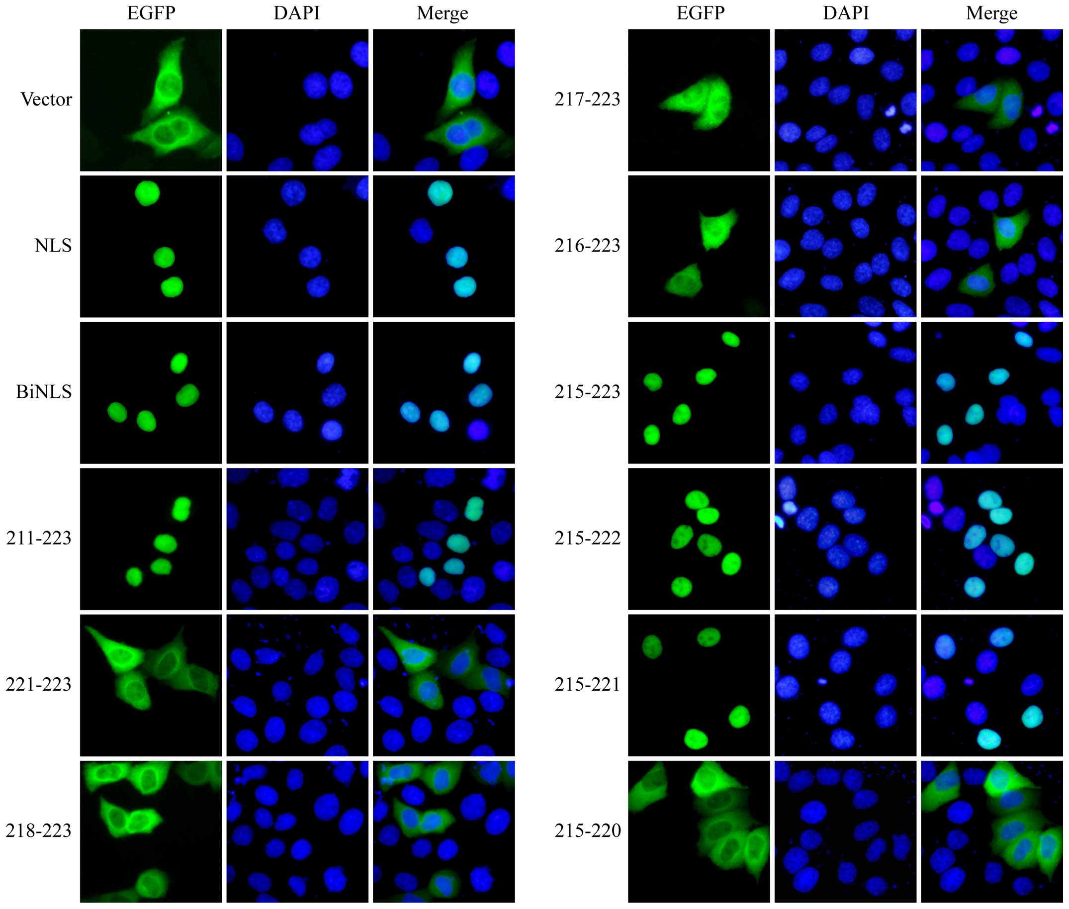
In order to accurately determine the NLS of Bel1, we
inserted into the EGFP-GST fusion protein the truncated fragments
of Bel1 that encompass the amino acid positions 211-223, 221-223,
218-223, 217-223, 216-223, 215-223, 215-222, 215-221 and 215-220
(Fig. 1) and observed their
subcellular distribution by performing indirect IFA. The
monopartite NLS of SV40 large T antigen (NLS) and the bipartite NLS
of Xenopus laevis nucleoplasmin (BiNLS) were also inserted
into EGFP-GST as positive controls for nuclear localization. As
illustrated in Fig. 2, similar to
the activity of SV40-NLS and the BiNLS, the 211-223 peptide of Bel1
enabled the nuclear localization of the fusion protein. In view of
the fact that residues
R221R222R223 are necessary for
Bel1 nuclear distribution (10,13–17), we extended the N-terminal of the
peptide segment to observe the effects. As shown in Fig. 2, the 221-223, 218-223, 217-223 and
216-223 fusion proteins still mainly distributed in the cytoplasm
with little nuclear distribution, although containing the residues
R221R222R223. Until the N-terminal
extended to residue P215, the fusion protein 215-223 localized to
the nucleus (Fig. 2). We then
shortened the C-terminal of 215-223 to continue our observation. As
shown in Fig. 2, the both
(215-222)- and (215-221)-containing EGFP-GST fusion proteins were
distributed in the nucleus, whereas 215-220 was distributed in the
cytoplasm. Taken together, these data suggest that peptide
215PRQKRPR221 is the NLS of Bel1 and is
essential for nuclear distribution.
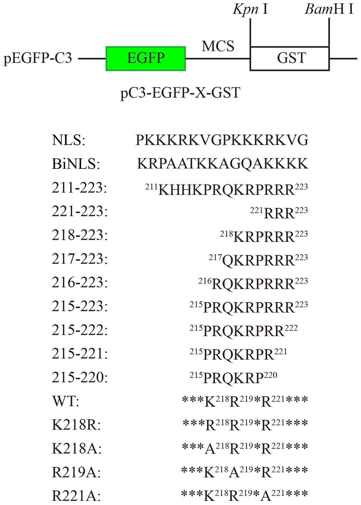 | Figure 1Schematic illustration of the
pC3-EGFP-X-GST vector and the inserted amino acid sequences. The
green rectangular box represents EGFP DNA; the blank rectangular
box represents GST DNA; black lines represent pEGFP-C3 vector DNA.
The name of the plasmid is shown underneath or on the left. MCS,
multiple cloning sites; NLS, SV40 T antigen monopartite NLS; BiNLS,
Xenopus laevis nucleoplasmin bipartite NLS; 211–223,
221–223, 218–223, 217–223, 216–223, 215–223, 215–222, 215–221 and
215–220: truncated Bel1, which are inserted at the MCS site; WT,
wild-type Bel1; K218R, K218A, R219A and R221A: Bel1 mutants.
Capitalized letters represent amino acid sequences; the numbers
denote the amino acid position in Bel1 protein; asterisks indicate
Bel1 amino acids. |
The NLS of Bel1 is monopartite
Sequence analysis indicated that
215PRQKRPR221 accords with the consensus
sequence K(K/R) X(K/R) of monopartite NLS, comprised primarily of
lysine (K) and arginine (R) residues (31), wherein the basic amino acids are
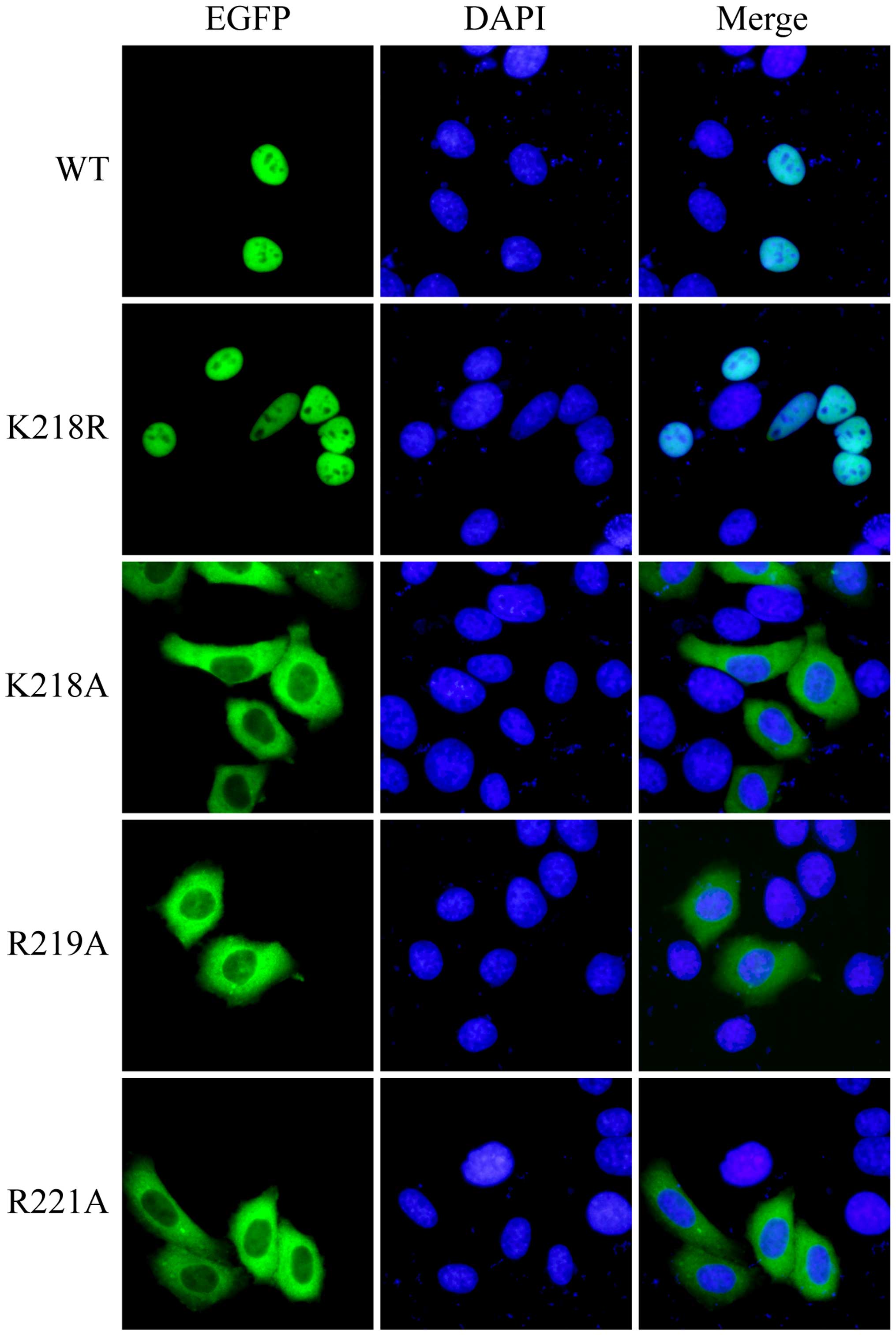
critical. To confirm this result, we generated four mutations of
the Bel1 protein sequence, named K218R that changed K218
to R218, K218A that turned K218 into
A218, R219A that changed R219 to
A219 and R221A that altered R221 to
A221, and then examined the subcellular distribution. As
shown in Fig. 3, the K218R mutant
and wild-type Bel1 (WT) were strictly localized to the nucleus in
contrast to the K218A, R219A and R221A mutants that were detected
predominantly in the cytoplasm. These results evidently prove that
the nuclear localization sequence
215PRQKRPR221 of Bel1 is monopartite and that
residues K218, R219 and R221 of
Bel1 are essential for its nuclear accumulation.
Bel1 interacts with KPNA1, KPNA2, KPNA6
and KPNA7
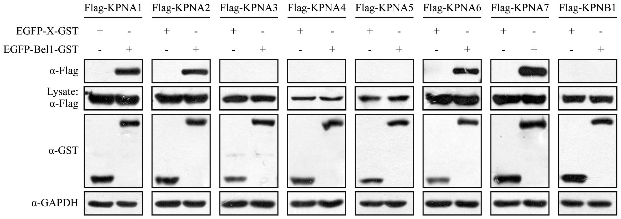
In the conventional nuclear transport pathway,
cargoes are recognized and bound by the transport receptor adaptor,
importin alpha, to translocate to the nucleus (32,33). In this study, in order to
determine which importins mediate the transportation of Bel1 into
the nucleus, we detected the interaction between Bel1 and 7
isoforms of importin alpha (KPNA1, KPNA2, KPNA3, KPNA4, KPNA5,
KPNA6 and KPNA7) and the most common importin beta protein, KPNB1,
in 293T cells by in vivo GST pull-down assay. The results of
western blot analysis revealed that Bel1 interacted with KPNA1,
KPNA2, KPNA6 and KPNA7 solidly, as opposed to other isoforms of
importin alpha or KPNB1 (Fig. 4).
This suggests that Bel1 may use KPNA1, KPNA2, KPNA6 and KPNA7 to
enter the nucleus.
The NLS peptide of Bel1 interacts with
KPNA1, KPNA6 and KPNA7 separately
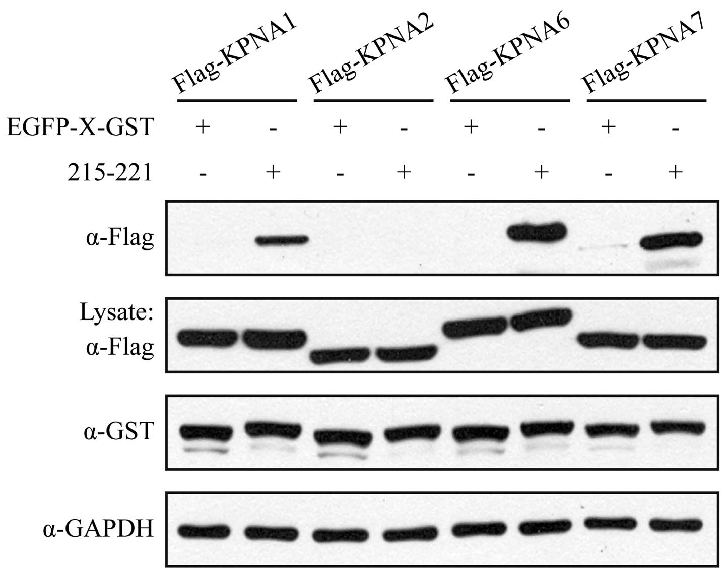
Classical NLSs (cNLS) are directly recognized and
bound by the adaptor protein importin alpha (34–36). To confirm this, we then determined
the interrelation between truncated mutant 215-221, the NLS peptide
of Bel1, and KPNA1, KPNA6 and KPNA7 in 293T cells by in vivo
GST pull-down assay. As shown in Fig.
5, although KPNA2 bound to WT Bel1 (Fig. 4), truncated 215-221 did not
interact with KPNA2. In accordance with the above findings, KPNA1,
KPNA6 and KPNA7 bound solidly to the NLS sequence 215-221 of Bel1.
These results further confirm that these three nuclear-import
receptors are involved in the translocation of Bel1 into the
nucleus.
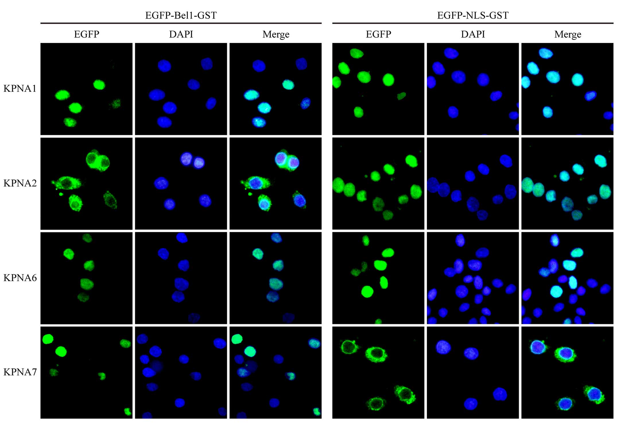
KPNA1, KPNA6 and KPNA7 mediate the
nuclear import of Bel1
To determine whether KPNA1, KPNA6 or KPNA7 can
indeed mediate the nuclear import of Bel1, we finally carried out
an in vitro nuclear import assay, but replaced the cytosol
with recombinant transport receptors (Fig. 6). As a control for active
transport, the SV40-NLS-containing EGFP-NLS-GST fusion protein was
included in the same experiment. In contrast to KPNA2, KPNA1, KPNA6
and KPNA7 were sufficient for the nuclear accumulation of Bel1
(Fig. 6). The subcellular
distribution of the positive control EGFP-NLS-GST was consistent
with that previously reported (26,27): KPNA1, KPNA2 or KPNA6 were able to
mediate SV40-NLS alone, while KPNA7 failed to do that. Taken
together, these findings indicate that the efficient nuclear import
of Bel1 in cells is mediated by KPNA1, KPNA6 and KPNA7 via the
importin alpha/beta transport pathway.
Discussion
As a key positive regulator of viral gene
expression, Bel1 contains a conventional NLS and is located in the
nucleus to conduct its transactivational activity (11,37). Previous studies have confirmed the
key role of R221R222R223 in Bel1
nuclear localization, yet the accurate nuclear localization
sequence is controversial and the adaptor-mediated Bel1 nuclear
transport is unclear.
In this study, with the purpose of defining the
peptide sequences that are essential for the nuclear distribution
of Bel1, we introduced an EGFP-GST fusion expression system that
has been widely utilized in studying the subcellular localization
of retrovirus transactivators (38,39). With the Bel1 shortened mutant
211-223, we finally confirmed that
215PRQKRPR221 is necessary and sufficient for
the nuclear localization of Bel1. Furthermore, we found that
residues K218, R219 and R221 of
Bel1 are indispensable for its nuclear accumulation by the results
of mutagenesis experiments. Comprehensive analysis of the consensus
sequence K(K/R)X(K/R) of monopartite NLS indicated that
218KRPR221 is the core sequence of Bel1 NLS
and the NLS of Bel1 is monopartite.
Consistent with the characteristics of the NLS
sequence of mammalian cells, the residue K218 in the
consensus sequence was replaced by the positive charge residue R,
which had no change in the subcellular distribution. This suggests
that the importance of the basic amino acid residues in the nuclear
protein is closely related to the positive charge. That is to say,
Bel1 may use a similar way to enter the nucleus as a host cell
transcription factor to complete its transactivational
function.
NLSs are categorized into cNLSs and non-classical
NLSs (ncNLS) (40). cNLSs are
characterized by either monopartite (e.g., PKKKRRV from SV40 large
T antigen) or bipartite (e.g., KRPAATKKAGQAKKKK from nucleoplasmin)
stretches of basic amino acids (41,42). There is no consensus on whether
different types of NLS have different biological functions. As RNA
virus, the genome fidelity of foamy virus is lower than that of DNA
genome. In the course of viral inheritance, monopartite NLS, less
conserved nucleic acid sequence, may have some certain evolutionary
advantages. In addition, monopartite NLS, shorter stretches of
basic amino acids, may be more conducive to efficiently use of
limited resources for virus.
Human KPNA isoforms are well conserved, with 26%
identity and 42% conservation in their amino acid sequences
(43,44). They can be divided into three
subfamilies according to phylogenetic analysis: i) the α1 subfamily
containing KPNA1, KPNA5 and KPNA6; ii) the α2 subfamily containing
KPNA2 and KPNA7; and iii) the α3 subfamily containing KPNA3 and
KPNA4. Although the α1 subfamily shares a maximum of 82.1% identity
and 82% sequence conservation (26), their affinity for Bel1 differed
markedly, which may due to the restriction of KPNA5 expression to
the testes (45), in our GST
pull-down experiments using 293T cells. The α2 subfamily is the
least conserved of the KPNA subfamilies, with 55% identity and 71%
conservation (24). In addition,
phylogenetic analysis of the ARM repeats, responsible for
identifying and combining with the NLS of cargo proteins (36), of the KPNAs shows that the KPNA7
ARM repeats is more divergent than that of KPNA2 (26). To a certain extent, this explains
the different performance of KPNA2 and KPNA7. Besides, the
combination between KPNA2 and Bel1 may be involved in the other
amino acids apart from the NLS, and/or the peptide
215PRQKRPR221 is not sufficient to mediate
the binding between the two. It is thus revealed that KPNA2 may
participate in other biological functions of Bel1 except nuclear
transport.
The present study provided evidence that KPNA1,
KPNA6 and KPNA7 may be 'hijacked' by PFV Bel1 for efficient nuclear
import and viral replication. Given this fact, the restricted
expression of KPNA isoforms may provide a mechanism for the
suppression of PFV replication and disease progression.
Acknowledgments
This study was supported by a grant from the
National Key Clinical Specialist Construction Programs of China
(grant no. 2013-544). We would like to thank Maxine L. Linial
(University of Washington and Fred Hutchinson Cancer Research
Center) for the PFV full-length infectious clone pCHFV.
References
|
1
|
Broussard SR, Comuzzie AG, Leighton KL,
Leland MM, Whitehead EM and Allan JS: Characterization of new
simian foamy viruses from African nonhuman primates. Virology.
237:349–359. 1997. View Article : Google Scholar : PubMed/NCBI
|
|
2
|
Hatama S, Otake K, Omoto S, Murase Y,
Ikemoto A, Mochizuki M, Takahashi E, Okuyama H and Fujii Y:
Isolation and sequencing of infectious clones of feline foamy virus
and a human/feline foamy virus Env chimera. J Gen Virol.
82:2999–3004. 2001. View Article : Google Scholar : PubMed/NCBI
|
|
3
|
Herchenröder O, Renne R, Loncar D, Cobb
EK, Murthy KK, Schneider J, Mergia A and Luciw PA: Isolation,
cloning, and sequencing of simian foamy viruses from chimpanzees
(SFVcpz): high homology to human foamy virus (HFV). Virology.
201:187–199. 1994. View Article : Google Scholar : PubMed/NCBI
|
|
4
|
Materniak M, Bicka L and Kuźmak J:
Isolation and partial characterization of bovine foamy virus from
Polish cattle. Pol J Vet Sci. 9:207–211. 2006.
|
|
5
|
Tobaly-Tapiero J, Bittoun P, Neves M,
Guillemin MC, Lecellier CH, Puvion-Dutilleul F, Gicquel B, Zientara
S, Giron ML, de Thé H, et al: Isolation and characterization of an
equine foamy virus. J Virol. 74:4064–4073. 2000. View Article : Google Scholar
|
|
6
|
Löchelt M, Zentgraf H and Flügel RM:
Construction of an infectious DNA clone of the full-length human
spumaretrovirus genome and mutagenesis of the bel 1 gene. Virology.
184:43–54. 1991. View Article : Google Scholar : PubMed/NCBI
|
|
7
|
He F, Blair WS, Fukushima J and Cullen BR:
The human foamy virus Bel-1 transcription factor is a
sequence-specific DNA binding protein. J Virol. 70:3902–3908.
1996.PubMed/NCBI
|
|
8
|
Kang Y, Blair WS and Cullen BR:
Identification and functional characterization of a high-affinity
Bel-1 DNA binding site located in the human foamy virus internal
promoter. J Virol. 72:504–511. 1998.PubMed/NCBI
|
|
9
|
Löchelt M, Flügel RM and Aboud M: The
human foamy virus internal promoter directs the expression of the
functional Bel 1 transactivator and Bet protein early after
infection. J Virol. 68:638–645. 1994.PubMed/NCBI
|
|
10
|
Chang J, Lee KJ, Jang KL, Lee EK, Baek GH
and Sung YC: Human foamy virus Bel1 transactivator contains a
bipartite nuclear localization determinant which is sensitive to
protein context and triple multimerization domains. J Virol.
69:801–808. 1995.PubMed/NCBI
|
|
11
|
Venkatesh LK, Theodorakis PA and
Chinnadurai G: Distinct cis-acting regions in U3 regulate
trans-activation of the human spumaretrovirus long terminal repeat
by the viral bel1 gene product. Nucleic Acids Res. 19:3661–3666.
1991. View Article : Google Scholar : PubMed/NCBI
|
|
12
|
Flügel RM: Spumaviruses: a group of
complex retroviruses. J Acquir Immune Defic Syndr. 4:739–750.
1991.PubMed/NCBI
|
|
13
|
He F, Sun JD, Garrett ED and Cullen BR:
Functional organization of the Bel-1 trans activator of human foamy
virus. J Virol. 67:1896–1904. 1993.PubMed/NCBI
|
|
14
|
Venkatesh LK and Chinnadurai G: The
carboxy-terminal transcription enhancement region of the human
spumaretrovirus transactivator contains discrete determinants of
the activator function. J Virol. 67:3868–3876. 1993.PubMed/NCBI
|
|
15
|
Venkatesh LK, Yang C, Theodorakis PA and
Chinnadurai G: Functional dissection of the human spumaretrovirus
transactivator identifies distinct classes of dominant-negative
mutants. J Virol. 67:161–169. 1993.PubMed/NCBI
|
|
16
|
Lee CW, Chang J, Lee KJ and Sung YC: The
Bel1 protein of human foamy virus contains one positive and two
negative control regions which regulate a distinct activation
domain of 30 amino acids. J Virol. 68:2708–2719. 1994.PubMed/NCBI
|
|
17
|
Ma Q, Tan J, Cui X, Luo D, Yu M, Liang C
and Qiao W: Residues R(199)H(200) of prototype foamy virus
transactivator Bel1 contribute to its binding with LTR and IP
promoters but not its nuclear localization. Virology. 449:215–223.
2014. View Article : Google Scholar : PubMed/NCBI
|
|
18
|
Görlich D, Prehn S, Laskey RA and Hartmann
E: Isolation of a protein that is essential for the first step of
nuclear protein import. Cell. 79:767–778. 1994. View Article : Google Scholar : PubMed/NCBI
|
|
19
|
van der Watt PJ, Stowell CL and Leaner VD:
The nuclear import receptor Kpnβ1 and its potential as an
anticancer therapeutic target. Crit Rev Eukaryot Gene Expr.
23:1–10. 2013. View Article : Google Scholar
|
|
20
|
Flores K and Seger R: Stimulated nuclear
import by β-like importins. F1000Prime Rep. 5:412013. View Article : Google Scholar
|
|
21
|
Zehorai E and Seger R: Beta-like importins
mediate the nuclear translocation of mitogen-activated protein
kinases. Mol Cell Biol. 34:259–270. 2014. View Article : Google Scholar :
|
|
22
|
Goldfarb DS, Corbett AH, Mason DA,
Harreman MT and Adam SA: Importin alpha: a multipurpose
nuclear-transport receptor. Trends Cell Biol. 14:505–514. 2004.
View Article : Google Scholar : PubMed/NCBI
|
|
23
|
Cimica V, Chen HC, Iyer JK and Reich NC:
Dynamics of the STAT3 transcription factor: nuclear import
dependent on Ran and importin-β1. PLoS One. 6:e201882011.
View Article : Google Scholar
|
|
24
|
Pumroy RA and Cingolani G: Diversification
of importin-α isoforms in cellular trafficking and disease states.
Biochem J. 466:13–28. 2015. View Article : Google Scholar : PubMed/NCBI
|
|
25
|
Mattaj IW and Englmeier L:
Nucleocytoplasmic transport: the soluble phase. Annu Rev Biochem.
67:265–306. 1998. View Article : Google Scholar : PubMed/NCBI
|
|
26
|
Kelley JB, Talley AM, Spencer A, Gioeli D
and Paschal BM: Karyopherin alpha7 (KPNA7), a divergent member of
the importin alpha family of nuclear import receptors. BMC Cell
Biol. 11:632010. View Article : Google Scholar : PubMed/NCBI
|
|
27
|
Köhler M, Speck C, Christiansen M,
Bischoff FR, Prehn S, Haller H, Görlich D and Hartmann E: Evidence
for distinct substrate specificities of importin alpha family
members in nuclear protein import. Mol Cell Biol. 19:7782–7791.
1999. View Article : Google Scholar : PubMed/NCBI
|
|
28
|
Life RB, Lee EG, Eastman SW and Linial ML:
Mutations in the amino terminus of foamy virus Gag disrupt
morphology and infectivity but do not target assembly. J Virol.
82:6109–6119. 2008. View Article : Google Scholar : PubMed/NCBI
|
|
29
|
Cassany A and Gerace L: Reconstitution of
nuclear import in permeabilized cells. Methods Mol Biol.
464:181–205. 2009. View Article : Google Scholar
|
|
30
|
Adam SA, Marr RS and Gerace L: Nuclear
protein import in permeabilized mammalian cells requires soluble
cytoplasmic factors. J Cell Biol. 111:807–816. 1990. View Article : Google Scholar : PubMed/NCBI
|
|
31
|
McLane LM and Corbett AH: Nuclear
localization signals and human disease. IUBMB Life. 61:697–706.
2009. View
Article : Google Scholar : PubMed/NCBI
|
|
32
|
Marfori M, Lonhienne TG, Forwood JK and
Kobe B: Structural basis of high-affinity nuclear localization
signal interactions with importin-α. Traffic. 13:532–548. 2012.
View Article : Google Scholar : PubMed/NCBI
|
|
33
|
Jans DA, Xiao CY and Lam MH: Nuclear
targeting signal recognition: a key control point in nuclear
transport? BioEssays. 22:532–544. 2000. View Article : Google Scholar : PubMed/NCBI
|
|
34
|
Stewart M: Molecular mechanism of the
nuclear protein import cycle. Nat Rev Mol Cell Biol. 8:195–208.
2007. View
Article : Google Scholar : PubMed/NCBI
|
|
35
|
Marfori M, Mynott A, Ellis JJ, Mehdi AM,
Saunders NF, Curmi PM, Forwood JK, Bodén M and Kobe B: Molecular
basis for specificity of nuclear import and prediction of nuclear
localization. Biochim Biophys Acta. 1813:1562–1577. 2011.
View Article : Google Scholar
|
|
36
|
Fontes MR, Teh T and Kobe B: Structural
basis of recognition of monopartite and bipartite nuclear
localization sequences by mammalian importin-alpha. J Mol Biol.
297:1183–1194. 2000. View Article : Google Scholar : PubMed/NCBI
|
|
37
|
Keller A, Partin KM, Löchelt M, Bannert H,
Flügel RM and Cullen BR: Characterization of the transcriptional
trans activator of human foamy retrovirus. J Virol. 65:2589–2594.
1991.PubMed/NCBI
|
|
38
|
Meertens L, Chevalier S, Weil R, Gessain A
and Mahieux R: A 10-amino acid domain within human T-cell leukemia
virus type 1 and type 2 tax protein sequences is responsible for
their divergent subcellular distribution. J Biol Chem.
279:43307–43320. 2004. View Article : Google Scholar : PubMed/NCBI
|
|
39
|
Gu L, Tsuji T, Jarboui MA, Yeo GP, Sheehy
N, Hall WW and Gautier VW: Intermolecular masking of the HIV-1 Rev
NLS by the cellular protein HIC: novel insights into the regulation
of Rev nuclear import. Retrovirology. 8:172011. View Article : Google Scholar : PubMed/NCBI
|
|
40
|
Korlimarla A, Bhandary L, Prabhu JS,
Shankar H, Sankaranarayanan H, Kumar P, Remacle J, Natarajan D and
Sridhar TS: Identification of a non-canonical nuclear localization
signal (NLS) in BRCA1 that could mediate nuclear localization of
splice variants lacking the classical NLS. Cell Mol Biol Lett.
18:284–296. 2013. View Article : Google Scholar : PubMed/NCBI
|
|
41
|
Soniat M and Chook YM: Nuclear
localization signals for four distinct karyopherin-β nuclear import
systems. Biochem J. 468:353–362. 2015. View Article : Google Scholar : PubMed/NCBI
|
|
42
|
Lange A, Mills RE, Lange CJ, Stewart M,
Devine SE and Corbett AH: Classical nuclear localization signals:
Definition, function, and interaction with importin alpha. J Biol
Chem. 282:5101–5105. 2007. View Article : Google Scholar
|
|
43
|
Larkin MA, Blackshields G, Brown NP,
Chenna R, McGettigan PA, McWilliam H, Valentin F, Wallace IM, Wilm
A, Lopez R, et al: Clustal W and Clustal X version 2.0.
Bioinformatics. 23:2947–2948. 2007. View Article : Google Scholar : PubMed/NCBI
|
|
44
|
Henikoff S and Henikoff JG: Amino acid
substitution matrices from protein blocks. Proc Natl Acad Sci USA.
89:10915–10919. 1992. View Article : Google Scholar : PubMed/NCBI
|
|
45
|
Köhler M, Ansieau S, Prehn S, Leutz A,
Haller H and Hartmann E: Cloning of two novel human importin-alpha
subunits and analysis of the expression pattern of the
importin-alpha protein family. FEBS Lett. 417:104–108. 1997.
View Article : Google Scholar : PubMed/NCBI
|