Introduction
Osteoporosis is a common postmenopausal disease that
markedly affects the quality of life of the patients (1). There is a known association between
alveolar bone loss and osteoporosis in women following pausimenia
(2). Furthermore, alveolar bone
provides essential tooth support through desmodontal fiber
anchoring. It was previously reported that alveolar bone mass maybe
reduced and alveolar bone structure may be altered in patients
suffering from osteoporosis (3).
During the early postmenopausal period, alveolar bone loss occurs
rapidly, but this process is leveled out in the ~6th postmenopausal
year, which likely results from postmenopausal estrogen reduction
(4). Alveolar bone loss causes
tooth loss or mobile teeth, severely compromising postmenopausal
quality of life (5).
Estrogen (6),
parathyroid hormone (PTH) (7) and
bisphosphonates (8) are currently
used for preventing postmenopausal alveolar bone loss. However, it
was indicated that long-term use of these agents may be associated
with side effects, including higher risk of endometrial and ovarian
cancer (9,10), nervous system disorders (11), osteonecrosis of the jaws (12) and venous thromboembolism (13). A substitutive method or drug with
verified safety and efficiency is urgently required for treating
alveolar bone loss. In recent decades, certain herbal medicines or
botanical drugs have been widely recognized as effective remedies
for relieving or treating alveolar bone loss (14–16).
Rhizoma Dioscoreae (RD), a Chinese medicinal
herb/medicinal food, has long been used to promote bone and tooth
strength in China (17). Our
previous study demonstrated that RD extract (RDE) exerts a
protective effect on maintaining alveolar bone among rats subjected
to ovariectomy (OVX) through the regulation of p38
mitogen-activated protein kinase (MAPK) and Wnt signaling pathway
(18). However, it has not been
fully elucidated whether this effect is associated with other
signaling pathways.
Previous studies have demonstrated that the
interleukin-6 (IL-6)/signal transducer and activator of
transcription 3 (STAT3) signaling pathway in osteoclasts and
osteoblasts plays a central role in osseous metabolism and
remodeling (19,20). Moreover, using bioinformatics
methods in our previous study, we predicted that RDE protection
against alveolar bone loss may be associated with the IL-6/STAT3
signaling pathway (21). The aim
of the present study was to analyze the inhibitory effect of RDE on
alveolar bone loss among OVX rats, and investigate the association
between this effect and the IL-6/STAT3 signaling pathway.
Materials and methods
Preparation of RDE
RDE was prepared as previously reported (22), and that same batch of extract was
used in the present study.
Grouping and treating animals
A total of 48 Wistar female virgin rats (aged 6
months and weighing ~310±20.0 g) were acquired from the
Experimental Animal Center in the Academy of Military Medical
Sciences [SCXK-(Military) 2014-005, Beijing, China]. The protocol
involving animals in the present study was authorized by the
Institutional Ethics Committee of China Academy of Chinese Medical
Sciences (approval no. 2015-009). Sham surgery (n=12) or bilateral
OVX (n=36) using a dorsal incision was conducted on the rats
following acclimatization. The rats undergoing OVX were divided in
three groups based on the treating agent, namely control (OVX), RDE
and 17β-estradiol (E2) groups. Each group contained 12 rats.
Subsequently, 17β-estradiol (Sigma-Aldrich, St. Louis, MO, USA) was
dissolved in ethanol and diluted with olive oil. The preparation
was used for daily subcutaneous injection in the E2 rats at a
dosage of 30 μg/kg body weight. RDE was dissolved with
distilled water and force-fed to RDE rats at a dosage of 1.3 g/kg
body weight/day, which was calculated using the human recommended
dosage (30 g/day) proposed by Chinese Pharmacopeia and the weight
ratio of rat and human. OVX and sham rats were force-fed equal
quantities of distilled water, and standardized rat food was
provided to all subjects throughout the study (Animal Center of the
Fourth Military Medical University, Xi'an, China). The rats were
treated with agents or distilled water for 7 days postoperatively
and the treatment was maintained until the 13th week, with body
weight monitoring of each subject once weekly.
Preparation of specimens
On the day after the final treatment, xylazine (12
mg/kg) and ketamine (80 mg/kg) were intraperitoneally injected to
anesthetize the animals, which were subsequently sacrificed by
exsanguination. The uteri were excised and immediately weighed
(23). The abdominal aorta was
punctured prior to death to collect blood specimens into
heparinized tubes. Subsequently, the blood specimens were separated
by centrifugation at 3,000 × g at a temperature of 4°C for 10 min,
and then aliquoted and preserved at −80°C until use. The left
mandibles were excised and preserved at −80°C for reverse
transcription-quantitative polymerase chain reaction (RT-qPCR) and
microarrays. The right mandibles were excised, immersed into normal
saline solution and preserved at −20°C, with the aim of measuring
bone mineral density (BMD) and studying the microscopic structure
using microscopic computed tomography (micro-CT).
Biomarkers of bone turnover
Enzyme-linked immunosorbent assays (ELISA; Sunbio,
Inc., Beijing, China) were conducted to assess plasma
concentrations of bone formation and bone absorption biomarkers,
such as alkaline phosphatase (ALP) and tartrate-resistant acid
phosphatase (TRAP), in control, standardized and duplicated
experiments. An ELISA reader (Bio-Tek, Colmar, France) was used to
read 450 nm absorption values.
Micro-CT analyses
Untreated right mandibles were scanned with
high-resolution micro-CT (SkyScan 1172 micro-CT system; SkyScan,
Antwerp, Belgium) which applied cone beam reconstruction for the
determination of the cone geometric construction of the X-ray
source. The desktop SkyScan micro-CT system was operated as
previously described (24). The
desirable resolution value of 6.8 μm was obtained by placing
a specimen on the rotating stage and translating it along the
persistently varying magnifying stage. The rotation angle of a
specimen was 185°, and an image was generated with the specimen
rotating for 0.9°. The repeatability of this protocol was verified
by performing repetitive scanning from the start of this
experiment. The resulting gray level images were denoised with a
low-pass filter, and a constant threshold value was used to
determine the trabeculae.
Following image capture (100 keV, 100 μA), a
quadrate region of interest (ROI) (1×1 mm) was constructed using CT
analyser, which was affiliated to SkyScan on the sagittal planes of
the first molar teeth. None of the selected ROIs overlapped with
any tooth roots. As a 3-D rebuilding software affiliated to
SkyScan, NRecon was applied for establishing a volume of interest
(VOI) with a cubical mass (1×1×1 mm) underneath the bottom of the
first molar crown, with a vertical distance of 1.5 mm, and the
trabeculae in VOI were measured morphologically by the standardized
SkyScan software package. Subsequently, degree of anisotropy (DA),
structure model index (SMI), trabecular number (Tb.N), trabecular
separation (Tb.Sp), trabecular thickness (Tb.Th), bone volume
fraction (BV/TV) and BMD were assessed for a certain VOI using 3-D
analysis (25).
RT-qPCR array assay
Alveolar bone from 6 OVX and 6 RDE rats was used for
RT-qPCR. The differential expression profiles of IL-6/STAT3
signaling pathway-related genes were analyzed by the rat IL-6/STAT3
signaling pathway PCR array (PARN-160Z; Qiagen, Valencia, CA, USA)
obtained from Kangchen Biotech (Shanghai, China). A total of 84
essential genes that may be involved in the activation of the
IL-6/STAT3 signaling pathway and downstream reactions were profiled
by this PCR array. RNA was extracted and utilized to synthesize
First-Strand cDNA by RT2 First Strand kit (Qiagen, Manchester, UK)
on the basis of standardized protocol and the cDNA template was
mixed with RT2 SYBR-Green qPCR Master mix (Qiagen, Germantown, MD,
USA), which was ready to be used in the specific kit. Subsequently,
mixed reagent and template was injected into wells on PCR array
plates, which contained cytokines and IL-6/STAT3
signaling-associated genes, at a dosage of 25 μl for 96-well
plates, to perform RT-qPCR. An instrument-specific software was
utilized to calculate quantification cycles (Cq) of all genes
throughout each PCR experiment and the 2−ΔΔCq approach
was applied for calculating fold-change in the gene-expressing
profiles for comparing between any two means.
Confirmation by RT-qPCR
Alveolar bone from another 6 OVX and 6 RDE subject
rats was used for RT-qPCR. The RNeasy mini kit (Qiagen, Valencia,
CA, USA) was utilized to purify total extracted RNA and SuperScript
First Strand Synthesis system (Invitrogen; Thermo Fisher
Scientific, Carlsbad, CA, USA) was used to reversely transcribe 4
μg RNA into cDNA. In an ABI-7500 Sequence Detection system
(Applied Biosystems, Foster City, CA, USA), cDNA synthesized in the
process of RT-qPCR was detected by SYBR-Green based on the
manufacturer's instructions. The primers of the RT-qPCR analyses
are listed in Table I. The
RT-qPCR conditions in the present study were as follows: 10 sec of
initiation at 95°C, 5 sec of thermal denaturation at 95°C, and 34
sec of annealing at 60°C for 40 cycles. The expressing quantities
of glyceraldehyde-3-phosphate dehydrogenase (GAPDH) were used to
normalize PCR results and 2-ΔΔCq was applied for data
analysis. The purity of the amplified products was assessed by
melting curves of each RT-qPCR assay containing negative control
without templates.
 | Table IPrimers used for RT-qPCR
analysis. |
Table I
Primers used for RT-qPCR
analysis.
| Transcript | Sequence
(5′-3′) |
|---|
| Gapdh | F:
GGAAAGCTGTGGCGTGAT |
| R:
AAGGTGGAAGAATGGGAGTT |
| Akt1 | F:
CACGACCGCCTCTGCTTT |
| R:
CACAGCCCGAAGTCCGTTA |
| Ccl4 | F:
TGCTGCTTCTCTTACACCTCC |
| R:
TCATTCACATACTCATTGACCCA |
| Cxcl3 | F:
CAGTGCCTGAAGACCCTACCA |
| R:
GATCGACTCGGACGTTATTTGA |
| Stat3 | F:
TTAACATTCTGGGCACGAACA |
| R:
TCAGTGACAATCAAGGAGGCA |
| Tnfsf11 | F:
TACCTGGATAACCCTTGATGACC |
| R:
TCTCCAGAAATCCCTACAACGG |
| Cd4 | F:
TCAGCCCGACAGCAACACTT |
| R:
AGCACGACAGCCAGGAACAT |
| Csf3r | F:
GGTTCCATTCAAGACCCCAG |
| R:
TGTTTCCCTCAGGACCAGTAGA |
| Hgf | F:
TATTGCCCTATTTCCCGTTGT |
| R:
CCATCCACCCTACTGTTGTTTG |
| Il12a | F:
CAGCACTTCAGAGCCACAATC |
| R:
GCCGCTGTGATTCAGAGACC |
| Il13 | F:
AGTCCTGGCTCTCGCTTGC |
| R:
TGTGTGATGTTGCTCAGCTCCT |
| Il1r1 | F:
AAGTGGAATGGGTCGGAAAT |
| R:
AAGCAGATGAACGGATAGCG |
| Il2 | F:
CACTTGGAAGACGCTGGAAAT |
| R:
CACAGTTGCTGGCTCATCATC |
| Il6st | F:
CGTGGCAGAAGTCCTCCTACA |
| R:
GGATCGCTTGAGCCTACATAAC |
| Jak2 | F:
AGAAGGGTGCCCAGACGA |
| R:
GGTTGACATTGTTGTTCCAGC |
| Lifr | F:
CCGCCCTCTTATCCATCTTT |
| R:
ACCAGTCCCGTTATCCTTCC |
| Mapk14 | F:
CTGTATTGTCAGGATTCTCGGA |
| R:
GCAGTGATGGGCTCTGGTTAG |
| Mapk1 | F:
CAGGAAAGCATTACCTTGACCAG |
| R:
CAGAGCCTGTTCAACTTCAATCC |
| Met | F:
GAAAATACCTCAACAGCGGCA |
| R:
AAAGATTTGGTCGGGTGGATT |
| Mtor | F:
CCAACTACCTTCGGAACCTC |
| R:
CTTCACTTCAAACTCCACATACTC |
| Nfkb1 | F:
ACTCAAGAACAGCAAGGCAGC |
| R:
GGTGTCGTCCCATCGTAGGT |
| Osm | F:
CAATGTTTACTGCATGGCTCG |
| R:
GGTCTGATTCTGTGGTCTCCCT |
| Osmr | F:
ACTGTCCCAACCTTTAGTCATCA |
| R:
GCGTCATCTACCATAGCCCTTA |
| Pias3 | F:
CTCCTTCCCAATACTCAGCG |
| R:
CAACCTTTATTGTAGGCGAGAA |
| Src | F:
TGCTTCATACTGGGTGACGAG |
| R:
TGGGTAGAGTGGGTTGAGGTT |
Statistical analysis
Data are presented as mean ± standardized difference
and were statistically analyzed by SPSS 13.0 software (SPSS, Inc.,
Chicago, IL, USA). Analysis of variance and the least significant
difference (LSD) test were used to determine inter-group
differences in the parameters under evaluation. The normality of
all data was proven by Kolmogorov-Smirnov tests, and a P-value of
<0.05 was set as the threshold of statistical significance.
Results
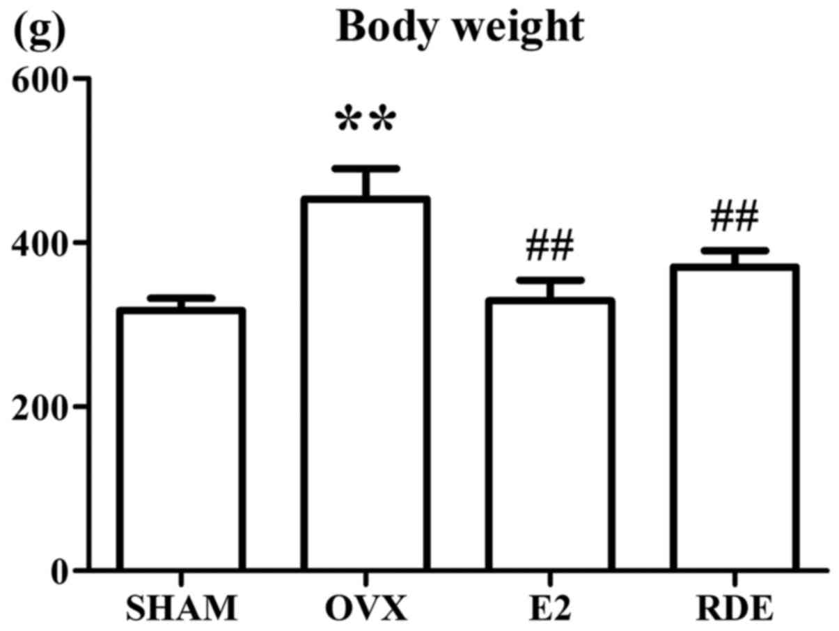
Effect of RDE on body and uterine
weight
The weight of sham rats was significantly lower
compared with that of OVX rats (Fig.
1). Administration of E2 or RDE markedly inhibited weight gain
induced by OVX during the 12-week treatment.
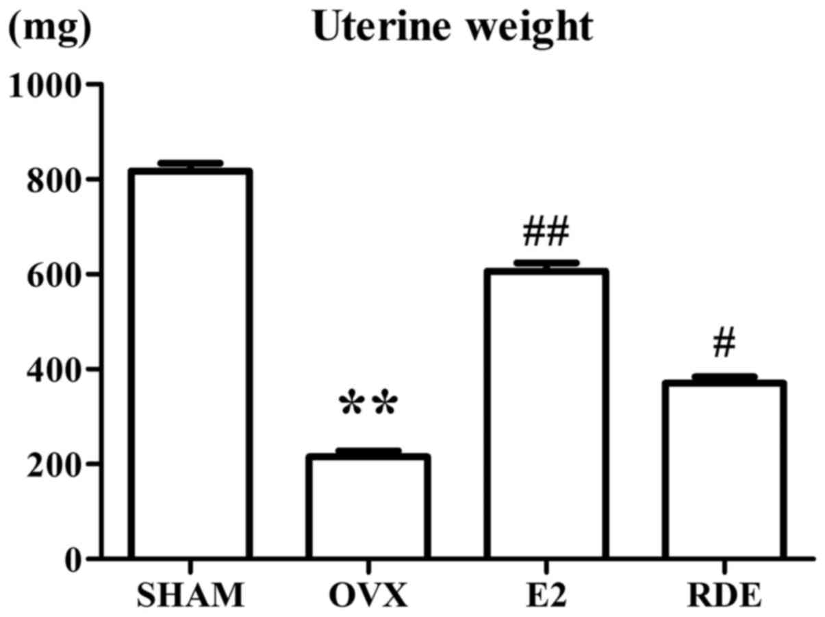
Compared with the sham group, OVX was
associated with marked atrophy of the uterus, which indicated a
successful surgery
E2 injection markedly reduced the atrophy of uterine
tissue compared with OVX rats, whereas administration of RDE
exerted a mild uterotrophic effect (Fig. 2).
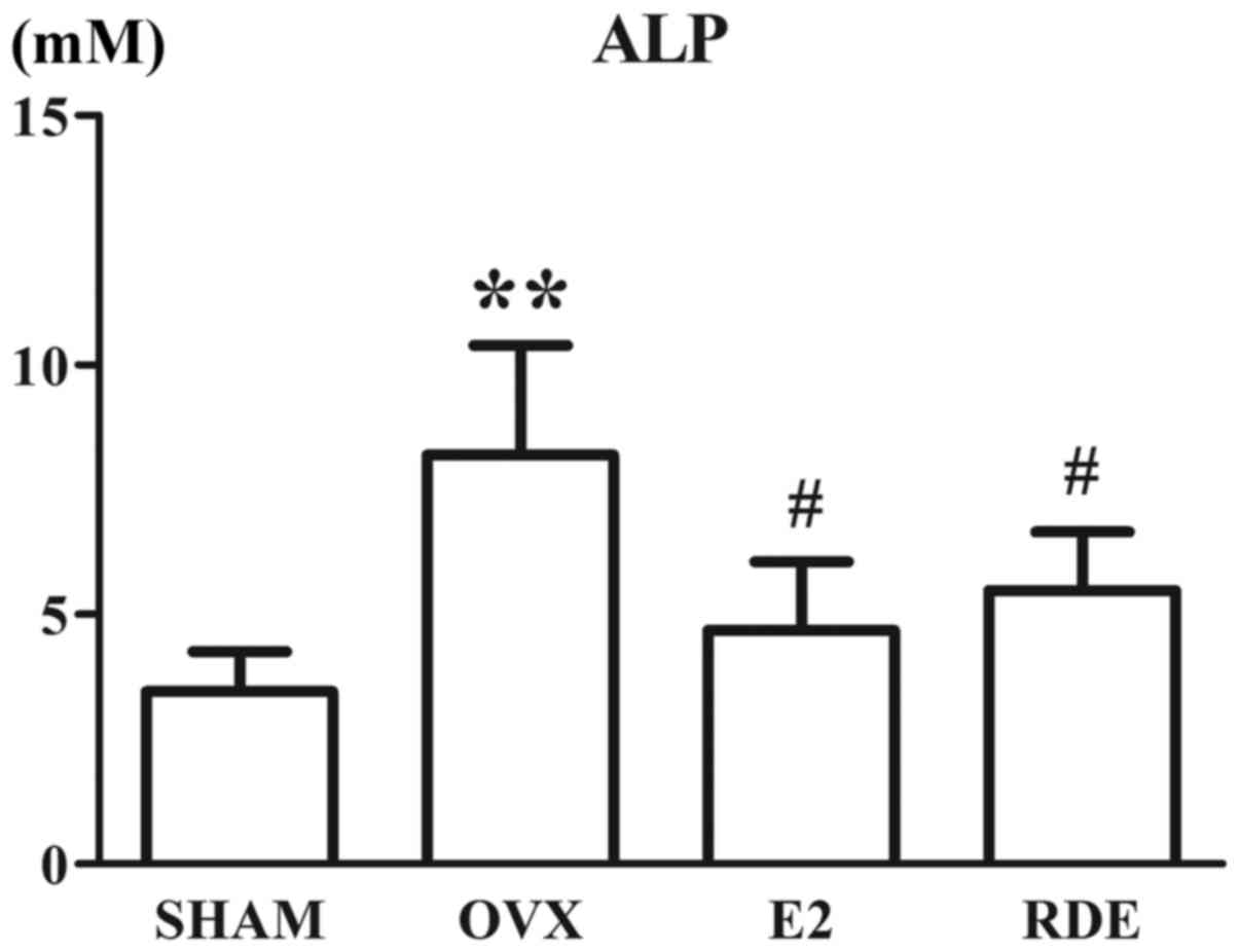
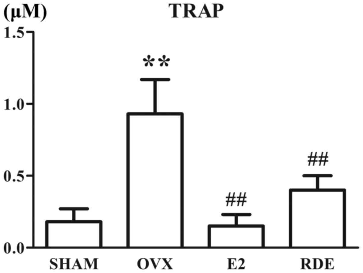
Effect of RDE on bone turnover
biomarkers
The plasma concentrations of ALP and TRAP in
different subject rats after a 12-week treatment are shown in
Figs. 3 and 4. After 12 weeks, the ALP and TRAP
levels in OVX rats were significantly higher compared with those in
sham rats (P<0.01). Moreover, the plasma ALP and TRAP levels in
RDE and E2 rats were significantly lower compared with those in OVX
rats (P<0.01).
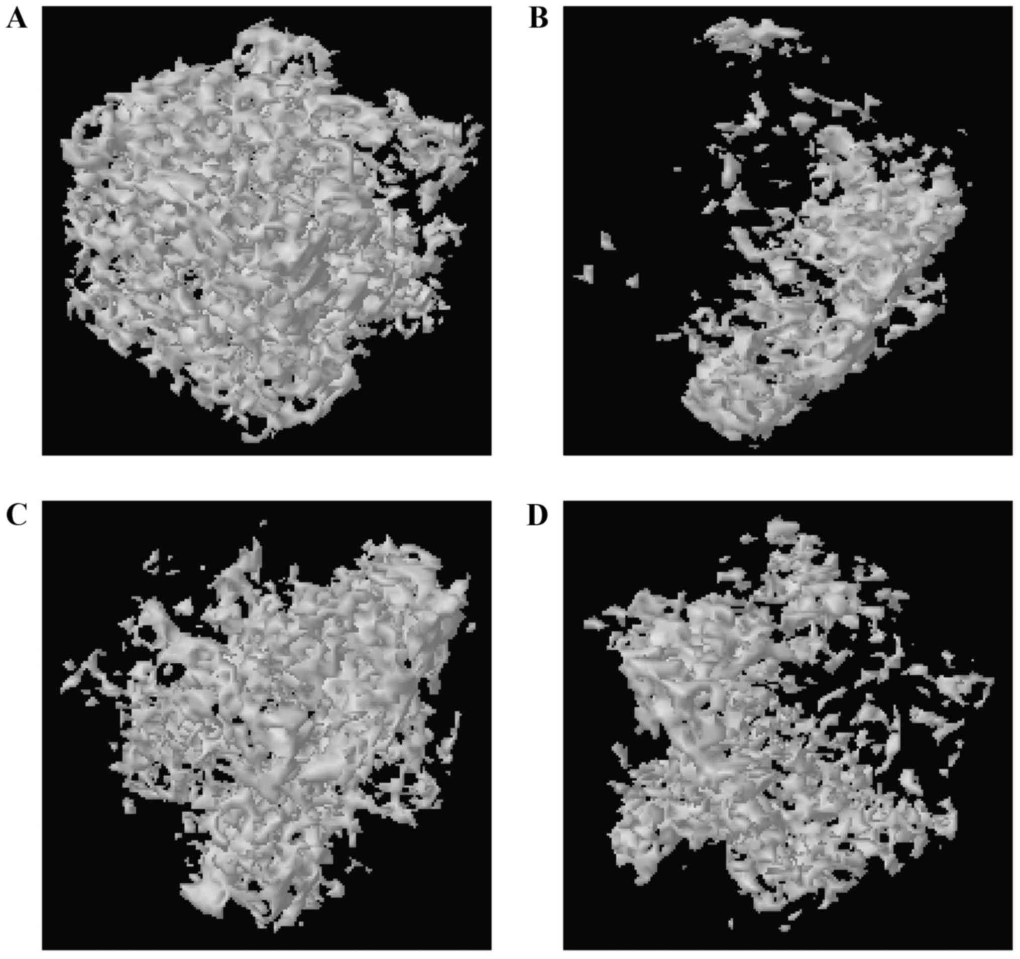
Effect of RDE on BMD and trabecular bone
microarchitecture
It was revealed by analyzing morphological
measurements that Tb.N, trabecular BV/TV and BMD were significantly
decreased (P<0.01), while DA, SMI and Tb.Sp were significantly
increased (P<0.01) in OVX rats compared with sham rats.
Treatment with RDE or E2 relieved OVX-induced bone loss and limited
the OVX-induced damage to alveolar bone trabeculae (Fig. 5 and Table II).
| Table IIEffect of RDE on BMD and trabecular
bone microarchitecture. |
Table II
Effect of RDE on BMD and trabecular
bone microarchitecture.
| SHAM | OVX | E2 | RDE |
|---|
| BMD
(g/cm3) | 0.861±0.105 | 0.344±0.017a | 0.601±0.004c | 0.499±0.089b |
| BV/TV (%) | 27.708±4.371 | 7.839±1.364a |
17.631±1.765c |
13.886±2.888b |
| Tb.Th
(μm) | 24.989±0.165 | 24.473±0.173 | 24.568±0.518 | 24.491±0.595 |
| Tb.Sp
(μm) | 48.646±12.107 |
93.224±7.745a |
60.646±12.360c |
66.763±12.060b |
| Tb.N (1/mm) | 0.011±0.002 | 0.003±0.000a | 0.007±0.001c | 0.006±0.001b |
| SMI | 1.206±0.118 | 2.090±0.122a | 1.523±0.115c | 1.680±0.147c |
| DA | 1.330±0.150 | 1.834±0.095a | 1.455±0.267b | 1.584±0.111 |
Effect of RDE on gene expression
profile
It was revealed by IL-6/STAT3 signaling pathway PCR
arrays that the expression of 24 genes from alveolar bone exhibited
differences of >3-fold between OVX and RDE rats (Table III). Specifically, 2 genes were
upregulated, while 22 were downregulated.
| Table IIIDifferential expression of genes
(≥2-fold) in alveolar bone from 6 RDE and 6 OVX rats. |
Table III
Differential expression of genes
(≥2-fold) in alveolar bone from 6 RDE and 6 OVX rats.
| Symbol | P-value | Fold-change |
|---|
| Akt1 | 0.000264 | −3.09 |
| Ccl4 | 0.001408 | −3.26 |
| Cd4 | 0.000581 | −2.34 |
| Csf3r | 0.001814 | −4.45 |
| Cxcl3 | 0.023671 | −11.72 |
| Hgf | 0.004032 | −3.30 |
| Il12a | 0.027687 | −2.87 |
| Il13 | 0.019136 | 2.06 |
| Il1r1 | 0.000045 | −3.85 |
| Il2 | 0.027486 | 5.58 |
| Il6st | 0.005906 | −2.88 |
| Jak2 | 0.000009 | −2.31 |
| Lifr | 0.009917 | −3.25 |
| Mapk14 | 0.000994 | −2.81 |
| Mapk1 | 0.040108 | −1.64 |
| Met | 0.000938 | −5.16 |
| Mtor | 0.010758 | −2.03 |
| Nfkb1 | 0.000017 | −3.05 |
| Osm | 0.040752 | −4.52 |
| Osmr | 0.016804 | −2.01 |
| Pias3 | 0.007814 | −2.75 |
| Src | 0.030115 | −2.34 |
| Stat3 | 0.004448 | −2.57 |
| Tnfsf11 | 0.007872 | −6.58 |
Confirmation of differential levels of
gene expression by RT-qPCR
With the aim of confirming the differential gene
expression determined using IL-6/STAT3 signaling pathway PCR
arrays, all 24 genes listed in Table III were verified using RT-qPCR
and the results are presented in Fig.
6. Alveolar bone from 6 OVX and 6 RDE rats was used for the
RT-qPCR assay. In the majority of the cases, the variations of
genes in microarray analyses conformed to the RT-qPCR results,
apart from 4 genes (Cxcl3, Hgf, Il2 and
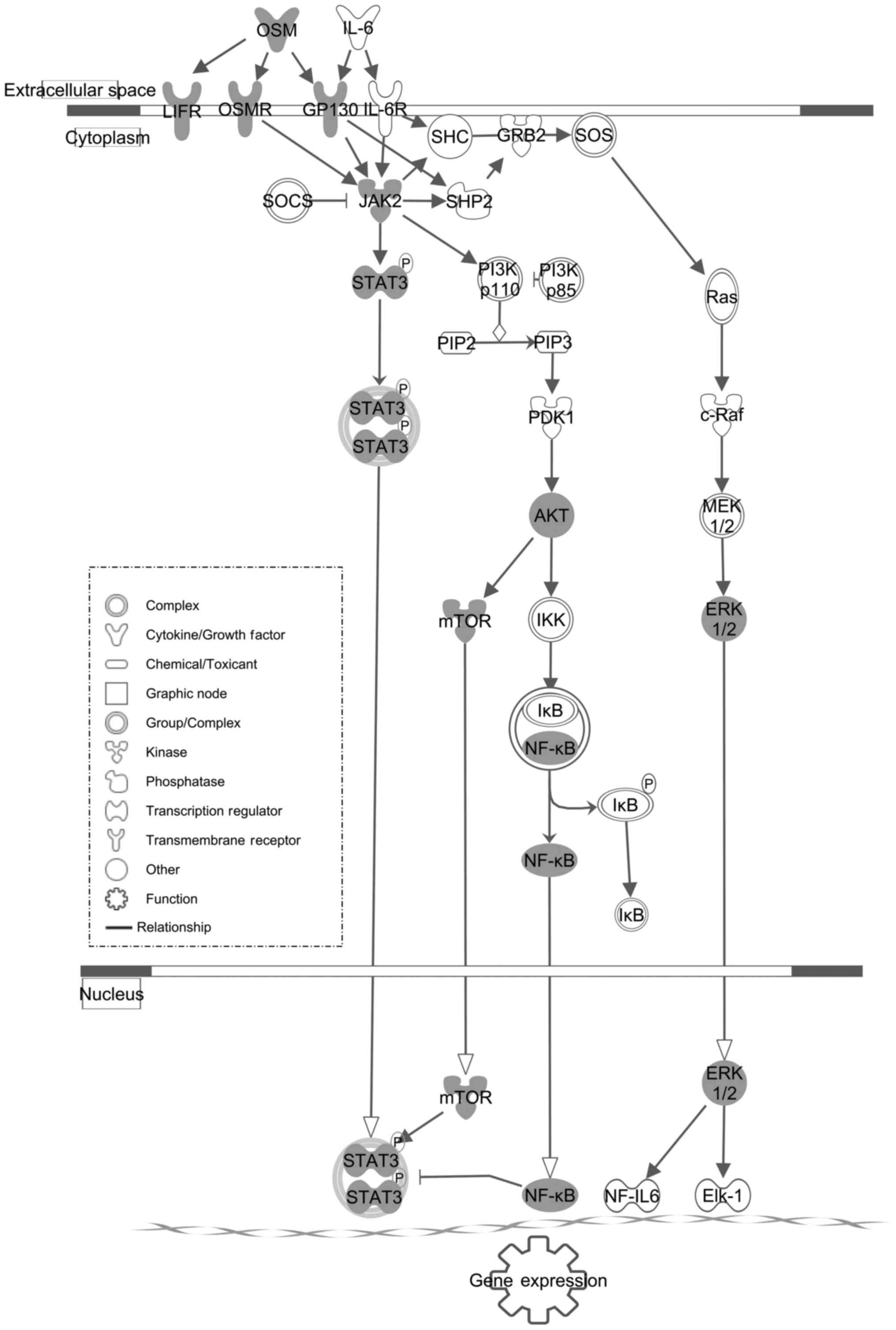
Il12a). The role of the IL-6/STAT3 signaling pathway in the
inhibitory effect of RDE on osteoporosis is schematically
represented in Fig. 7.
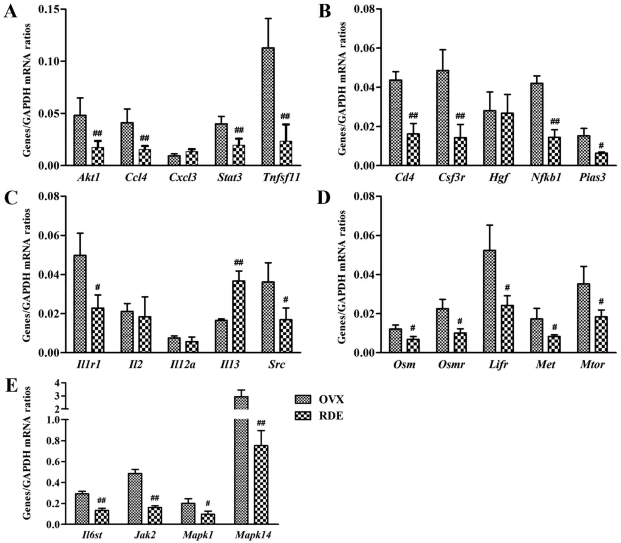 | Figure 6Validation of 24 differential
expression genes identified by microarray and IPA in a replicated
experiment by RT-qPCR. (A-E) Effect of RDE on the expressions of
Akt1, Ccl4, Cd4, Csf3r, Cxcl3,
Hgf, Il12a, Il13, Il1r1, Il2,
Il6st, Jak2, Lifr, Mapk14,
Mapk1, Met, Mtor, Nfkb1, Osm,
Osmr, Pias3, Src, Stat3 and
Tnfsf11. #P<0.05 and ##P<0.01
compared with the OVX group. Alveolar bone prepared from 6 RDE and
6 OVX rats was used. IPA, Ingenuity Pathway Analysis; RDE,
Rhizoma Dioscoreae extract; OVX, ovariectomy; RT-qPCR,
reverse transcription-quantitative polymerase chain reaction. |
Discussion
In our previous study, using bioinformatics and PCR
or western blotting to confirm the expression of Stat3, it
was demonstrated that RDE inhibited osteoporosis in OVX rats, an
effect which was associated with the IL-6/oncostatin M (OSM)/STAT3
pathway via miRNA regulation (21). The putative miRNAs targets' gene
prediction and pathway analysis are both bioinformatics methods,
and the results occasionally do not fully reflect the true effects
of RDE; however, the results raise the question whether RDE exerts
an anti-osteopenic effect via another IL-6 family cytokine pathway.
The IL-6 cytokine family includes 10 members: IL-6, IL-31, IL-27,
IL-11, neuropoietin, cardiotrophin-like cytokine, cardiotrophin-1
(CT-1), ciliary neurotrophic factor (CNTF), OSM and leukemia
inhibitory factor (LIF). All IL-6 family cytokines employ the
transducing receptor β-subunit gp130 as part of a multimeric
receptor complex (26). The
present study aimed to determine the association between the IL-6
family of cytokines pathway and the anti-osteopenic effect of
RDE.
Our results demonstrated that treatment with RDE for
12 weeks prevented loss of uterine wet weight and body weight gain
resulting from lack of estrogen in rats of the OVX group (Figs. 1 and 2). It was deduced that RDE exerts a mild
estrogen-like effect, which may slow down OVX-induced uterine
atrophy and weight gain.
Following ovary removal, OVX rats exhibited markedly
reduced BMD, resulting from increasing alveolar bone metabolism,
compared with the sham rats. By contrast, treatment with RDE or E2
increased the BMD of alveolar bone.
The significant and coincident increases in plasma
ALP and TRAP verified mature female OVX rat as an appropriate and
reliable animal model for studies on early postmenopausal
osteoporosis characterized by high-turnover bone loss. Treating
rats with RDE or E2 for 12 weeks inhibited the excessive bone
metabolism, as shown by significantly decreased ALP and TRAP levels
(Figs. 3 and 4).
From analyses of 3-D bone microarchitecture by
micro-CT, it was indicated that the loss of alveolar bone among
rats treated with E2 or RDE was less severe compared with that in
OVX rats, which was inferred through significant variations in SMI,
Tb.Sp, Tb.N and BV/TV. The inhibition of alveolar bone loss with E2
was superior to that of RDE (Table
II and Fig. 5).
The results of bone metabolic biomarkers, micro-CT
and BMD demonstrated that RDE significantly inhibited alveolar bone
loss.
To investigate the association between the
IL-6/STAT3 signaling pathway and the anti-osteopenic effect of RDE,
the differential expression of genes by was screened using
IL-6/STAT3 signaling pathway array. A total of 20 genes (19
downregulated and 1 upregulated) from alveolar bone were
successfully identified and validated; their expression profiles
differed significantly among RDE rats compared with the OVX group.
The IL-6/STAT3 signaling pathway was downregulated following RDE
treatment (Fig. 7).
In the IL-6 family cytokine signaling cascade,
oligomerization of receptor subunits induced by a specific ligand
may activate janus protein-tyrosine kinases (JAKs), which further
activate the MAPKs or the STATs (mainly STAT1 and STAT3). Another
signaling cascade activated by cytokines of the IL-6 family is the
phosphoinositide-3-kinase/AKT (27,28). Receptor complexes that are capable
of signaling after being activated by cytokines of the IL-6 family
may provide specificity in a given signal transduction pathway. For
example, OSM is considered to be a unique gp130-binding cytokine,
as it binds first to gp130, and then develops a signaling complex
with either the OSM receptor (OSMR) or the LIF receptor (LIFR),
which are both capable of intracellular signaling (29,30).
Cytokines of the IL-6 family exert a major effect on
osseous remodeling through osteoclasts or osteoblasts, despite
seemingly functioning as a double-edged sword. These cytokines
maintain bone generation while driving bone absorption induced by a
variety of osteolytic factors. This dual effect is apparent in two
main signaling pathways (STAT and MAPK signaling) bidirectionally
affecting osteocytes (19).
Certain members of the IL-6 family (OSM, CNTF, LIF,
IL-11 and IL-6) may promote bone formation. This effect was mainly
reflected in three aspects: First, these cytokines promote the
differentiation of osteoblast precursors or maturing osteoblasts
isolated from the bone marrow or calvaria. During this process,
STAT3 must be activated (31,32). Thus, the expression of
osteoblastic biomarkers, such as bone sialoprotein, osteocalcin or
ALP, was increased, while extracellular matrix mineralization and
bone nodule formation were enhanced. Second, the biomarkers inhibit
certain osteoblasts from proliferating via activating gp130/STAT3
and increasing the expression of p21WAF1, which is a
cell cycle inhibitor and is also required for cytokines of the IL-6
family to induce ALP (33).
Finally, these cytokines may protect osteoblasts from apoptosis
caused by tumor necrosis factor-α (TNF-α) or serum depletion
(34).
As regards bone absorption, cytokines of the IL-6
family promote osteoclast differentiation and bone absorption by
accelerating the interactions between osteoclasts and osteoblasts.
In co-cultures of osteoclast precursors and stromal cells or
osteoblasts, certain IL-6-type cytokines (OSM, CNTF, LIF, IL-11 and
IL-6) are able to promote osteoclast differentiation and bone
absorption. In fact, these cytokines induce production of various
osteoblastic downstream effectors that, in turn, promote osteoclast
differentiation or activity, such as IL-1, receptor activator of
nuclear factor-κB (NF-κB) ligand (RANKL), prostaglandin E2 and
PTH-related protein. Recent studies implicated activation of
IL-6-type cytokines/STAT3 signaling as the pivotal event in the
induction of RANKL in osteoblasts, which leads to pro-resorption
action of osteoclasts (35,36).
It was observed that the gene expression of certain
signaling molecules (Osm, Osmr, Lifr,
Il6st, Jak2, Stat3, Mtor, Nfkb1
and Mapk1) in the IL-6/STAT3 pathway were downregulated
following RDE treatment (Fig. 7),
which subsequently inhibited IL-6/STAT3 signaling. The RT-qPCR
analysis of Osm, Osmr, Lifr, Il6st,
Jak2, Stat3, Mtor, Nfkb1 and
Mapk1 expression (Fig. 6)
proved that RDE effectively reduced excessive alveolar bone
formation and bone absorption synchronously caused by attenuated
canonical IL-6/STAT3 signaling following OVX.
Further studies are required for RDE to be developed
into a novel promising drug for the prevention or treatment of
alveolar bone loss in postmenopausal women.
In conclusion, RDE was effective in inhibiting rat
alveolar bone loss caused by OVX, by simultaneously inhibiting bone
formation as well as bone resorption through regulation of the
IL-6/STAT3 signaling pathway. The present study verified that RDE
may be used as an oral agent for the treatment of alveolar
osteopenia in postmenopausal women.
Acknowledgments
The present study was supported by the National
Natural Science Foundation of China (grant no. 81473450) and the
Fundamental Research Funds for the Beijing Administration of
Traditional Chinese Medicine (grant no. JJ2015-54).
References
|
1
|
Gallagher JC and Levine JP: Preventing
osteoporosis in symptomatic postmenopausal women. Menopause.
18:109–118. 2011. View Article : Google Scholar
|
|
2
|
Sultan N and Rao J: Association between
periodontal disease and bone mineral density in postmenopausal
women: A cross sectional study. Med Oral Patol Oral Cir Bucal.
16:e440–e447. 2011. View Article : Google Scholar : PubMed/NCBI
|
|
3
|
Lee BD and White SC: Age and trabecular
features of alveolar bone associated with osteoporosis. Oral Surg
Oral Med Oral Pathol Oral Radiol Endod. 100:92–98. 2005. View Article : Google Scholar : PubMed/NCBI
|
|
4
|
Streckfus CF, Johnson RB, Nick T, Tsao A
and Tucci M: Comparison of alveolar bone loss, alveolar bone
density and second metacarpal bone density, salivary and gingival
crevicular fluid interleukin-6 concentrations in healthy
premenopausal and postmenopausal women on estrogen therapy. J
Gerontol A Biol Sci Med Sci. 52:M343–M351. 1997. View Article : Google Scholar : PubMed/NCBI
|
|
5
|
Tezal M, Wactawski-Wende J, Grossi SG,
Dmochowski J and Genco RJ: Periodontal disease and the incidence of
tooth loss in postmenopausal women. J Periodontol. 76:1123–1128.
2005. View Article : Google Scholar : PubMed/NCBI
|
|
6
|
Civitelli R, Pilgram TK, Dotson M,
Muckerman J, Lewandowski N, Armamento-Villareal R,
Yokoyama-Crothers N, Kardaris EE, Hauser J, Cohen S, et al:
Alveolar and postcranial bone density in postmenopausal women
receiving hormone/estrogen replacement therapy: A randomized,
double-blind, placebo-controlled trial. Arch Intern Med.
162:1409–1415. 2002. View Article : Google Scholar : PubMed/NCBI
|
|
7
|
Liu J, Cao Z and Li C: Intermittent PTH
administration: A novel therapy method for periodontitis-associated
alveolar bone loss. Med Hypotheses. 72:294–296. 2009. View Article : Google Scholar
|
|
8
|
Palomo L, Bissada NF and Liu J:
Periodontal assessment of postmenopausal women receiving
risedronate. Menopause. 12:685–690. 2005. View Article : Google Scholar : PubMed/NCBI
|
|
9
|
Strom BL, Schinnar R, Weber AL, Bunin G,
Berlin JA, Baumgarten M, DeMichele A, Rubin SC, Berlin M, Troxel
AB, et al: Case-control study of postmenopausal hormone replacement
therapy and endometrial cancer. Am J Epidemiol. 164:775–786. 2006.
View Article : Google Scholar : PubMed/NCBI
|
|
10
|
Rossing MA, Cushing-Haugen KL, Wicklund
KG, Doherty JA and Weiss NS: Menopausal hormone therapy and risk of
epithelial ovarian cancer. Cancer Epidemiol Biomarkers Prev.
16:2548–2556. 2007. View Article : Google Scholar : PubMed/NCBI
|
|
11
|
Rizzoli R, Reginster JY, Boonen S, Bréart
G, Diez-Perez A, Felsenberg D, Kaufman JM, Kanis JA and Cooper C:
Adverse reactions and drug-drug interactions in the management of
women with postmenopausal osteoporosis. Calcif Tissue Int.
89:91–104. 2011. View Article : Google Scholar : PubMed/NCBI
|
|
12
|
Woo SB, Hellstein JW and Kalmar JR:
Narrative [corrected] review: Bisphosphonates and osteonecrosis of
the jaws. Ann Intern Med. 144:753–761. 2006. View Article : Google Scholar : PubMed/NCBI
|
|
13
|
Clemett D and Spencer CM: Raloxifene: A
review of its use in postmenopausal osteoporosis. Drugs.
60:379–411. 2000. View Article : Google Scholar : PubMed/NCBI
|
|
14
|
Sugimoto H1, Watanabe K, Toyama T,
Takahashi SS, Sugiyama S, Lee MC and Hamada N: Inhibitory effects
of French pine bark extract, Pycnogenol®, on alveolar
bone resorption and on the osteoclast differentiation. Phytother
Res. 29:251–259. 2015. View
Article : Google Scholar
|
|
15
|
Sağlam M, Köseoğlu S, Hatipoğlu M, Esen HH
and Köksal E: Effect of sumac extract on serum oxidative status,
RANKL/OPG system and alveolar bone loss in experimental
periodontitis in rats. J Appl Oral Sci. 23:33–41. 2015. View Article : Google Scholar
|
|
16
|
Guimarães MV, Melo IM, Adriano Araújo VM,
Tenazoa Wong DV, Roriz Fonteles CS, Moreira Leal LK, Ribeiro RA and
Lima V: Dry extract of Matricaria recutita L. (Chamomile) prevents
ligature-induced alveolar bone resorption in rats via inhibition of
tumor necrosis factor-α and interleukin-1β. J Periodontol.
87:706–715. 2016. View Article : Google Scholar
|
|
17
|
Feng XF, Huang LQ, Ge XG, Yang LJ and Yang
JY: Textual research on origin and development of genuine medicinal
herbs of Shanyao. Zhongguo Zhong Yao Za Zhi. 33:859–862. 2008.In
Chinese. PubMed/NCBI
|
|
18
|
Zhang Z, Xiang L, Bai D, Wang W, Li Y, Pan
J, Liu H, Wang S, Xiao GG and Ju D: The protective effect of
Rhizoma Dioscoreae extract against alveolar bone loss in
ovariectomized rats via regulating Wnt and p38 MAPK signaling.
Nutrients. 6:5853–5870. 2014. View Article : Google Scholar : PubMed/NCBI
|
|
19
|
Blanchard F, Duplomb L, Baud' huin M and
Brounais B: The dual role of IL-6-type cytokines on bone remodeling
and bone tumors. Cytokine Growth Factor Rev. 20:19–28. 2009.
View Article : Google Scholar
|
|
20
|
Sims NA and Walsh NC: GP130 cytokines and
bone remodelling in health and disease. BMB Rep. 43:513–523. 2010.
View Article : Google Scholar : PubMed/NCBI
|
|
21
|
Zhang Z, Song C, Zhang F, Xiang L, Chen Y,
Li Y, Pan J, Liu H, Xiao GG and Ju D: Rhizoma Dioscoreae extract
protects against alveolar bone loss in ovariectomized rats via
microRNAs regulation. Nutrients. 7:1333–1351. 2015. View Article : Google Scholar : PubMed/NCBI
|
|
22
|
Zhang Z, Xiang L, Bai D, Fu X, Wang W, Li
Y, Liu H, Pan J, Li Y, Xiao GG, et al: Treatment with Rhizoma
Dioscoreae extract has protective effect on osteopenia in
ovariectomized rats. ScientificWorldJournal.
2014:6459752014.PubMed/NCBI
|
|
23
|
Hidaka S, Okamoto Y, Yamada Y, Kon Y and
Kimura T: A Japanese herbal medicine, Chujo-to, has a beneficial
effect on osteoporosis in rats. Phytother Res. 13:14–19. 1999.
View Article : Google Scholar : PubMed/NCBI
|
|
24
|
Yang J, Pham SM and Crabbe DL:
High-resolution micro-CT evaluation of mid- to long-term effects of
estrogen deficiency on rat trabecular bone. Acad Radiol.
10:1153–1158. 2003. View Article : Google Scholar : PubMed/NCBI
|
|
25
|
Bouxsein ML, Boyd SK, Christiansen BA,
Guldberg RE, Jepsen KJ and Müller R: Guidelines for assessment of
bone microstructure in rodents using micro-computed tomography. J
Bone Miner Res. 25:1468–1486. 2010. View
Article : Google Scholar : PubMed/NCBI
|
|
26
|
Garbers C, Hermanns HM, Schaper F,
Müller-Newen G, Grötzinger J, Rose-John S and Scheller J:
Plasticity and cross-talk of interleukin 6-type cytokines. Cytokine
Growth Factor Rev. 23:85–97. 2012. View Article : Google Scholar : PubMed/NCBI
|
|
27
|
Grant SL and Begley CG: The oncostatin M
signalling pathway: Reversing the neoplastic phenotype? Mol Med
Today. 5:406–412. 1999. View Article : Google Scholar : PubMed/NCBI
|
|
28
|
Heinrich PC, Behrmann I, Haan S, Hermanns
HM, Müller-Newen G and Schaper F: Principles of interleukin
(IL)-6-type cytokine signalling and its regulation. Biochem J.
374:1–20. 2003. View Article : Google Scholar : PubMed/NCBI
|
|
29
|
Mosley B, De Imus C, Friend D, Boiani N,
Thoma B, Park LS and Cosman D: Dual oncostatin M (OSM) receptors.
Cloning and characterization of an alternative signaling subunit
conferring OSM-specific receptor activation. J Biol Chem.
271:32635–32643. 1996. View Article : Google Scholar : PubMed/NCBI
|
|
30
|
Lindberg RA, Juan TS, Welcher AA, Sun Y,
Cupples R, Guthrie B and Fletcher FA: Cloning and characterization
of a specific receptor for mouse oncostatin M. Mol Cell Biol.
18:3357–3367. 1998. View Article : Google Scholar : PubMed/NCBI
|
|
31
|
Bellido T, Borba VZ, Roberson P and
Manolagas SC: Activation of the Janus kinase/STAT (signal
transducer and activator of transcription) signal transduction
pathway by interleukin-6-type cytokines promotes osteoblast
differentiation. Endocrinology. 138:3666–3676. 1997. View Article : Google Scholar : PubMed/NCBI
|
|
32
|
Itoh S, Udagawa N, Takahashi N, Yoshitake
F, Narita H, Ebisu S and Ishihara K: A critical role for
interleukin-6 family-mediated Stat3 activation in osteoblast
differentiation and bone formation. Bone. 39:505–512. 2006.
View Article : Google Scholar : PubMed/NCBI
|
|
33
|
Bellido T, O' Brien CA, Roberson PK and
Manolagas SC: Transcriptional activation of the p21(WAF1, CIP1,
SDI1) gene by interleukin-6 type cytokines. A prerequisite for
their pro-differentiating and anti-apoptotic effects on human
osteoblastic cells. J Biol Chem. 273:21137–21144. 1998. View Article : Google Scholar : PubMed/NCBI
|
|
34
|
Jilka RL, Weinstein RS, Bellido T, Parfitt
AM and Manolagas SC: Osteoblast programmed cell death (apoptosis):
modulation by growth factors and cytokines. J Bone Miner Res.
13:793–802. 1998. View Article : Google Scholar : PubMed/NCBI
|
|
35
|
Palmqvist P, Persson E, Conaway HH and
Lerner UH: IL-6, leukemia inhibitory factor, and oncostatin M
stimulate bone resorption and regulate the expression of receptor
activator of NF-kappa B ligand, osteoprotegerin, and receptor
activator of NF-kappa B in mouse calvariae. J Immunol.
169:3353–3362. 2002. View Article : Google Scholar : PubMed/NCBI
|
|
36
|
Kim S, Yamazaki M, Shevde NK and Pike JW:
Transcriptional control of receptor activator of nuclear
factor-kappaB ligand by the protein kinase A activator forskolin
and the transmembrane glycoprotein 130-activating cytokine,
oncostatin M, is exerted through multiple distal enhancers. Mol
Endocrinol. 21:197–214. 2007. View Article : Google Scholar
|