Introduction
Liver cancer ranks sixth for incidence and third for
mortality among the most common cancers worldwide (1). In 2014, it was reported that among
87,988 malignant liver and intrahepatic cancer diagnoses reported
during 2000–2010 in SEER 18 registries, 63,735 (72%) were
classified as HCCs in USA. Between 2006 and 2010, US liver cancer
mortality rates increased with age in all racial/ethnic groups
(2). A significant variance in
the incidence rate of liver cancer has been observed among
different nations and regions, and 55% of new cases of liver cancer
worldwide are reported to occur in China (3). In the USA, the incidence of liver
cancer has been rising faster than that of other malignant cancers
(4). The occurrence of liver
cancer is closely correlated with infection by hepatitis B virus
(HBV), and 50% of cases result from HBV infection, the infection
rate of which is high (5,6). Although great advances in the
surgical therapy and other treatments for liver cancer have been
achieved, the 5-year survival rate is only 5–6% and the poor
prognosis is a cause of severe economic loss and a heavy burden for
society (7–9).
Liver cancer is usually occult in onset and no
effective screening measure is able to screen for the disease
validly and reliably; therefore, two-thirds of cases are not
diagnosed until an advanced stage (10,11). Furthermore, liver cancer is often
resistant to chemotherapeutic drugs because it usually originates
from pre-existing liver diseases (12,13). These factors largely contribute to
the high mortality rate of liver cancer. The initiation and
progression of liver cancer is an interactive process involving
multiple genes and a variety of environmental factors, which
include multiple pathological stages and a variety of molecular
events. These factors induce normal liver cells to transform into
liver cancer cells and may even cause the occurrence of metastasis
(14,15).
Recent studies of the molecular mechanism of liver
cancer have mainly focused on the expression of specific tumor
genes, differentially expressed genes and their proteomics
(16,17). Numerous non-coding sequences in
the human genome have been detected and their proportion is far
greater than that of protein-encoding sequences (>97%). These
sequences may be transcribed into multiple types of RNA that serve
an important role in the development of diseases (18–20). Non-coding small RNA (length from
18 to 200 nucleotides), particularly microRNA, has attracted
considerable attention (21,22). Studies on the expression profile
and functions of microRNA are advancing the understanding of liver
cancer. The association between microRNA and the incidence of liver
cancer and its potential value for the diagnosis and treatment are
being increasingly highlighted (23–25). MicroRNAs are single stranded,
non-coding small molecular weight endogenous RNAs ~22 nucleotides
in length. They reduce the expression of mRNA by complementary
pairing with the 3′-untranslated region (3′-UTR) of targeted mRNA,
which participates in multiple physiological processes, including
cell proliferation, differentiation, apoptosis, metabolism and
development, as well as multiple pathological processes such as
cardiovascular disease, nervous system diseases and tumor diseases
(22,26–28). More than one-third of human genes
are considered to be the targets of conservative microRNAs and
these microRNAs have received growing attention in the development
of human diseases, particularly in cancers (29,30).
A number of studies have shown that miRNAs are
closely associated with the occurrence, development and invasion of
tumors by regulating the cell cycle, cell apoptosis, cell migration
and the angiogenesis of tumors (31–33). The epidermal growth factor
receptor has a tumor-promoting role on cell proliferation,
survival, metastasis and tumor-induced vascularization) during
hepatocellular carcinoma (HCC) formation, which is regulated by
miR-302b (34–36). Wang et al (37) reported that hsa-miR-613 prohibits
the proliferation and invasion of HCC by regulating the expression
of doublecortin like kinase 1. In addition, the proliferation of
liver cancer cells is accelerated by the reduction in NFAIP3
interacting protein 2 expression induced by interference with
miR-1180 (38). In gastric
cancer, hsa-microRNA-493 has been shown to reduce cell
proliferation and invasion (39).
In addition, Sakai et al (40) substantiated that binding of
mitogen-activated protein kinase kinase 7 by miR-493 inhibits the
hepatic metastasis of colon cancer cells. However, the role that
miR-493-5p serves in liver cancer remains unclear.
The present study investigated the expression of
miR-493-5p in liver cancer tissues and cell lines, and explored the
effect of miR-493-5p on the biological behavior of the HepG2 cell
line. Furthermore, whether the expression of vesicle associated
membrane protein 2 (VAMP2) is regulated by miR-493-5p was also
explored. The results of these investigations may provide a new
method or strategy for the therapy of liver cancer.
Materials and methods
Samples
Tissue samples, comprising 15 fresh HCC tissues and
6 adjacent normal tissues from patients (age, 40–70 years old; 12
male and 3 female; 10 at cancer stage I–II and 5 at cancer stage
III–IV) with HCC were collected in Anhui Provincial Hospital
Affiliated to Anhui Medical University (Hefei, China) from 2012 to
2015. The samples were immediately placed in liquid nitrogen for
analysis. All patients had not undergone radiotherapy, chemotherapy
or other treatments and had no other complications such as
inflammatory diseases. The study was approved by the Biomedical
Ethics Committee of Anhui Medical University (approval no.
20140272). The patients provided written informed consent to
participate in this study and cooperated fully with the
researchers.
Reagents
The TaqMan miRNA isolation kit and TaqMan microRNA
assay kit were purchased from Applied Biosystems (Thermo Fisher
Scientific, Inc., Waltham, MA, USA). The primers for miR-493-5p
were: 5′-TCC TAC GGA GAG GCT CAG-3′ and 5′-TCC TCG TAG TCC AAC
ACG-3′. The miR-493-5p mimic and negative control (mimic control)
were synthesized by Shanghai GenePharma Co., Ltd. (Shanghai,
China).
Fetal bovine serum (FBS), Dulbecco's modified
Eagle's medium (DMEM) culture medium, RPMI-1640 culture medium and
Lipofectamine® 2000 were all purchased from Invitrogen
(Thermo Fisher Scientific, Inc.). The culture dishes, plates and
Transwell chambers were from Corning Incorporated (Corning, NY,
USA).
3-(4,5-Dimethylthiazol-2-yl)-2,5-diphenyltetrazolium
bromide (MTT) and radioimmunoprecipitation assay (RIPA) protein
lysis buffer were obtained from Beyotime Institute of Biotechnology
(Shanghai, China). Trypsin and Hoechst 33342 were obtained from
Sigma-Aldrich (Merck KGaA, Darmstadt, Germany). Matrigel was from
BD Biosciences (Franklin Lakes, NJ, USA). Annexin V-fluorescein
isothiocyanate (FITC) and propidium iodide (PI) were from Roche
Diagnostics (Basel, Switzerland). The rabbit anti-human VAMP2
polyclonal antibody (ab3347) and mouse anti-human β-actin
monoclonal antibody (ab8226) were from Abcam (Cambridge, UK). The
horseradish peroxidase (HRP)-conjugated affinity purified goat
anti-mouse (A1146) and goat anti-rabbit (A0545) IgG secondary
antibodies were from Sigma-Aldrich (Merck KGaA). The bicinchoninic
acid (BCA) protein quantification kit was from Bio-Rad
Laboratories, Inc. (Hercules, CA, USA).
The vectors including pcDNA3.1 (Thermo Fisher
Scientific, Inc.), pGEM-T (Promega Corporation, Madison, WI, USA)
and pGL3-Basic (Promega Corporation) were preserved in the present
authors' laboratory. The competent cells (DH5α) and TRIzol reagent
were purchased from Invitrogen Thermo Fisher Scientific, Inc.). The
DNA marker and restriction endonucleases BamHI and
XhoI were purchased from Takara Bio, Inc. (Otsu, Japan). The
T4 DNA ligase was acquired from Promega Corporation. The QIAGEN
OneStep RT-PCR kit (cat. no. 210210) was acquired from Qiagen, Inc.
(Valencia, CA, USA).
Methods
Cell culture
The HCCC-9810 cell line (an intrahepatic
cholangiocarcinoma cell line) was cultured in RPMI-1640 medium
containing 10% FBS. The HepG2 cell line (a hepatoblastoma cell
line) was cultured in DMEM containing 1.0 mM sodium pyruvate and
10% FBS. The HL-7702 cell line (a normal human liver cell line) was
cultured in RPMI-1640 medium containing 20% FBS. The HuH-7 cell
line (a HCC cell line) was cultured in DMEM containing 10% FBS.
The four cell lines were incubated at 37°C with 5%
CO2 and saturated humidity. The status of the cells was
observed under an inverted phase microscope and the passage of the
cells was conducted by 0.25% trypsinization when their confluence
reached 70–80%. The culture medium was changed every other day.
The 293 cells were seeded in a 6-well plate at a
density of 3×10⁵/ml with a volume of 1,000 μl in
each well. When the cell confluence reached 80%, the cells were
transfected with the miR-493-5p mimic or negative control (final
concentration, 50 nM; synthesized by Shanghai GenePharma Co.,
Ltd.), with or without the recombinant plasmid pcDNA3.1-VAMP2 (500
ng/well) using Lipofectamine 2000 according to the manufacturer's
protocol. The cells transfected with the negative control and
pcDNA3.1-blank plasmid served as a control. Untransfected cells
served as the normal control.
Construction of recombinant plasmid
pcDNA3.1-VAMP2
According to the mRNA sequence of VAMP2 (GenBank no.
NM-014232) and restriction site analysis, primers were designed
using Primer Premier 5 software (Premier Biosoft International,
Palo Alto, CA, USA) for a location flanking the open reading frame
of the VAMP2 gene. The primer sequences were: Forward, 5′-CG
GGATCC ATG TCT GCT
ACC GCT GCC AC-3′ (containing a BamHI site) and reverse,
5′-CC CTCGAG AGT
GCT GAA GTA AAC TAT GA-3′ (containing a XhoI site) (the
underlined sections of the primer sequences indicate the
restriction endonuclease site). The primers were synthesized by
Shanghai Invitrogen (Thermo Fisher Scientific, Inc.).
RNA extraction from the HepG2 cell line was
performed using TRIzol reagent and first-strand cDNA synthesis was
conducted using the QIAGEN OneStep RT-PCR kit. Amplification of the
VAMP2 gene was conducted with the above primers and the PCR
products were inserted into the pGEM-T vector. The positive clone
was then sequenced and subcloned into the vector pcDNA3.1 for the
establishment of the pcDNA3.1-VAMP2 recombinant plasmid.
Expression of miR-493-5p and VAMP2 in
the liver cancer tissues and cell lines
The expression of miR-493-5p and VAMP2 was examined
by RT-qPCR (temperature protocol: 16°C for 30 min, 42°C for 30 min,
85°C for 5 min, in the end hold in 4°C) in the cell lines and
tissue samples. The thermocycling conditions were as follows: 95°C
for 10 min, 95°C for 10 sec, 60°C for 60 sec, 40 cycles.
Quantification was conducted using the 2−ΔΔCq method
(41). The microRNA was extracted
using the TaqMan miRNA isolation kit and the expression of
miR-493-5p was detected using a TaqMan microRNA assay kit with U6
as an internal reference. RNA was extracted from the tissues and
cell lines using TRIzol and first-strand synthesis was conducted
using the RT kit. The expression of VAMP2 mRNA was detected by qPCR
and GAPDH served as an internal reference. All the experiments were
replicated three times.
Effect of miR-493-5p on VAMP2
expression
The miR-493-5p mimic and negative control were
transfected into the HepG2 cells and 48 h later, the microRNA was
extracted using a TaqMan miRNA isolation kit. The expression of
miR-493-5p was detected by RT-qPCR and the expression of VAMP2 was
confirmed by western blot analysis. For the latter, protein
extraction was conducted by lysis using RIPA buffer solution and
the total protein concentration was measured by the BCA kit. The
proteins (50 μg per lane) were subjected to electrophoresis
using 5% SDS-PAGE, transferred onto a polyvinylidene difluoride
membrane (Bio-Rad Laboratories, Inc.) and blocked at 37°C in 5%
skimmed milk TBS with Tween 20 (TBST) solution for 1 h. The VAMP2
polyclonal rabbit anti-human antibody was then added at a dilution
of 1:500 and β-actin rabbit anti-human monoclonal antibody was used
as internal reference (dilution 1:1,000). The membrane was
incubated at 4°C overnight, washed with TBST three times, and then
probed with HRP-conjugated goat anti-rabbit IgG (1:2,000) at 37°C
for 1 h. Following washing with TBST, the membrane was treated with
an ECL chemiluminescence reagent (Thermo Fisher Scientific, Inc.)
and autoradiography was conducted. The relative expression of the
target protein was evaluated as the gray value ratio of the target
protein to β-actin (target protein/β-actin) content. The software
used for quantification of the western blotting results was
Quantity One (Bio-Rad Laboratories, Inc.).
Effect of miR-493-5p on cell
viability
The miR-493-5p mimic and negative control were
transfected into HepG2 cells and 48 h later, 100 μl MTT (0.5
mg/ml) solution was added to each well. The plate was then
incubated at 37°C, 5% CO2 and saturated humidity for
another 4 h. Following this, 100 μl 20% sodium dodecyl
sulfate (containing 50% dimethylformamide) was added to dissolve
the formazan crystals. The optical density value was measured using
a microplate reader (BioTek Instruments, Inc., Winooski, VT, USA)
at a wavelength of 570 nm. The experiments were conducted in
triplicate.
Effect of miR-493-5p on cell
proliferation
The miR-493-5p mimic and negative control were
transfected into HepG2 cells and 48 h later, these cells were
collected by trypsinization. The cells were then fixed using 75%
ethanol and dyed with PI solution for 30 min in the absence of
light. Afterwards, these cells were analyzed by flow cytometry (BD
Biosciences), using FCM CellQuest software (BD Biosciences) for
cell counting and FACsuite software for data analysis.
Effect of miR-493-5p on cell
apoptosis
The miR-493-5p mimic and negative control were
transfected into HepG2 cells and 48 h later, the cells were
collected by trypsinization. The cells were then dyed with Annexin
V-FITC and PI solution for 15 min in the dark. Finally, the cells
were subjected to flow cytometric analysis for the detection of
apoptosis.
Effect of miR-493-5p on cell
invasion
The cell treatment and transfection were conducted
as described above. At 24 h after transfection, the cells were
seeded in a Transwell chamber and incubated for another 24 h. The
cells were then washed with phosphate-buffered saline three times
and stained with Hoechst 33342. The stained cells that had migrated
through the Transwell polycarbonate membrane were counted under a
microscope in six randomly selected fields. Matrigel was used in
invasion assay.
Identification and confirmation of the
targeting of VAMP2 by miR-493-5p
TargetScan (http://www.targetscan.org/) was used to predict the
gene targeted by miR-493-5p, and VAMP2 was predicted as a potential
target. To establish the fluorescent reporter vector, the 3′-UTR of
VAMP2 and the 3′-UTR of VAMP2 containing a mutation in the seed
region were synthesized and inserted into the pGL3-Basic vector at
the XbaI restriction site. The HepG2 cells were seeded in a
6-well plate at a density of 3×10⁵/ml in 1,000 μl
culture medium. The cells were transfected with plasmid VAMP2
3′-UTR (wild-type) or mutant VAMP2 3′-UTR, in combination with
either miR-493-5p mimic or negative control. At 48 h following
transfection, the cells were lysed and detected using a
Dual-Luciferase® Reporter Assay system (Promega
Corporation).
Evaluation of the VAMP2-dependence of
miR-493-5p
In order to verify that the effect of miR-493-5p on
cell biological behavior is mediated via VAMP2, the overexpression
vector pcDNA3.1-VAMP2 was established. HepG2 cells were divided
into five groups: Control, pcDNA3.1 control, pcDNA3.1-VAMP2
transfection, miR-493-5p mimic + pcDNA3.1 and miR-493-5p mimic +
pcDNA3.1-VAMP2 groups. The transfection procedure was conducted as
described above. Cell behavior characteristics, including cell
viability, cell proliferation and cell migration, were then
measured by MTT, flow cytometry and Transwell chamber assays.
Statistical analysis
Data are presented as the means ± SD (3 replicates).
SPSS 17.0 statistical software (SPSS, Inc., Chicago, IL, USA) was
used to analyze the data with one-way analysis of variance.
Multiple comparison between the groups was performed using
Student-Newman-Keuls method. All P-values were two-sided and
P<0.05 was considered to indicate a statistically significant
result.
Results
Expression of miR-493-5p and VAMP2 in
liver cancer tissues and cell lines
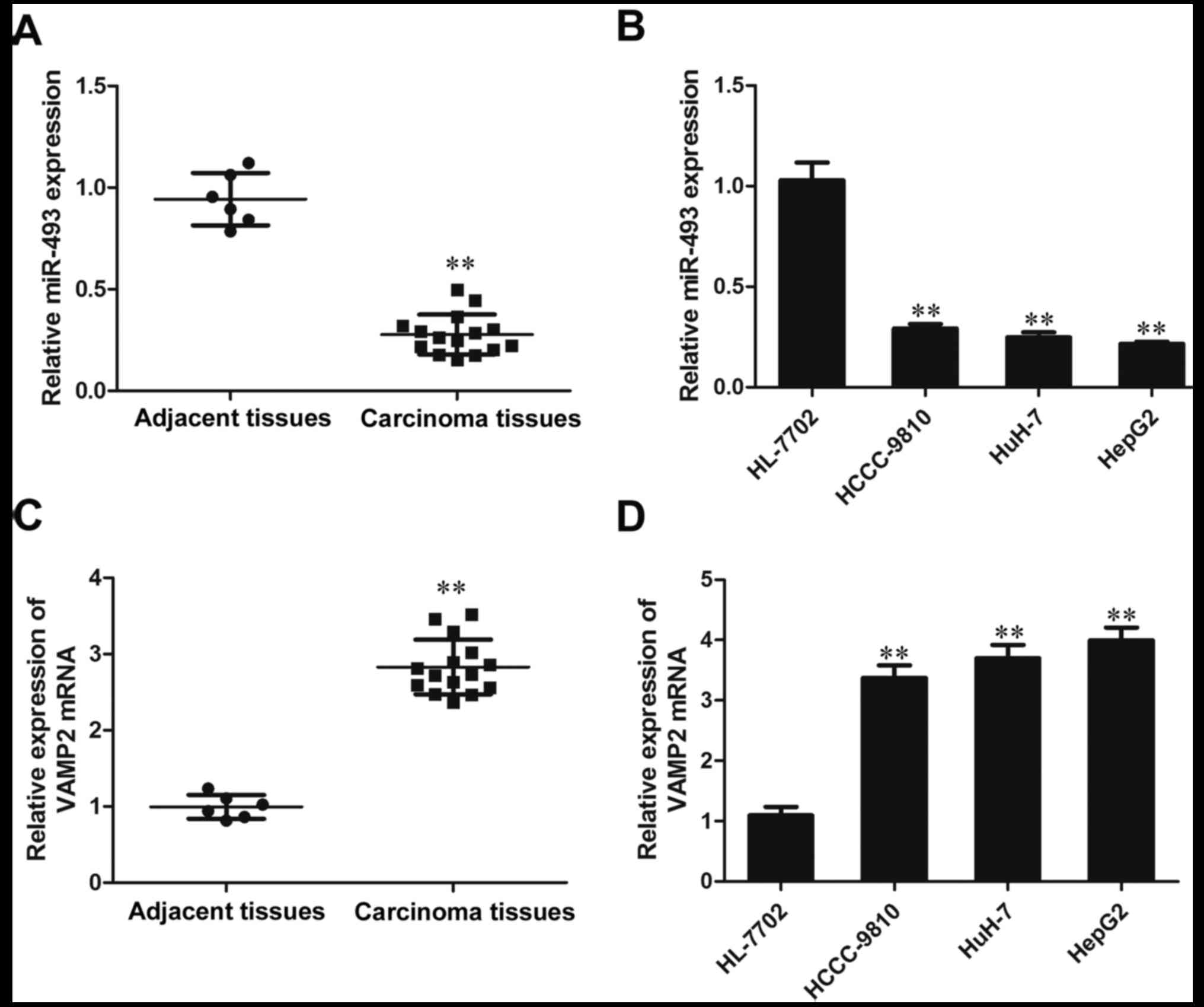
The results of RT-qPCR analysis indicated that the
expression of miR-493-5p in the HCC tissue samples was
significantly lower than that in the adjacent normal tissues
(P<0.01). The expression of miR-493-5p in the liver cancer cell
lines HCCC-9810, HuH-7 and HepG2 was reduced significantly compared
with that in the HL-7702 hepatic cells (P<0.01; Fig. 1A and B). These results indicate
that the expression of miR-493-5p was inhibited in the liver cancer
tissues and cell lines. However, in contrast to miR-493-5p, the
expression of VAMP2 was observed to be increased in the liver
cancer tissues and cell lines compared with those in the adjacent
normal tissues and HL-7702 cells, respectively (P<0.01; Fig. 1C and D). These results demonstrate
that the expression of VAMP2 is elevated in liver cancer tissues
and cell lines.
Effect of miR-493-5p mimic on the
expression of miR-493-5p in HepG2 cells
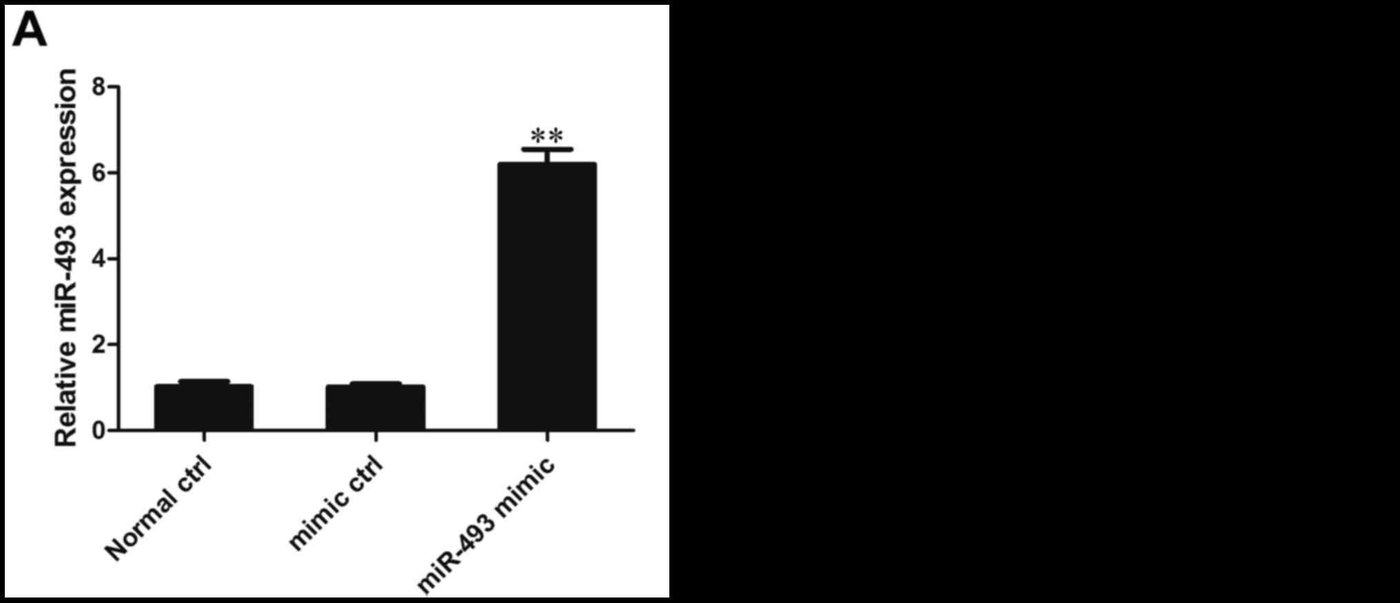
The expression of miR-493-5p in HepG2 cells was
detected following their transfection with miR-493-5p mimic. The
results of RT-qPCR analysis demonstrated that the expression of
miR-493-5p in the miR-493-5p mimic transfection group was
significantly higher than that in the negative control and normal
control groups, indicating that the transfection was effective in
inducing the overexpression of miR-493-5p (P<0.01; Fig. 2A).
Effect of miR-493-5p on cell
viability
In an MTT assay conducted 2 days after transfection
of the HepG2 cells with the miR-493-5p mimic, the cell viability of
the miR-493-5p mimic transfection group was significantly lower
than that of the normal control and negative control groups (both
P<0.01; Fig. 2B). These
results indicate that the overexpression of miR-493-5p reduced the
viability of the HepG2 cells.
Effect of miR-493-5p on the cell cycle
and apoptosis
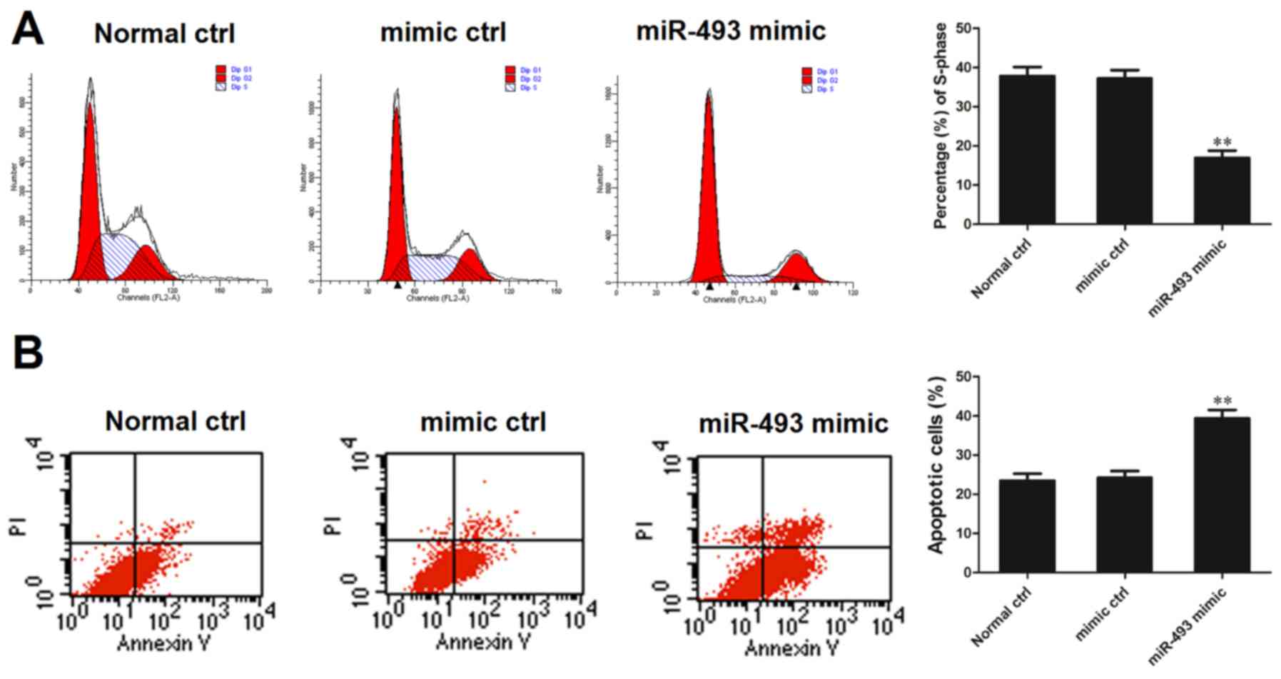
The HepG2 cell line was transfected with miR-493-5p
mimic or negative control and 48 h later, the cells were subjected
to flow cytometric analysis of the cell cycle and apoptosis. The
percentage of cells in the S phase was determined relative to the
total number of cells in the G0/G1, S and G2M phases. The flow
cytometric analysis demonstrated that the percentage of cells in
the S phase in the miR-493-5p mimic transfection group was
significantly smaller compared with that in the normal control and
negative control groups (both P<0.01; Fig. 3A). On the basis of these results,
it may be speculated that miR-493-5p is an inhibitor of HepG2 cell
replication. Analysis of cell apoptosis indicated that the
proportion of apoptotic cells in the miR-493-5p mimic transfection
group was significantly greater than that in the normal control and
negative control groups (both P<0.01; Fig. 3B). This assay indicates that
miR-493-5p promoted cell apoptosis.
Effect of miR-493-5p on the invasion of
HepG2 cells
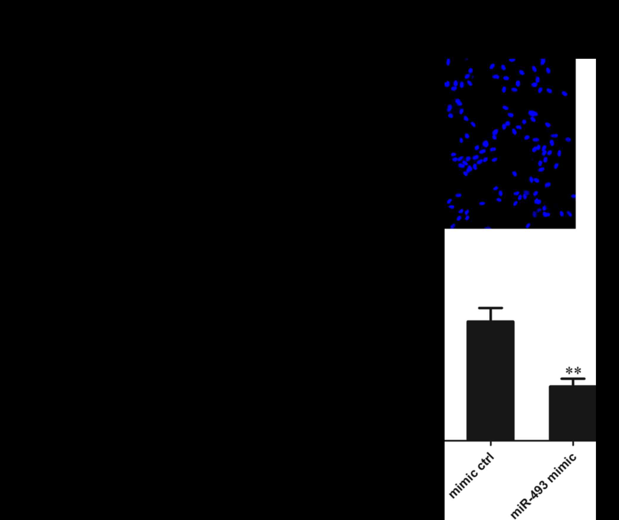
The HepG2 cells were transfected with miR-493-5p
mimic or negative control and 48 h later, they were subjected to
Transwell invasion experiments. The results of Hoechst 33342
staining revealed that the number of cells migrating through the
polycarbonate membrane in the miR-493-5p mimic transfection group
was significantly reduced compared with that in the mimic and
normal control groups (both P<0.01). These results suggest that
miR-493-5p inhibits the migration of HepG2 cells (Fig. 4).
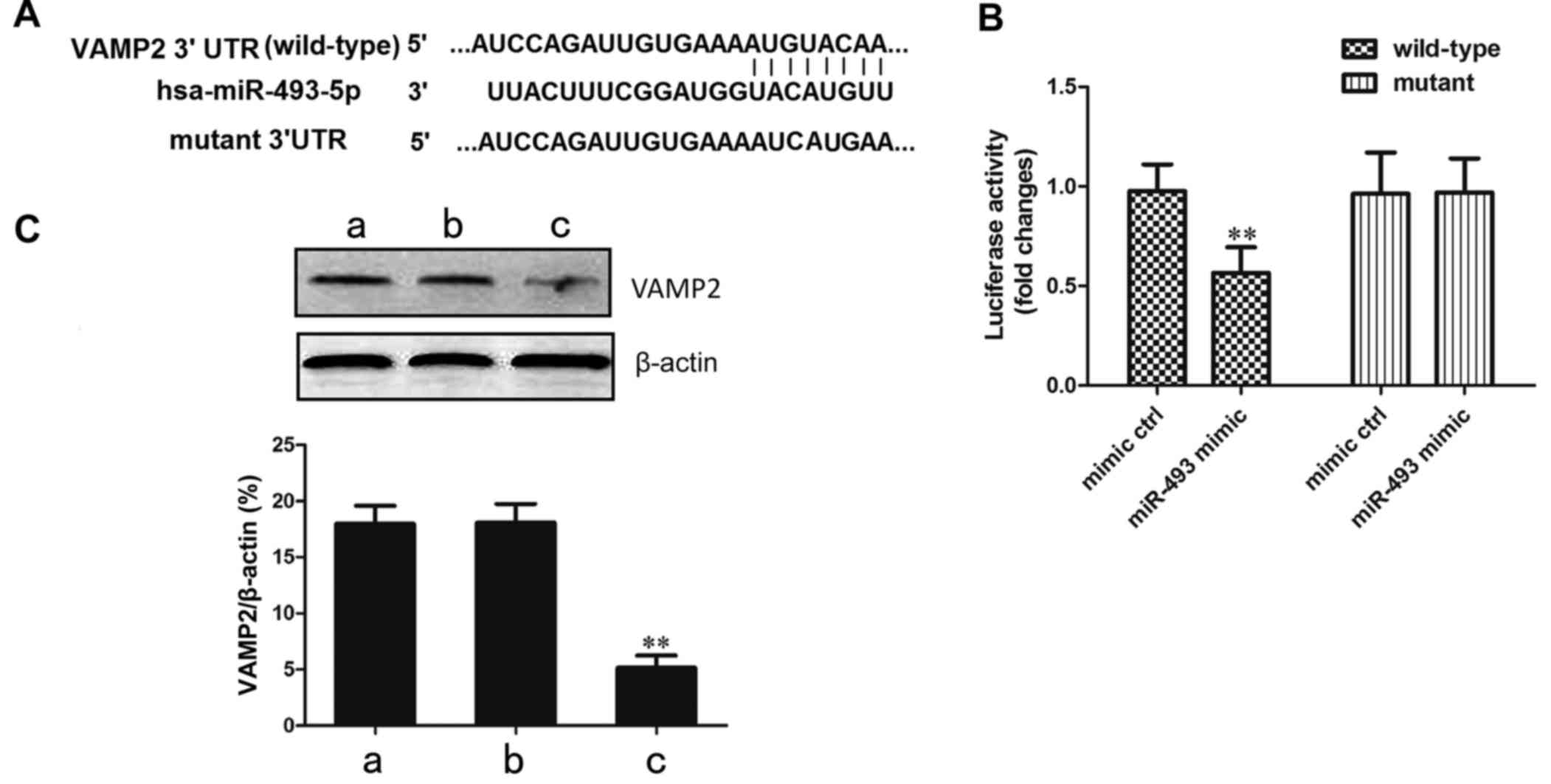
Confirmation that the VAMP2 gene is
targeted by miR-493-5p
In order to confirm that the target site of
miR-493-5p is located in the 3′-UTR of the VAMP2 gene, recombinant
vectors containing wild-type and mutant VAMP2 3′-UTR in the seed
region were established (Fig.
5A). These plasmids were transfected together with miR-493-5p
mimic or negative control into the 293 cell line. In the cells
transfected with wild-type VAMP2, the luciferase activity in the
miR-493-5p mimic transfection group was significantly decreased
compared with that in the negative control group (P<0.01;
Fig. 5B). This indicates that the
predicted site of VAMP2 was directly bound by miR-493-5p.
The regulation of the VAMP2 gene by miR-493-5p was
verified by western blot analysis. The results demonstrated that
the protein level of VAMP2 in the miR-493-5p mimic transfection
group was significantly lower than those in the mimic control and
normal control groups (P<0.01; Fig. 5C). These results of the luciferase
and western blot assays confirm that the overexpression of
miR-493-5p resulted in a reduction of VAMP2 protein levels.
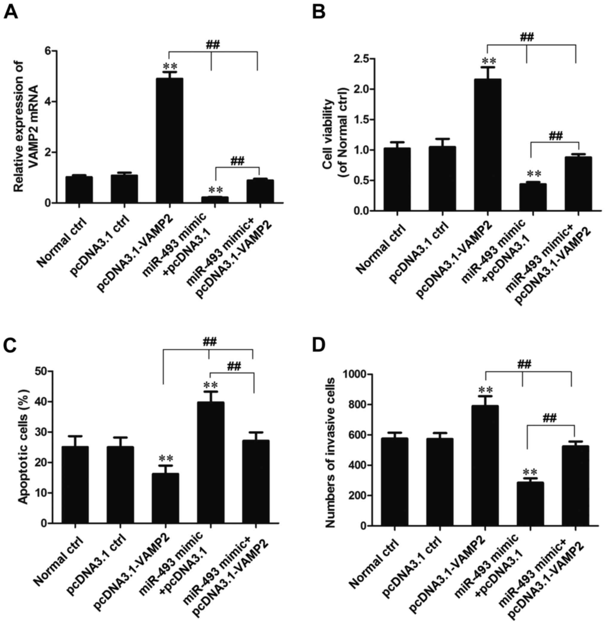
miR-493-5p affects cell behavior by
decreasing the expression of VAMP2
HepG2 cells were cotransfected with miR-493-5p mimic
and pcDNA3.1-VAMP2. Whether the effects of miR-493-5p on cell
behavior are dependent upon the regulation of VAMP2 expression was
evaluated by MTT, flow cytometric and Transwell chamber migration
assays (Fig. 6).
The expression of VAMP2 mRNA was detected using
RT-qPCR, and the results indicated that the expression of VAMP2
mRNA in the pcDNA3.1-VAMP2 transfection group was increased
significantly compared with that in the normal and pcDNA3.1 control
groups (P<0.01). However, the expression of VAMP2 mRNA in the
miR-493-5p mimic + pcDNA3.1-VAMP2 transfection group was
significantly reduced compared with that in the pcDNA3.1-VAMP2
transfection group (P<0.01), significantly increased compared
with that in the miR-493-5p mimic + pcDNA3.1 transfection group
(P<0.01), and slightly reduced compared with that in the normal
control and pcDNA3.1 control group (Fig. 6A).
Analysis using MTT and Transwell experiments
demonstrated that the cell viability and invasion ability of cells
in the miR-493-5p mimic + pcDNA3.1-VAMP2 group was significantly
lower than that in the pcDNA3.1-VAMP2 transfection group
(P<0.01), significantly higher than that in the miR-493-5p mimic
+ pcDNA3.1 transfection group (P<0.01), and slightly lower than
that in the normal control and pcDNA3.1 control groups (Fig. 6B and D). Flow cytometric analysis
indicated that the percentage of apoptotic cells in the miR-493-5p
mimic + pcDNA3.1-VAMP2 transfection group was significantly higher
than that in the pcDNA3.1-VAMP2 transfection group (P<0.01),
significantly lower than that in the miR-493-5p mimic + pcDNA3.1
transfection group (P<0.01) and slightly higher than that in the
normal control and pcDNA3.1 control groups (Fig. 6C). These results indicate that the
overexpression of VAMP2 reverses the effect of miR-493-5p as an
inhibitor of proliferation and inducer of apoptosis.
Discussion
Liver cancer is a highly malignant tumor with an
insidious onset, invasive properties, and high recurrence and
fatality rates. Liver cancer is often detected at a late stage
because it is difficult to diagnose at an early stage and the
disease lacks sensitivity to radiation and chemotherapy, which
makes surgical removal more challenging. This results in a poor
prognosis and endangers human life (42,43). As with other cancers, multiple
genes and environmental factors are involved in the initiation and
progression of liver cancer. These are associated with numerous
pathological processes and molecular events, and result in the
transformation of normal liver cells into cancer cells and the
occurrence of metastasis (44).
Previous studies have shown that non-coding RNA is involved in
tumorigenesis (45,46). While certain microRNAs, including
miR-21 and miR-765 increase tumor invasion and metastasis and thus
function as carcinogenic miRNAs (47), other microRNAs, such as miR-335,
miR-146a and miR-543, inhibit tumor occurrence, development and
invasion, and function as suppressor miRNAs (48). Notably, miR-493-5p has been
reported to serve a tumor suppressor role and prohibit the
proliferation and invasion of gastric carcinoma (39). In the present study, in HCC
specimens, adjacent normal tissues and liver cancer cell lines, the
expression of miRNA-493-5p was measured by RT-qPCR and the results
revealed that the level of miR-493-5p in the HCC tissues was
significantly lower than that in the adjacent normal tissues.
Furthermore, the expression of miR-493-5p in the liver cancer cell
lines HCCC-9810, HuH-7 and HepG2 was significantly reduced compared
with that in the HL-7702 hepatic cell line. This suggests that
miR-493-5p functions as a suppressor miRNA in liver cancer.
A recent study revealed that miR-493-5p was able to
reduce the expression of mitotic arrest deficient-2 and was
involved in the regulation of cancer cell mitosis as well as the
response to paclitaxel chemotherapy (49). Okamoto et al (50) reported that the apoptosis of colon
cancer cells induced by miR-493-5p blocked their metastasis from
the colon to the liver. In the present study, the role of
miR-493-5p in the cell behavior of HepG2 cells was analyzed using
MTT, flow cytometry and Transwell assays. The results demonstrate
that the overexpression of miR-493-5p decreased cell viability,
attenuated cell proliferation and migration and increased apoptosis
in HepG2 cells. These data suggest that miR-493-5p may act as a
tumor suppressor miRNA in liver cancer.
VAMP2, a member of the soluble N-ethylmaleimide
sensitive factor attachment protein receptor family, is responsible
for the intracellular transportation and extracellular secretion of
vesicle, for example, in the transport of membrane proteins and the
secretion of cytokines and hormones (51,52). A number of events in vesicle
transport, including tethering, anchoring and membrane fusion,
involve the participation of VAMPs (53). A recent study demonstrated that
extracellular vesicles and their contents are potential biomarkers
for liver cancer (54). In the
present study, the expression of VAMP2 was examined by RT-qPCR in
HCC tissues and adjacent normal tissues as well as in liver cancer
cell lines. The results revealed that the expression of VAMP2 in
HCC tissue was significantly higher than that in the adjacent
normal tissue and that the expression of VAMP2 in liver cancer cell
lines (HCCC-9810, HuH-7 and HepG2) was significantly higher than
that in the HL-7702 normal hepatic cell line. The initiation and
progression of certain types of cancer is closely associated with
compositional changes in the nervous system. For example, a study
revealed that acetylcholine is involved in the occurrence and
progression of hepatocarcinoma (55), and acetylcholine is transported by
vesicles (56). The results of
the present study demonstrate that VAMP2 expression is increased in
liver cancer tissues, which suggests that VAMP2 serves an important
role in the occurrence and development of liver cancer.
Using the online software TargetScan, it was
predicted that the VAMP2 gene is a potential target of miR-493-5p.
The luciferase reporter system was used to verify the interaction
between miR-493-5p and the 3′-UTR of the VAMP2 gene. The
interaction was further confirmed by western blot analysis, and the
results indicated that the protein level of VAMP2 in the miR-493-5p
mimic transfection group was significantly lower than that in the
untransfected and negative control groups, which indicates that the
expression of VAMP2 was regulated by miR-493-5p. In order to verify
whether the effect exerted on cell behavior by miR-493-5p was
dependent upon VAMP2 participation, the recombinant vector
pcDNA3.1-VAMP2 was established. The results of MTT, flow cytometry
and Transwell experiments supported the conclusion that miR-493-5p
exerted its effects on cell behavior by regulating the expression
of VAMP2. These results demonstrate that the overexpression of
VAMP2 reversed the impact of miR-493-5p as an inhibitor of
proliferation and accelerator of apoptosis in liver cancer
cells.
In conclusion, the present study revealed that
miR-493-5p expression was reduced in liver cancer tissues and cell
lines, and the expression of VAMP2 was negatively regulated by
miR-493-5p. In addition, it demonstrates that miR-493-5p suppresses
cell proliferation and cell migration, and promotes cell apoptosis
in liver cancer by decreasing the expression of VAMP2. Therefore,
the induction of miR-493-5p may be a therapeutic option for
patients with liver cancer.
References
|
1
|
Ingle PV, Samsudin SZ, Chan PQ, Ng MK,
Heng LX, Yap SC, Chai AS and Wong AS: Development and novel
therapeutics in hepatocellular carcinoma: A review. Ther Clin Risk
Manag. 12:445–455. 2016. View Article : Google Scholar : PubMed/NCBI
|
|
2
|
Altekruse SF, Henley SJ, Cucinelli JE and
McGlynn KA: Changing hepatocellular carcinoma incidence and liver
cancer mortality rates in the United States. Am J Gastroenterol.
109:542–553. 2014. View Article : Google Scholar : PubMed/NCBI
|
|
3
|
Li H, Wei Y, Li B and Peng B: The first
case of total laparoscopic living donor right hemihepatectomy in
mainland China and literature review. Surg Laparosc Endosc Percutan
Tech. 26:172–175. 2016. View Article : Google Scholar : PubMed/NCBI
|
|
4
|
Wu L, Shen F, Xia Y and Yang YF: Evolving
role of radiopharmaceuticals in hepatocellular carcinoma treatment.
Anticancer Agents Med Chem. 16:1155–1165. 2016. View Article : Google Scholar : PubMed/NCBI
|
|
5
|
Navadgi S, Chang CC, Bartlett A, McCall J
and Pandanaboyana S: Systematic review and meta-analysis of
outcomes after liver resection in patients with hepatocellular
carcinoma (HCC) with and without bile duct thrombus. HPB (Oxford).
18:312–316. 2016. View Article : Google Scholar
|
|
6
|
Li YW, Yang FC, Lu HQ and Zhang JS:
Hepatocellular carcinoma and hepatitis B surface protein. World J
Gastroenterol. 22:1943–1952. 2016. View Article : Google Scholar : PubMed/NCBI
|
|
7
|
Yu SJ: A concise review of updated
guidelines regarding the management of hepatocellular carcinoma
around the world: 2010–2016. Clin Mol Hepatol. 22:7–17. 2016.
View Article : Google Scholar : PubMed/NCBI
|
|
8
|
Dost S, Baichoo E and Dieterich DT:
Chronic hepatitis B and C infection in the United States: A review
of current guidelines, disease burden and cost effectiveness of
screening. Expert Rev Anti Infect Ther. 14:511–521. 2016.
View Article : Google Scholar
|
|
9
|
Moirangthem A and Patel T: Mesenchymal
stem cell derived extracellular vesicles: A promising new
therapeutic approach for hepatic injury. Biotarget. 1:122017.
View Article : Google Scholar
|
|
10
|
Pascual S, Herrera I and Irurzun J: New
advances in hepatocellular carcinoma. World J Hepatol. 8:421–438.
2016. View Article : Google Scholar : PubMed/NCBI
|
|
11
|
Taketomi A: Development and future
directions of antiangiogenic therapy in hepatocellular carcinoma.
Int J Clin Oncol. 21:2052016. View Article : Google Scholar : PubMed/NCBI
|
|
12
|
Griffith OL, Griffith M, Krysiak K,
Magrini V, Ramu A, Skidmore ZL, Kunisaki J, Austin R, McGrath S,
Zhang J, et al: A genomic case study of mixed fibrolamellar
hepatocellular carcinoma. Ann Oncol. 27:1148–1154. 2016. View Article : Google Scholar : PubMed/NCBI
|
|
13
|
Li R, Yang D, Tang CL, Cai P, Ma KS, Ding
SY, Zhang XH, Guo DY and Yan XC: Combined hepatocellular carcinoma
and cholangiocarcinoma (biphenotypic) tumors: Clinical
characteristics, imaging features of contrast-enhanced ultrasound
and computed tomography. BMC Cancer. 16:1582016. View Article : Google Scholar : PubMed/NCBI
|
|
14
|
Lu SC, Mato JM, Espinosa-Diez C and Lamas
S: Micro-RNA-mediated regulation of glutathione and methionine
metabolism and its relevance for liver disease. Free Radic Biol
Med. 100:66–72. 2016. View Article : Google Scholar : PubMed/NCBI
|
|
15
|
Tornesello ML, Buonaguro L, Izzo F and
Buonaguro FM: Molecular alterations in hepatocellular carcinoma
associated with hepatitis B and hepatitis C infections. Oncotarget.
7:25087–25102. 2016. View Article : Google Scholar : PubMed/NCBI
|
|
16
|
Parasramka MA, Maji S, Matsuda A, Yan IK
and Patel T: Long non-coding RNAs as novel targets for therapy in
hepatocellular carcinoma. Pharmacol Ther. 161:67–78. 2016.
View Article : Google Scholar : PubMed/NCBI
|
|
17
|
D'Costa N, Chavez-Munoz C and So A: Role
of fibroblast growth factor receptor in sunitinib-resistant renal
cell carcinoma. Biotarget. 1:32017. View Article : Google Scholar
|
|
18
|
Liu YR, Tang RX, Huang WT, Ren FH, He RQ,
Yang LH, Luo DZ, Dang YW and Chen G: Long noncoding RNAs in
hepatocellular carcinoma: Novel insights into their mechanism.
World J Hepatol. 7:2781–2791. 2015. View Article : Google Scholar : PubMed/NCBI
|
|
19
|
Gonzalez-Rodriguez A and Valverde AM: RNA
interference as a therapeutic strategy for the treatment of liver
diseases. Curr Pharm Des. 21:4574–4586. 2015. View Article : Google Scholar : PubMed/NCBI
|
|
20
|
Xie QY, Almudevar A, Whitney-Miller CL,
Barry CT and McCall MN: A microRNA biomarker of hepatocellular
carcinoma recurrence following liver transplantation accounting for
within-patient heterogeneity. BMC Med Genomics. 9:182016.
View Article : Google Scholar : PubMed/NCBI
|
|
21
|
Lee CH, Kim JH and Lee SW: The role of
microRNA in pathogenesis and as markers of HCV chronic infection.
Curr Drug Targets. 17:12016.
|
|
22
|
Fang Q, Xu T, Wu C, Zhou S and Sun H:
Biotargets in neural regeneration. Biotarget. 1:62017. View Article : Google Scholar
|
|
23
|
Afonso MB, Rodrigues PM, Simão AL and
Castro RE: Circulating microRNAs as potential biomarkers in
non-alcoholic fatty liver disease and hepatocellular carcinoma. J
Clin Med. 5:52016. View Article : Google Scholar
|
|
24
|
Wang L, Yue Y, Wang X and Jin H: Function
and clinical potential of microRNAs in hepatocellular carcinoma.
Oncol Lett. 10:3345–3353. 2015. View Article : Google Scholar
|
|
25
|
Mizuguchi Y, Takizawa T, Yoshida H and
Uchida E: Dysregulated miRNA in progression of hepatocellular
carcinoma: a systematic review. Hepatol Res. 46:391–406. 2016.
View Article : Google Scholar
|
|
26
|
Wang Q, Wei L, Guan X, Wu Y, Zou Q and Ji
Z: Briefing in family characteristics of microRNAs and their
applications in cancer research. Biochim Biophys Acta. 1844(1 Pt
B): 191–197. 2013. View Article : Google Scholar : PubMed/NCBI
|
|
27
|
Kaplan BB, Kar AN, Gioio AE and Aschrafi
A: MicroRNAs in the axon and presynaptic nerve terminal. Front Cell
Neurosci. 7:1262013. View Article : Google Scholar : PubMed/NCBI
|
|
28
|
Li M, Fu W, Wo L, Shu X, Liu F and Li C:
miR-128 and its target genes in tumorigenesis and metastasis. Exp
Cell Res. 319:3059–3064. 2013. View Article : Google Scholar : PubMed/NCBI
|
|
29
|
Leite-Moreira AM, Lourenço AP,
Falcão-Pires I and Leite-Moreira AF: Pivotal role of microRNAs in
cardiac physiology and heart failure. Drug Discov Today.
18:1243–1249. 2013. View Article : Google Scholar : PubMed/NCBI
|
|
30
|
Piva R, Spandidos DA and Gambari R: From
microRNA functions to microRNA therapeutics: novel targets and
novel drugs in breast cancer research and treatment (Review). Int J
Oncol. 43:985–994. 2013. View Article : Google Scholar : PubMed/NCBI
|
|
31
|
Palumbo S, Miracco C, Pirtoli L and
Comincini S: Emerging roles of microRNA in modulating cell-death
processes in malignant glioma. J Cell Physiol. 229:277–286. 2014.
View Article : Google Scholar
|
|
32
|
Zhang WC, Liu J, Xu X and Wang G: The role
of microRNAs in lung cancer progression. Med Oncol. 30:6752013.
View Article : Google Scholar : PubMed/NCBI
|
|
33
|
Yu JJ and Xia SJ: Novel role of microRNAs
in prostate cancer. Chin Med J (Engl). 126:2960–2964. 2013.
|
|
34
|
Wu Y and Jiang M: The revolution of lung
cancer treatment: From vaccines, to immune checkpoint inhibitors,
to chimeric antigen receptor T therapy. Biotarget. 1:72017.
View Article : Google Scholar
|
|
35
|
Lanaya H, Natarajan A, Komposch K, Li L,
Amberg N, Chen L, Wculek SK, Hammer M, Zenz R, Peck-Radosavljevic
M, et al: EGFR has a tumour-promoting role in liver macrophages
during hepatocellular carcinoma formation. Nat Cell Biol.
16:972–977. 2014. View
Article : Google Scholar : PubMed/NCBI
|
|
36
|
Wang L, Yao J, Shi X, Hu LL, Li ZF, Song
TS and Huang C: MicroRNA-302b suppresses cell proliferation by
targeting EGFR in human hepatocellular carcinoma SMMC-7721 cells.
BMC Cancer. 13:4482013. View Article : Google Scholar : PubMed/NCBI
|
|
37
|
Wang W, Zhang H, Wang L, Zhang S and Tang
M: miR-613 inhibits the growth and invasiveness of human
hepatocellular carcinoma via targeting DCLK1. Biochem Biophys Res
Commun. 473:987–992. 2016. View Article : Google Scholar : PubMed/NCBI
|
|
38
|
Zhou X, Zhu HQ, Ma CQ, Li HG, Liu FF,
Chang H and Lu J: miR-1180 promoted the proliferation of
hepatocellular carcinoma cells by repressing TNIP2 expression.
Biomed Pharmacother. 79:315–320. 2016. View Article : Google Scholar : PubMed/NCBI
|
|
39
|
Zhou W, Zhang C, Jiang H, Zhang Z, Xie L
and He X: miR-493 suppresses the proliferation and invasion of
gastric cancer cells by targeting RhoC. Iran J Basic Med Sci.
18:1027–1033. 2015.
|
|
40
|
Sakai H, Sato A, Aihara Y, Ikarashi Y,
Midorikawa Y, Kracht M, Nakagama H and Okamoto K: MKK7 mediates
miR-493-dependent suppression of liver metastasis of colon cancer
cells. Cancer Sci. 105:425–430. 2014. View Article : Google Scholar : PubMed/NCBI
|
|
41
|
Livak KJ and Schmittgen TD: Analysis of
relative gene expression data using real-time quantitative PCR and
the 2(-Delta Delta C(T)) Method. Methods. 25:402–408. 2001.
View Article : Google Scholar
|
|
42
|
Tzeng CW, Tzeng WS, Lin LT, Lee CW, Yen FL
and Lin CC: Enhanced autophagic activity of artocarpin in human
hepatocellular carcinoma cells through improving its solubility by
a nanoparticle system. Phytomedicine. 23:528–540. 2016. View Article : Google Scholar : PubMed/NCBI
|
|
43
|
Mouli S, Hickey R, Thornburg B, Sato KT,
Desai K, Gabr A, Kallini JR, Niemeri H, Kircher S, Mulcahy MF, et
al: Single-versus triple-drug chemoembolization for hepatocellular
carcinoma: Comparing outcomes by toxicity, imaging response, and
survival. J Vasc Interv Radiol. 27:1279–1287. 2016. View Article : Google Scholar : PubMed/NCBI
|
|
44
|
Ren Y, Qiu L, Lü F, Ru X, Li S, Xiang Y,
Yu S and Zhang Y: TALENs-directed knockout of the full-length
transcription factor Nrf1α that represses malignant behaviour of
human hepatocellular carcinoma (HepG2) cells. Sci Rep. 6:237752016.
View Article : Google Scholar
|
|
45
|
Zhang D, Cao C, Liu L and Wu D:
Up-regulation of lncRNA SNHG20 predicts poor prognosis in
hepatocellular carcinoma. J Cancer. 7:608–617. 2016. View Article : Google Scholar : PubMed/NCBI
|
|
46
|
Gao JZ, Li J, Du JL and Li XL: Long
non-coding RNA HOTAIR is a marker for hepatocellular carcinoma
progression and tumor recurrence. Oncol Lett. 11:1791–1798. 2016.
View Article : Google Scholar : PubMed/NCBI
|
|
47
|
Vira D, Basak SK, Veena MS, Wang MB, Batra
RK and Srivatsan ES: Cancer stem cells, microRNAs, and therapeutic
strategies including natural products. Cancer Metastasis Rev.
31:733–751. 2012. View Article : Google Scholar : PubMed/NCBI
|
|
48
|
Link A, Kupcinskas J, Wex T and
Malfertheiner P: Macro-role of microRNA in gastric cancer. Dig Dis.
30:255–267. 2012. View Article : Google Scholar : PubMed/NCBI
|
|
49
|
Tambe M, Pruikkonen S, Mäki-Jouppila J,
Chen P, Elgaaen BV, Straume AH, Huhtinen K, Cárpen O, Lønning PE,
Davidson B, et al: Novel Mad2-targeting miR-493-3p controls mitotic
fidelity and cancer cells' sensitivity to paclitaxel. Oncotarget.
7:12267–12285. 2016. View Article : Google Scholar : PubMed/NCBI
|
|
50
|
Okamoto K, Ishiguro T, Midorikawa Y, Ohata
H, Izumiya M, Tsuchiya N, Sato A, Sakai H and Nakagama H: miR-493
induction during carcinogenesis blocks metastatic settlement of
colon cancer cells in liver. EMBO J. 31:1752–1763. 2012. View Article : Google Scholar : PubMed/NCBI
|
|
51
|
Han Q, Hong Y, Fu Z, Zhang M, Cao X, Liu
Y, Ma S, Guo Y, Lu K, Zhu C, et al: Characterization of VAMP2 in
Schistosoma japonicum and the evaluation of protective efficacy
induced by recombinant SjVAMP2 in mice. PLoS One. 10:e01445842015.
View Article : Google Scholar : PubMed/NCBI
|
|
52
|
Ropert N, Jalil A and Li D: Expression and
cellular function of vSNARE proteins in brain astrocytes.
Neuroscience. 323:76–83. 2016. View Article : Google Scholar
|
|
53
|
Kosiorek M, Zylinska L, Zablocki K and
Pikula S: Calcineurin/NFAT signaling represses genes Vamp1 and
Vamp2 via PMCA-dependent mechanism during dopamine secretion by
pheochromocytoma cells. PLoS One. 9:e921762014. View Article : Google Scholar : PubMed/NCBI
|
|
54
|
Mohankumar S and Patel T: Extracellular
vesicle long noncoding RNA as potential biomarkers of liver cancer.
Brief Funct Genomics. 15:249–256. 2016. View Article : Google Scholar :
|
|
55
|
Nie H, Cao Q, Zhu L, Gong Y, Gu J and He
Z: Acetylcholine acts on androgen receptor to promote the migration
and invasion but inhibit the apoptosis of human hepatocarcinoma.
PLoS One. 8:e616782013. View Article : Google Scholar : PubMed/NCBI
|
|
56
|
Rodrigues HA, Fonseca Mde C, Camargo WL,
Lima PM, Martinelli PM, Naves LA, Prado VF, Prado MA and Guatimosim
C: Reduced expression of the vesicular acetylcholine transporter
and neurotransmitter content affects synaptic vesicle distribution
and shape in mouse neuromuscular junction. PloS One. 8:e783422013.
View Article : Google Scholar : PubMed/NCBI
|