Introduction
Coronary heart disease, including acute myocardial
infarction, is considered to be one of the leading causes of
morbidity and mortality worldwide (1). Early and effective reperfusion
therapy for acute myocardial infarction can reduce infarct size and
improve the clinical outcomes of patients (2). However, reperfusion itself causes
ischemia/reperfusion (I/R) injury, which unexpectedly induces
additional cellular injury and paradoxically reduces the beneficial
effects of reperfusion therapy, leading to irreversible myocardial
tissue damage and increased infarct size (2). Therefore, it is necessary to protect
against myocardial I/R injury as an adjuvant therapy for the
treatment of acute myocardial infarction (3).
Multiple factors such as inflammation, oxidative
stress, calcium overload and apoptosis have been identified to
contribute to the process of myocardial I/R injury (2,4,5).
Autophagy has also been demonstrated to serve an important role in
regulating cardiomyocyte death induced by I/R (6). Autophagy is a dynamic process that
turns over damaged proteins and organelles through a
lysosome-associated degradation system and physiologically
maintains cellular homeostasis at basal level (7). However, over-activated autophagy
induces excessive self-digestion and degradation of essential
cellular constituents, which triggers non-apoptotic programmed cell
death (8). Emerging evidence has
reported that autophagy is upregulated during myocardial I/R injury
and contributes to cardiomyocyte death; whereas downregulation of
autophagy attenuates I/R-induced cardiomyocyte death and protects
against myocardial I/R injury (9–11).
The results of the aforementioned previous studies indicate that
modulation of autophagy may be a target strategy for treatment of
myocardial I/R injury.
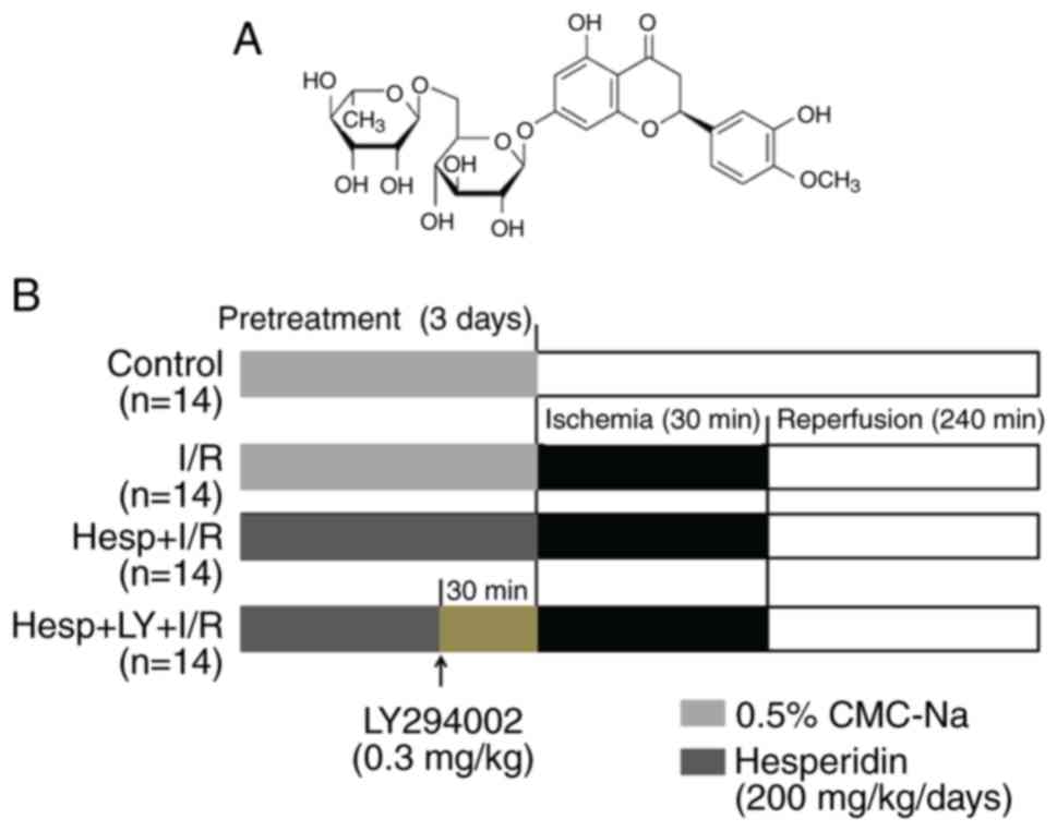
Hesperidin
[3′,5,7-trithydroxy-4′-methoxy-flavanone-7-(6-α-L-rhamnopyranosyl-β-D-glucopyranoside)]
is a natural flavanone glycoside abundantly present in citrus
fruits. It has a molecular formula of
C28H34O15 and a molecular weight
of 610.57 Da (12) (Fig. 1A). Hesperidin possesses good
radical scavenging activity and chelating metal ion ability due to
its structure, which readily donates electrons from the free
hydroxyls on the flavonoid nucleus (13). Furthermore, hesperidin has been
reported to exert a wide range of biological effects, including
anti-inflammatory (14),
anti-cancer (15),
radio-protective (16),
anti-allergic (17), and
antioxidant properties (18).
Gandhi et al (19)
demonstrated that hesperidin attenuates myocardial I/R
injury-induced arrhythmias and apoptosis. Our previous study
reported that hesperidin reduced myocardial I/R injury by
inhibiting inflammation and oxidative stress (20), however, its effect on autophagy
during myocardial I/R and the underlying mechanisms have not been
completely elucidated.
The phosphoinositide 3-kinase (PI3K)/protein kinase
B (Akt) pathway is known to serve an important role in the control
of cell growth, proliferation, survival and metabolism (21). Mammalian target of rapamycin
(mTOR), a major intracellular repressor of autophagy, is positively
regulated by the PI3K/Akt pathway (22). Our previous study reported that
hesperidin could activate the PI3K/Akt pathway (20). Therefore, it was hypothesized that
the PI3K/Akt/mTOR pathway may contribute to the regulatory effect
of hesperidin on autophagy induced by myocardial I/R injury.
In the present study, the effect of hesperidin on
autophagy induced by I/R was investigated, and its underlying
mechanisms were explored, with particular focus on the potential
involvement of the PI3K/Akt/mTOR pathway in the regulation of
myocardial autophagy.
Materials and methods
Animals
Adult male Sprague-Dawley rats (6–8 weeks old;
weighing 200–250 g; n=56) were supplied by the animal experiment
center of Wuhan University (Wuhan, China), and were housed in an
air-conditioned room (temperature, 25±3°C; humidity, 50–60%) with
free access to food and water on a 12 h light/12 h dark cycle. All
experimental protocols conformed to the Guideline for the Care and
Use of Laboratory Animals published by the US National Institutes
of Health and were approved by the Institutional Animal Care and
Use Committee of Wuhan University (Approval no. 2016-0318).
Experimental protocol and myocardial I/R
model
Hesperidin (HPLC>98%) was purchased from
Sinopharm Chemical Reagent Co., Ltd. (Shanghai, China) and
dissolved in 0.5% sodium carboxymethyl-cellulose (CMC-Na) to
produce a 100 mg/ml solution. Then, the solution of hesperidin or
0.5% CMC-Na was administered orally to rats by gavage for a total
of 3 days (20,23). After being anesthetized with an
intraperitoneal injection of sodium pentobarbital (45 mg/kg;
Sigma-Aldrich; Merck KGaA, Darmstadt, Germany), all animals were
artificially ventilated using a volume-controlled rodent respirator
and monitored with an electrocardiogram using a computer-based EP
system (LEAD2000B; Jinjiang Ltd., Chengdu, China). A thoracotomy
through a left parasternal incision was performed to expose the
heart. A 4–0 silk was passed under the left anterior descending
coronary artery (LAD). The myocardial I/R model was induced by
ligation and release of the silk. A successful myocardial I/R model
was verified by changes of ST segment elevation in Lead-II and
regional cyanosis of the myocardial surface.
A total of 56 rats were randomly divided into the
following four groups (n=14 per group): i) Sham operated control
(Control): 0.5% CMC-Na; ii) Ischemia/reperfusion (I/R): 0.5%
CMC-Na; iii) Hesperidin (200 mg/kg/day) + I/R (Hesp+I/R); or iv)
Hesperidin (200 mg/kg/day) + LY294002 + I/R (Hesp+LY+I/R):
Following a 3 day pretreatment with hesperidin (200 mg/kg/day)
(20), rats in the Hesp+LY+I/R
group were administered LY294002 [a specific PI3K inhibitor, 0.3
mg/kg (24), Sigma-Aldrich; Merck
KGaA] via a caudal vein 30 min prior to LAD occlusion. Rats in the
Control group were subjected to surgical manipulation without
ligaturing the LAD; however, rats in the remaining groups were
subjected to 30 min of LAD occlusion followed by a 4 h reperfusion.
At the end of the experiment, the rats were sacrificed, and their
blood samples and hearts were harvested for subsequent analysis.
The study protocol is illustrated in Fig. 1B.
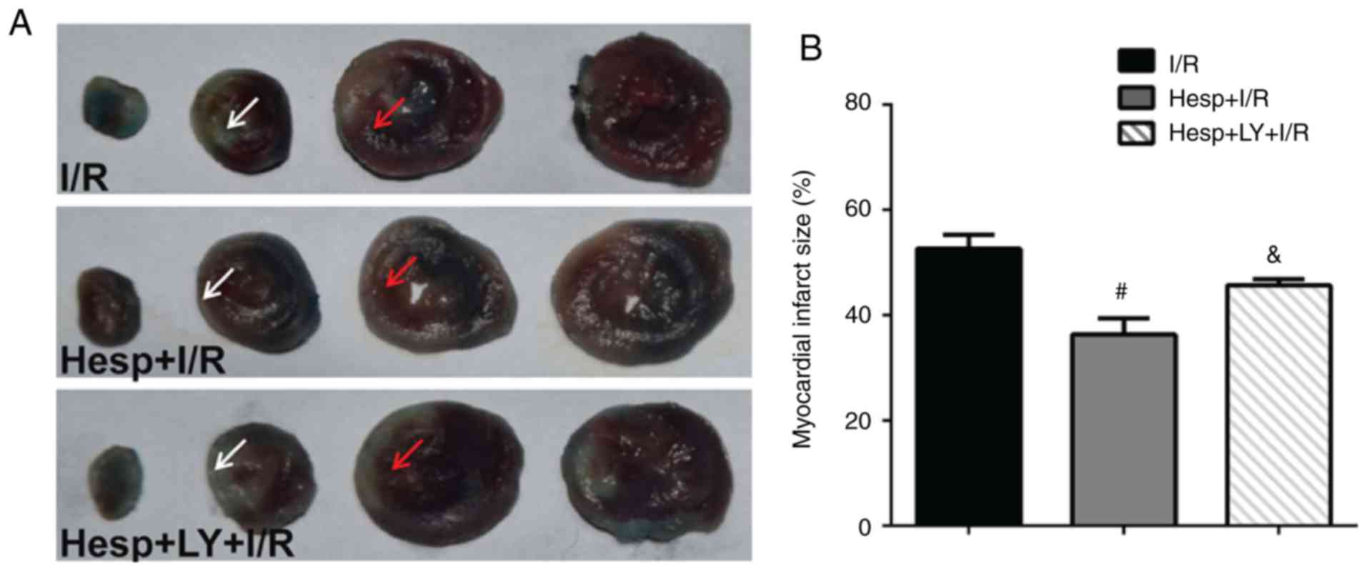
Myocardial infarct size
Myocardial infarct size was assessed by
2,3,5-triphenyltetrazolium chloride (TTC; Sigma-Aldrich; Merck
KGaA) as previously described (19,25). Briefly, following reperfusion, the
LAD was occluded again and 2 ml of 1% Evans blue dye
(Sigma-Aldrich; Merck KGaA) was infused via the femoral vein.
Hearts were removed and cut (~2 mm) from apex to base. The slices
were incubated in 1% TTC at 37°C for 20 min and fixed in 4%
paraformaldehyde at room temperature overnight. The infarct area
(white) and the risk area (red) in each section were determined by
Image-Pro Plus 6.0 (Media Cybernetics, Inc., Rockville, MD, USA). A
total of 6 animals in each group were used for myocardial infarct
size measurement. Myocardial infarct size was expressed as a
percentage of the risk area volume [%, infarct area / (risk +
infarct area)].
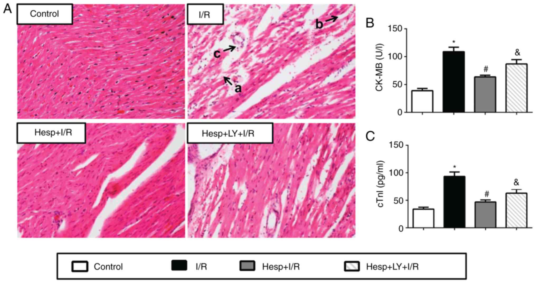
Myocardial injury
Serum levels of creatine kinase-MB (CK-MB) and
cardiac troponin I (cTnI) were used as indicators of myocardial
injury. Blood samples were centrifuged at 1,000 × g for 15 min at
room temperature, and the serum was collected for analysis.
Standard techniques were performed to determine the serum levels of
CK-MB and cTnI by using commercial kits (cat. no. E006; Nanjing
Jiancheng Bioengineering Institute, Nanjing, China) and
enzyme-linked immunosorbent assay kits (cat. no. E-EL-R0055c;
Elabscience, Wuhan, China), each procedure was conducted according
to the manufacturer's protocol.
Western blot analysis
Total protein extracts from the rat heart tissue
were prepared as previously described (26). Protein extraction was performed
using a radioimmunoprecipitation assay lysis buffer (cat. no.
P0013B; Beyotime Institute of Biotechnology, Jiangsu, China), and
the concentration of the protein was measured by a bicinchoninic
acid protein assay kit (cat. no. P0010; Beyotime Institute of
Biotechnology). A total of 50 µg protein per lane was
electrophoresed on 10% sodium dodecyl sulfate-polyacrylamide gel
and transferred to a nitrocellulose membrane. The membranes were
blocked with 5% non-fat dry milk in Tris-buffer saline-0.05% Tween
for 2 h at room temperature and incubated respectively at 4°C
overnight with the following primary antibodies including:
Anti-light chain (LC)3 (cat. no. 2775; 1:1,000), Beclin1 (cat. no.
3738; 1:1,000), mTOR (cat. no. 2972S; 1:1,000), phosphorylated
(p)-mTOR (cat. no. 2971S; 1:1,000), Akt (cat. no. 4691; 1:1,000),
p-Akt (cat. no. 4060; 1:2,000), p-PI3K (cat. no. 4228; 1:1,000) and
PI3K (cat. no. 4257; 1:800) (Cell Signaling Technology, Inc.,
Danvers, MA, USA). The membrane was then washed and incubated with
a horseradish peroxidase conjugated secondary antibody (cat. no.
BA1054; 1:50,000, Wuhan Boster Biological Technology, Ltd., Wuhan,
China) at 37°C for 2 h. The protein bands were visualized by an
enhanced chemiluminescence system (Thermo Fisher Scientific, Inc.,
Waltham, MA, USA), and glyceraldehydes-3-phosphate dehydrogenase
(GAPDH; cat. no. BM3896; 1:400; Wuhan Boster Biological Technology,
Ltd.) was used as an internal control to correct the variation
between samples. The densitometry of each band was quantified by
Image-Pro Plus 6.0 (Media Cybernetics, Inc.). A total of 5 hearts
in each group were used for the western blot analysis.
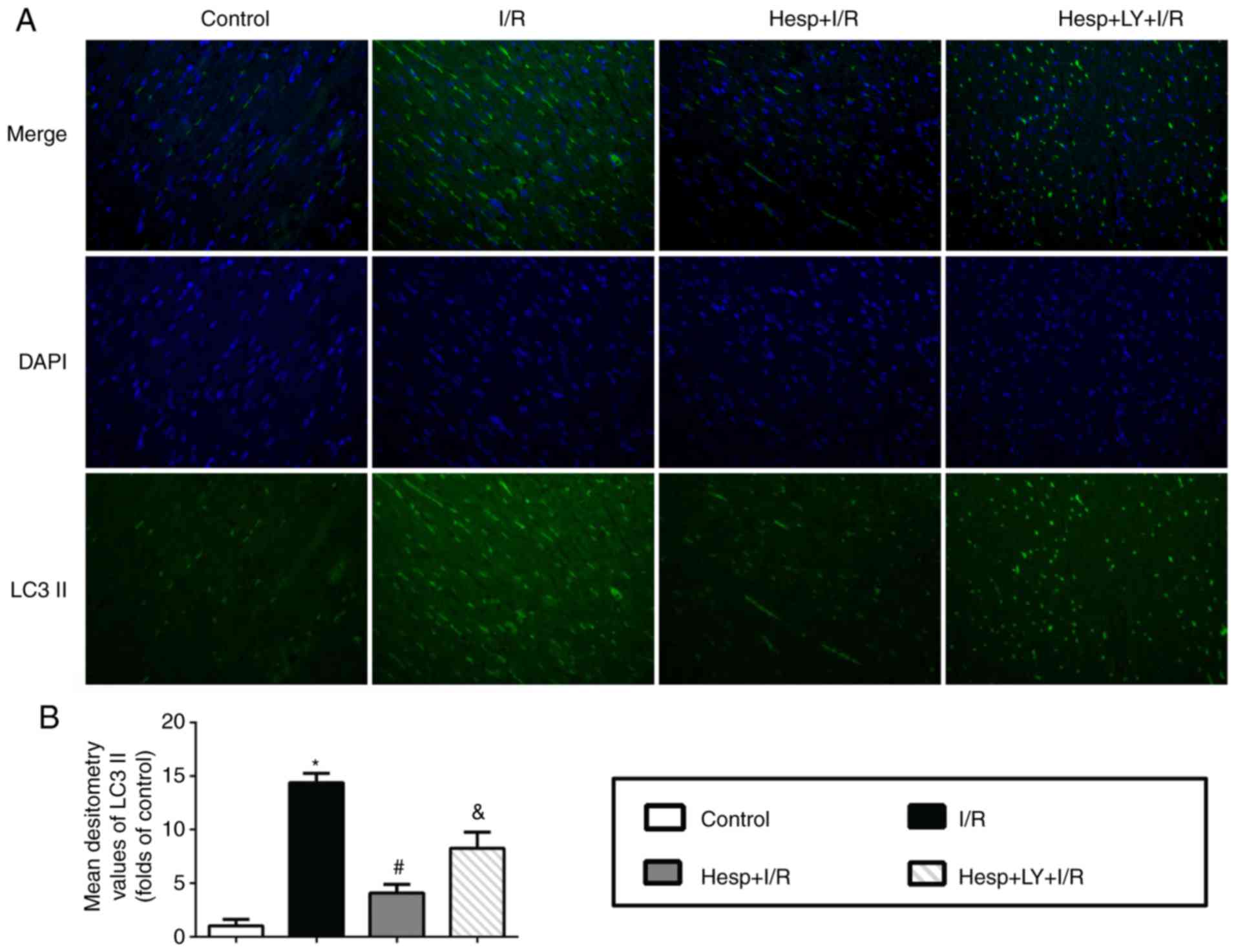
Immunofluorescence staining
To observe the LC3 expression in the myocardial
tissue, immunofluorescence staining was performed as described
previously (27). Following
deparaffinization, the paraffin-embedded sections (5 µm)
were replaced in the citrate buffer (pH 6.0) and boiled with
microwave on full power for antigen retrieval. After cooling, the
sections were washed with PBS (pH 7.4) three times (3 min each),
and blocked with 3% goat serum (cat. no. AR1009; Wuhan Boster
Biological Technology, Ltd.) for 30 min at room temperature. The
sections were then incubated with anti-LC3 antibody (1:100; Cell
Signaling Technology, Inc.) at 4°C overnight. Then, the sections
were incubated with a FITC conjugated secondary antibody (cat. no.
BA1105; 1:40; Wuhan Boster Biological Technology, Ltd.) at 37°C for
1 h. Nuclei were stained with 4′,6-diamidino-2-phenylindole (DAPI;
Sigma-Aldrich; Merck KGaA) for 5 min at room temperature in the
dark. A total of 6 fields at ×200 magnification were randomly
selected from 3 sections in each group and visualized with a
fluorescence microscope (excitation wavelength: 495 nm; DX51;
Olympus Corporation, Tokyo, Japan). The mean densitometry of the
fluorescence signal of LC3 in each field was analyzed by Image-pro
plus 6.0 software (Media Cybernetics, Inc.).
Hematoxylin and Eosin staining
Myocardial tissue was fixed in 4% paraformaldehyde,
routinely processed and embedded in paraffin. Paraffin-embedded
sections were stained with hematoxylin for 7 min, and eosin for 15
sec at room temperature. A total of 6 fields at ×200 magnification
were randomly selected from 3 sections in each group and visualized
with a light microscope to observe the myocardium damage.
Statistical analysis
All continuous data are expressed as the mean ±
standard deviation and analyzed using SPSS software, version 19.0
(IBM SPSS, Armonk, NY, USA). A one-way analysis of variance was
used for comparisons among groups with Bonferroni's adjustment for
multiple comparisons. Each experiment was repeated 3 times. All
plotting graphics were created using the GraphPad Prism 6.0
software (GraphPad Software, Inc., La Jolla, CA, USA). P<0.05
was considered to indicate a statistically significant
difference.
Results
Hesperidin decreases myocardial infarct
size during I/R
To investigate the cardioprotective effects of
hesperidin against I/R, the myocardial infarct size was determined.
As presented in Fig. 2, when
compared with the I/R group, the Hesp+I/R group exhibited a marked
decrease in the myocardial infarct size. However, the addition of
LY294002 partially inhibited the cardioprotective effect of
hesperidin (P<0.05).
Hesperidin alleviates cardiac injury
induced by I/R
The damage of myocardium induced by I/R was further
confirmed by hematoxylin and eosin staining and the serum levels of
CK-MB and cTnI. As presented in Fig.
3A, the myocardial structure in the I/R group exhibited
ruptured cardiac muscle fibers, necrosis with inflammatory cell
infiltration and interstitium edema; however, no such damage was
observed in the Control group. Hesperidin treatment prevented
myocardial damage after I/R, and the myocardium exhibited rescued
fiber disruption, necrosis and inflammatory cell infiltration.
However, the administration of LY294002 abolished these effects.
The serum CK-MB and cTnI levels were also determined to assess
myocardial injury (Fig. 3B and
C). Compared with the Control group, the serum CK-MB and cTnI
levels were significantly increased in the I/R group (P<0.05).
The serum CK-MB and cTnI levels were significantly lower in the
Hesp+I/R group compared with the I/R group. However, LY294002
partially reversed these effects (P<0.05).
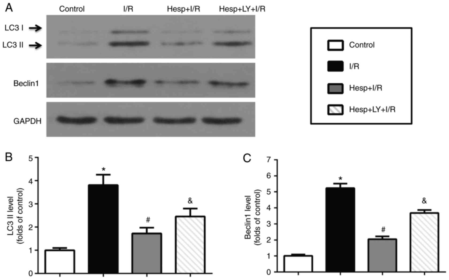
Hesperidin decreases autophagy in hearts
subjected to I/R
The present study first detected the regulatory
effect of hesperidin on LC3 and Beclin1 using immunofluorescence
staining and western blot analysis. As presented in Fig. 4A, the LC3 (green) signals
increased in the I/R group when compared with the Control group.
Hesperidin substantially decreased the I/R-induced LC3 signal
accumulation; however, its effect was abrogated by LY294002. The
mean densitometry values of LC3 and expression levels of LC3II and
Beclin1 were significantly increased in the I/R group when compared
with those in the Control group (P<0.05; Figs. 4B and 5). Hesperidin markedly suppressed the
increased mean densitometry values of LC3 and expression levels of
LC3II and Beclin1 induced by I/R (P<0.05), which indicated that
hesperidin could inhibit I/R-induced autophagy. However,
administration of LY294002 partially abolished these effects
(P<0.05).
Hesperidin increases expression levels of
p-mTOR, p-Akt and p-PI3K during myocardial I/R
To investigate the possible mechanism underlying the
inhibitory effect of hesperidin on autophagy during myocardial I/R
injury, the expression levels of p-mTOR, mTOR, p-Akt, Akt, p-PI3K
and PI3K in the myocardium were examined using western blot
analysis. As presented in Fig. 6,
compared with the Control group, the p-mTOR, p-Akt and p-PI3K
expression levels were significantly decreased in the I/R group
(P<0.05). Hesperidin treatment markedly inhibited the decrease
in the p-mTOR, p-Akt and p-PI3K expression levels, demonstrating
that hesperidin could activate the PI3K/Akt/mTOR pathway; however,
LY294002 significantly reversed these effects (P<0.05).
Additionally, no significant difference in mTOR, Akt and PI3K
expression levels were observed among the four groups
(P>0.05).
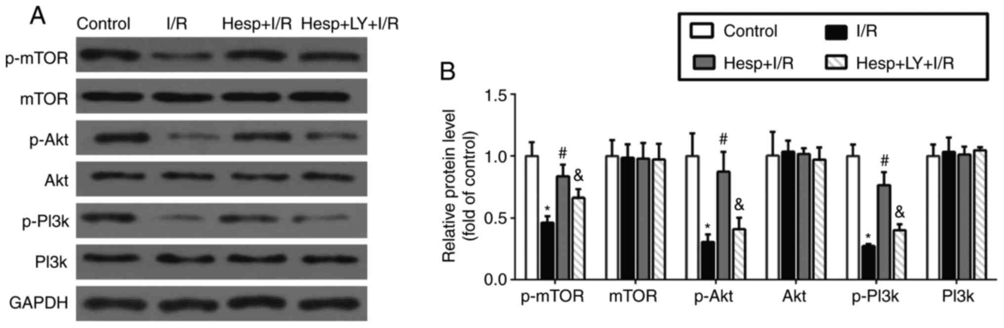 | Figure 6Effect of hesperidin on p-mTOR, mTOR,
p-Akt, Akt, p-PI3K and PI3K expression levels in the myocardium
during I/R. (A) Representative examples of western blot analysis
demonstrating the expression levels of p-mTOR, mTOR, p-Akt, Akt,
p-PI3K and PI3K. (B) Quantification of the expression levels of
p-mTOR, mTOR, p-Akt, Akt, p-PI3K and PI3K (n=5).
*P<0.05 vs. Control group; #P<0.05 vs.
I/R group; &P<0.05 vs. the Hesp+I/R group. P-,
phosphorylated; mTOR, mammalian target of rapamycin; PI3K,
phosphatidylinositol 3-kinase; Akt, protein kinase B; Hesp,
hesperidin; I/R, ischemia/reperfusion; LY, LY294002. |
Discussion
The present study aimed to investigate the potential
mechanism of cardioprotection of hesperidin against
ischemia/reperfusion (I/R), with a particular focus on autophagy.
To the best of our knowledge, the present study was the first to
demonstrate that hesperidin inhibited excessive autophagy following
I/R, which was displayed by the downregulation of LC3II and Beclin1
expression levels. Furthermore, hesperidin was demonstrated to
activate the PI3K/Akt/mTOR pathway, and LY294002, a specific PI3K
inhibitor, significantly reversed the effects of hesperidin. These
results suggested that inhibition of autophagy via activation of
the PI3K/Akt/mTOR pathway contributes to the cardioprotective
effect of hesperidin during I/R.
Irreversible loss of cardiomyocytes is the principal
cause of myocardial injury induced by myocardial infarction or I/R,
which leads to persistent cardiac dysfunction and heart failure
(28). Necrosis, apoptosis, and
autophagy, the three main mechanisms of cell death, are known to
participate in the loss of cardiomyocytes induced by I/R (28). Of these, autophagy, an
intracellular lysosomal degradative pathway, serves a key role in
cellular homeostasis (7). LC3 and
Beclin1 are important and reliable markers of autophagy. LC3 exists
in the cytosolic form (LC3I) and membrane bound form (LC3II)
(29). Increased LC3II is
considered to be closely associated with the extent of
autophagosome formation (30).
Beclin1 is essential in the recruitment of other autophagic
proteins during expansion of the pre-autophagosomal membrane and
structure (31).
Under basal conditions, autophagy functions as a
cell survival mechanism and maintains the cell size, construction
and function of cardiomyocytes (32). However, under the condition of
I/R, autophagy is upregulated and its role in modulating
cardiomyocyte survival and death remains controversial. Previous
studies have reported that autophagy promotes cell survival by
purging damaged proteins and organelles to generate the
intracellular building blocks required to maintain vital functions
during ischemia (33,34); whereas other studies demonstrated
that autophagy also promotes cell death via excessive
self-digestion and degradation of essential cellular constituents
(9,34). In the present study, the
immunofluorescence staining results demonstrated that the LC3
signals were markedly increased in the I/R group. The expression
levels of LC3II and Beclin1 in the myocardium, myocardial infarct
size and serum levels of CK-MB and cTnI were also significantly
upregulated following reperfusion, indicating that autophagy is
over-activated during I/R. These findings suggested that excessive
autophagy contributes to the process of myocardial I/R injury.
A growing number of epidemiological studies have
reported that diets rich in herbs, fruits and spices can attenuate
the risk of cardiovascular diseases (35,36). Hesperidin, a bioactive flavanone
glycoside, is highly abundant in citrus fruit (12). In a randomized crossover study,
Morand et al (37)
reported that hesperidin (294 mg in 500 ml of orange juice)
contributes to the cardioprotective effect of orange juice in
overweight men by decreasing diastolic blood pressure and
increasing endothelium-dependent microvascular reactivity. Another
double-blind, crossover study also reported that hesperidin
treatment (500 mg/day) improves endothelial function and reduces
inflammatory markers in patients with metabolic syndrome (38). Using the dose conversion of body
surface area comparisons between humans and rats (1:6.17) (39), 200 mg/kg/day of hesperidin used in
rats is equal to 1,944 mg/day for a 60-kg man, which is 6-fold
greater than 294 mg/day and 3-fold greater than 500 mg/day. Hence,
the dose of hesperidin used in the present study far exceeded a
nutritional dose; however, it functioned as a therapeutic dose.
The study of Huang et al (40) reported that hesperidin protects
against amyloid β-induced neuronal injury through suppression of
neuronal autophagy. In the present study, the data presented
similar results, which revealed that hesperidin significantly
downregulated the expression levels of LC3II and Beclin1 in the
heart of rats, which was accompanied with low myocardial infarct
size and low serum levels of CK-MB and cTnI. Combined, the
aforementioned results indicated that excessive autophagy
contributes to the process of myocardial I/R injury, and the
findings of the present study suggested that hesperidin could
reduce myocardial I/R injury by inhibiting excessive autophagy.
Autophagy can be modulated by multiple signaling
pathways, including the PI3K/Akt pathway (22). The PI3K/Akt pathway, a major
regulator of cell growth and survival, serves an essential role in
the mediation of myocardial cell survival in many situations
(27). Previous studies
demonstrated that activation of PI3K/Akt ameliorates myocardial I/R
injury (20,24). Furthermore, activation of the
PI3K/Akt pathway can phosphorylate mTOR, which is a key regulator
of autophagy (22).
Phosphorylated mTOR has been reported to protect against I/R injury
by reducing autophagy and enhancing recovery in the heart (41). It has been reported that
hesperidin activates the Akt pathway in a rat model of
neuroinflammation induced by aluminum chloride (42). Saiprasad et al (43) also demonstrated that hesperidin
regulates the PI3K/Akt/mTOR pathway in a mouse model of colon
carcinogenesis. Similar results were obtained in the present study,
which demonstrated that hesperidin significantly increased the
expression levels of p-PI3K, p-Akt and p-mTOR and decreased the
expression levels of LC3II and Beclin1, while LY294002, a specific
inhibitor of PI3K, markedly reversed the aforementioned effects of
hesperidin. These findings suggested that hesperidin inhibited
excessive autophagy via activating the PI3K/Akt/mTOR pathway.
Although the present study suggested that hesperidin
may be a novel drug for protection of myocardial I/R injury, there
are still various limitations. Firstly, the dose of hesperidin used
in the present study is a therapeutic dose; the effective range of
hesperidin dose and related safety evaluation need to be
investigated in further studies. Second, the effects of hesperidin
on longer term myocardial remodeling following myocardial I/R
injury also requires further exploration.
In conclusion, the present study demonstrated that
hesperidin reduced myocardial I/R injury by suppressing excessive
autophagy, and activation of the PI3K/Akt/mTOR pathway contributed
to the inhibitory effect of hesperidin on excessive autophagy.
Acknowledgments
Not applicable.
Funding
No funding was received.
Availability of data and materials
The data used and/or analyzed during the present
study are available from the corresponding author on reasonable
request.
Authors' contributions
XL, XH, and HJ conceived and designed the
experiment. XL, RM, CY and WX carried out the experiments. RM, CY
and WX acquired the reagents and materials. XL and JW analyzed the
data. XL, XH, JW and HJ wrote the manuscript. All authors approved
the final version of the manuscript.
Ethics approval and consent to
participate
The present study was approved by Institutional
Animal Care and Use Committee of Wuhan University (approval no.
2016-0318).
Patient consent for publication
Not applicable.
Competing interests
The authors declare that they have no competing
interests.
References
|
1
|
Roger VL, Go AS, Lloyd-Jones DM, Benjamin
EJ, Berry JD, Borden WB, Bravata DM, Dai S, Ford ES, Fox CS, et al:
Executive Summary: Heart disease and stroke statistics-2012 update:
A report from the american heart association. Circulation.
125:188–197. 2012. View Article : Google Scholar : PubMed/NCBI
|
|
2
|
Yellon DM and Hausenloy DJ: Myocardial
reperfusion injury. N Engl J Med. 357:1121–1135. 2007. View Article : Google Scholar : PubMed/NCBI
|
|
3
|
Ogura Y, Ouchi N, Ohashi K, Shibata R,
Kataoka Y, Kambara T, Kito T, Maruyama S, Yuasa D, Matsuo K, et al:
Therapeutic impact of follistatin-like 1 on myocardial ischemic
injury in preclinical models. Circulation. 126:1728–1738. 2012.
View Article : Google Scholar : PubMed/NCBI
|
|
4
|
Frangogiannis NG, Smith CW and Entman ML:
The inflammatory response in myocardial infarction. Cardiovasc Res.
53:31–47. 2002. View Article : Google Scholar
|
|
5
|
Gottlieb RA and Engler RL: Apoptosis in
myocardial ischemia-reperfusion. Ann N Y Acad Sci. 874:412–426.
1999. View Article : Google Scholar : PubMed/NCBI
|
|
6
|
Przyklenk K, Dong Y, Undyala VV and
Whittaker P: Autophagy as a therapeutic target for
ischaemia/reperfusion injury? Concepts, controversies, and
challenges. Cardiovasc Res. 94:197–205. 2012. View Article : Google Scholar : PubMed/NCBI
|
|
7
|
Marino G, Madeo F and Kroemer G: Autophagy
for tissue homeostasis and neuroprotection. Curr Opin Cell Biol.
23:198–206. 2011. View Article : Google Scholar
|
|
8
|
Loos B, Engelbrecht AM, Lockshin RA,
Klionsky DJ and Zakeri Z: The variability of autophagy and cell
death susceptibility: Unanswered questions. Autophagy. 9:1270–1285.
2013. View Article : Google Scholar : PubMed/NCBI
|
|
9
|
Ke J, Yao B, Li T, Cui S and Ding H: A2
Adenosine receptor-mediated cardioprotection against reperfusion
injury in rat hearts is associated with autophagy downregulation. J
Cardiovasc Pharmacol. 66:25–34. 2015. View Article : Google Scholar : PubMed/NCBI
|
|
10
|
Cao X, Chen A, Yang P, Song X, Liu Y, Li
Z, Wang X, Wang L and Li Y: Alpha-lipoic acid protects
cardiomyocytes against hypoxia/reoxygenation injury by inhibiting
autophagy. Biochem Biophys Res Commun. 441:935–940. 2013.
View Article : Google Scholar : PubMed/NCBI
|
|
11
|
Cheng BC, Huang HS, Chao CM, Hsu CC, Chen
CY and Chang CP: Hypothermia may attenuate
ischemia/reperfusion-induced cardiomyocyte death by reducing
autophagy. Int J Cardiol. 168:2064–2069. 2013. View Article : Google Scholar : PubMed/NCBI
|
|
12
|
Garg A, Garg S, Zaneveld LJ and Singla AK:
Chemistry and pharmacology of the Citrus bioflavonoid hesperidin.
Phytother Res. 15:655–669. 2001. View
Article : Google Scholar : PubMed/NCBI
|
|
13
|
Kuntic V, Brboric J, Holclajtner-Antunovic
I and Uskokovic-Markovic S: Evaluating the bioactive effects of
flavonoid hesperidin-a new literature data survey. Vojnosanit
Pregl. 71:60–65. 2014. View Article : Google Scholar
|
|
14
|
Wei D, Ci X, Chu X, Wei M, Hua S and Deng
X: Hesperidin suppresses ovalbumin-induced airway inflammation in a
mouse allergic asthma model. Inflammation. 35:114–121. 2012.
View Article : Google Scholar
|
|
15
|
Yumnam S, Hong GE, Raha S, Saralamma VV,
Lee HJ, Lee WS, Kim EH and Kim GS: Mitochondrial dysfunction and
Ca(2+) overload contributes to hesperidin induced paraptosis in
hepatoblastoma cells, HepG2. J Cell Physiol. 231:1261–1268. 2016.
View Article : Google Scholar
|
|
16
|
Petrova A, Davids LM, Rautenbach F and
Marnewick JL: Photoprotection by honeybush extracts, hesperidin and
mangiferin against UVB-induced skin damage in SKH-1 mice. J
Photochem Photobiol B. 103:126–139. 2011. View Article : Google Scholar : PubMed/NCBI
|
|
17
|
Kim SH, Kim BK and Lee YC: Antiasthmatic
effects of hesperidin, a potential Th2 cytokine antagonist, in a
mouse model of allergic asthma. Mediators Inflamm. 2011:4854022011.
View Article : Google Scholar : PubMed/NCBI
|
|
18
|
Selvaraj P and Pugalendi KV: Hesperidin, a
flavanone glycoside, on lipid peroxidation and antioxidant status
in experimental myocardial ischemic rats. Redox Rep. 15:217–223.
2010. View Article : Google Scholar : PubMed/NCBI
|
|
19
|
Gandhi C, Upaganalawar A and Balaraman R:
Protection against in vivo focal myocardial ischemia/reperfusion
injury-induced arrhythmias and apoptosis by hesperidin. Free Radic
Res. 43:817–827. 2009. View Article : Google Scholar : PubMed/NCBI
|
|
20
|
Li X, Hu X, Wang J, Xu W, Yi C, Ma R and
Jiang H: Short-term hesperidin pretreatment attenuates rat
myocardial ischemia/reperfusion injury by inhibiting high mobility
group box 1 protein expression via the PI3K/Akt pathway. Cell
Physiol Biochem. 39:1850–1862. 2016. View Article : Google Scholar : PubMed/NCBI
|
|
21
|
Wymann MP, Zvelebil M and Laffargue M:
Phosphoinositide 3-kinase signalling-which way to target? Trends
Pharmacol Sci. 24:366–376. 2003. View Article : Google Scholar : PubMed/NCBI
|
|
22
|
Shao X, Lai D, Zhang L and Xu H: Induction
of autophagy and apoptosis via PI3K/AKT/TOR pathways by
azadirachtin a in spodoptera litura cells. Sci Rep. 6:354822016.
View Article : Google Scholar : PubMed/NCBI
|
|
23
|
Rong Z, Pan R, Xu Y, Zhang C, Cao Y and
Liu D: Hesperidin pretreatment protects hypoxia-ischemic brain
injury in neonatal rat. Neuroscience. 255:292–299. 2013. View Article : Google Scholar : PubMed/NCBI
|
|
24
|
Wang J, Yang H, Hu X, Fu W, Xie J, Zhou X,
Xu W and Jiang H: Dobutamine-mediated heme oxygenase-1 induction
via PI3K and p38 MAPK inhibits high mobility group box 1 protein
release and attenuates rat myocardial ischemia/reperfusion injury
in vivo. J Surg Res. 183:509–516. 2013. View Article : Google Scholar : PubMed/NCBI
|
|
25
|
Ruisong M, Xiaorong H, Gangying H,
Chunfeng Y, Changjiang Z, Xuefei L, Yuanhong L and Hong J: The
protective role of interleukin-33 in myocardial ischemia and
reperfusion is associated with decreased HMGB1 expression and
up-regulation of the P38 MAPK signaling pathway. PloS One.
10:e01430642015. View Article : Google Scholar : PubMed/NCBI
|
|
26
|
Yang J, Jiang H, Yang J, Ding JW, Chen LH,
Li S and Zhang XD: Valsartan preconditioning protects against
myocardial ischemia-reperfusion injury through TLR4/NF-kappaB
signaling pathway. Mol Cell Biochem. 330:39–46. 2009. View Article : Google Scholar : PubMed/NCBI
|
|
27
|
Wang ZG, Wang Y, Huang Y, Lu Q, Zheng L,
Hu D, Feng WK, Liu YL, Ji KT, Zhang HY, et al: bFGF regulates
autophagy and ubiquitinated protein accumulation induced by
myocardial ischemia/reperfusion via the activation of the
PI3K/Akt/mTOR pathway. Sci Rep. 5:92872015. View Article : Google Scholar : PubMed/NCBI
|
|
28
|
Xu Q, Li X, Lu Y, Shen L, Zhang J, Cao S,
Huang X, Bin J and Liao Y: Pharmacological modulation of autophagy
to protect cardiomyocytes according to the time windows of
ischaemia/reperfusion. Br J Pharmacol. 172:3072–3085. 2015.
View Article : Google Scholar : PubMed/NCBI
|
|
29
|
Luo T, Liu G, Ma H, Lu B, Xu H, Wang Y, Wu
J, Ge P and Liang J: Inhibition of autophagy via activation of
PI3K/Akt pathway contributes to the protection of ginsenoside Rb1
against neuronal death caused by ischemic insults. Int J Mol Sci.
15:15426–15442. 2014. View Article : Google Scholar : PubMed/NCBI
|
|
30
|
Kabeya Y, Mizushima N, Ueno T, Yamamoto A,
Kirisako T, Noda T, Kominami E, Ohsumi Y and Yoshimori T: LC3, a
mammalian homologue of yeast Apg8p, is localized in autophagosome
membranes after processing. EMBO J. 19:5720–5728. 2000. View Article : Google Scholar : PubMed/NCBI
|
|
31
|
Liang XH, Jackson S, Seaman M, Brown K,
Kempkes B, Hibshoosh H and Levine B: Induction of autophagy and
inhibition of tumorigenesis by beclin 1. Nature. 402:672–676. 1999.
View Article : Google Scholar : PubMed/NCBI
|
|
32
|
Godar RJ, Ma X, Liu H, Murphy JT,
Weinheimer CJ, Kovacs A, Crosby SD, Saftig P and Diwan A:
Repetitive stimulation of autophagy-lysosome machinery by
intermittent fasting preconditions the myocardium to
ischemia-reperfusion injury. Autophagy. 11:1537–1560. 2015.
View Article : Google Scholar : PubMed/NCBI
|
|
33
|
Kuma A, Hatano M, Matsui M, Yamamoto A,
Nakaya H, Yoshimori T, Ohsumi Y, Tokuhisa T and Mizushima N: The
role of autophagy during the early neonatal starvation period.
Nature. 432:1032–1036. 2004. View Article : Google Scholar : PubMed/NCBI
|
|
34
|
Hamacher-Brady A, Brady NR, Logue SE,
Sayen MR, Jinno M, Kirshenbaum LA, Gottlieb RA and Gustafsson AB:
Response to myocardial ischemia/reperfusion injury involves Bnip3
and autophagy. Cell Death Differ. 14:146–157. 2007. View Article : Google Scholar
|
|
35
|
Mink PJ, Scrafford CG, Barraj LM, Harnack
L, Hong CP, Nettleton JA and Jacobs DR Jr: Flavonoid intake and
cardiovascular disease mortality: A prospective study in
postmenopausal women. Am J Clin Nutr. 85:895–909. 2007. View Article : Google Scholar : PubMed/NCBI
|
|
36
|
Banerjee SK and Maulik SK: Effect of
garlic on cardiovascular disorders: A review. Nutr J. 1:42002.
View Article : Google Scholar
|
|
37
|
Morand C, Dubray C, Milenkovic D, Lioger
D, Martin JF, Scalbert A and Mazur A: Hesperidin contributes to the
vascular protective effects of orange juice: A randomized crossover
study in healthy volunteers. Am J Clin Nutr. 93:73–80. 2011.
View Article : Google Scholar
|
|
38
|
Rizza S, Muniyappa R, Iantorno M, Kim JA,
Chen H, Pullikotil P, Senese N, Tesauro M, Lauro D, Cardillo C and
Quon MJ: Citrus polyphenol hesperidin stimulates production of
nitric oxide in endothelial cells while improving endothelial
function and reducing inflammatory markers in patients with
metabolic syndrome. J Clin Endocrinol Metab. 96:E782–E792. 2011.
View Article : Google Scholar : PubMed/NCBI
|
|
39
|
Reagan-Shaw S, Nihal M and Ahmad N: Dose
translation from animal to human studies revisited. FASEB J.
22:659–661. 2008. View Article : Google Scholar
|
|
40
|
Huang SM, Tsai SY, Lin JA, Wu CH and Yen
GC: Cytoprotective effects of hesperetin and hesperidin against
amyloid β-induced impairment of glucose transport through
downregulation of neuronal autophagy. Mol Nutr Food Res.
56:601–609. 2012. View Article : Google Scholar : PubMed/NCBI
|
|
41
|
Sciarretta S, Volpe M and Sadoshima J:
Mammalian target of rapamycin signaling in cardiac physiology and
disease. Circ Res. 114:549–564. 2014. View Article : Google Scholar : PubMed/NCBI
|
|
42
|
Justin-Thenmozhi A, Dhivya Bharathi M,
Kiruthika R, Manivasagam T, Borah A and Essa MM: Attenuation of
aluminum chloride-induced neuroinflammation and caspase activation
through the AKT/GSK-3β pathway by hesperidin in wistar rats.
Neurotox Res. 2018. View Article : Google Scholar
|
|
43
|
Saiprasad G, Chitra P, Manikandan R and
Sudhandiran G: Hesperidin induces apoptosis and triggers autophagic
markers through inhibition of aurora-A mediated
phosphoinositide-3-kinase/Akt/mammalian target of rapamycin and
glycogen synthase kinase-3 beta signalling cascades in experimental
colon carcinogenesis. Eur J Cancer. 50:2489–2507. 2014. View Article : Google Scholar : PubMed/NCBI
|