Introduction
Retinal neuronal cells undergo functional
alterations and cell death under diabetic conditions (1–3).
Diabetic retinopathy is associated with the loss of retinal
ganglion cells (RGCs), and RGC death causes permanent impairment of
visual function (2,3). Previous studies have suggested that
neurodegeneration undergoes two phases: Direct damage to the RGCs
and secondary damage to the RGCs by the responses of non-neuronal
cells. This secondary damage is considered to be the major cause of
RGC loss, which occurs in diabetic retinopathy (4).
Advanced glycation end products (AGEs) are the late
products of non-enzymatic glycation. The levels of these products
are significantly higher in patients with diabetes (5). AGEs are important in the mechanism of
diabetic retinopathy (6,7) and are accumulated in high levels in
the neural retina of diabetic rats (8). AGEs can induce apoptotic cell death
in retinal neuronal cells (9, and AGE-induced-apoptosis is mediated
by increasing oxidative stress or via the induction of
pro-apoptotic cytokines by interaction between the AGEs and their
receptor (RAGE) (10–12). RAGE can recognize multiple ligands,
including amyloid-β and AGEs. The over-expression of RAGE can
activate the membrane-transporting system of AGE, resulting in
accumulation of AGEs in the parenchyma (13).
Aminoguanidine (AG) is a selective inhibitor of
AGEs, and has been found to prevent the development of diabetic
retinopathy in experimental animals (14–16).
In a multicenter trial, AG slowed the progression of diabetic
retinopathy (17). Some medicinal
plants also have an inhibitory effect on the formation of AGEs
(18). Litsea japonica
(Thunb) Jussieu is a valuable species of native Korean plant
(19). This herb has been utilized
as a vegetable food in Korea, however, the pharmacological
activities of L. japonica have not yet been investigated.
Chemical constituents of this plant include several types of
essential oils, fatty acids, lactones, alkaloids and terpenoids
(19,20). In our previous study, L.
japonica extract (LJE) exhibited 2.9-fold higher inhibitory
activity against AGE formation compared with aminoguanidine, and
prevented the development of diabetic nephropathy in diabetic mice
(21). Therefore, the present
study examined the preventive effect of ethanol extract of L.
japonica on diabetes-induced retinal neuronal apoptosis in
db/db mouse, an animal model of type II diabetes. The present study
also aimed to investigate the possible mechanism underlying the
effect of L. japonica extract on the formation of AGE and
expression of RAGE associated with the loss of retinal ganglion
cells in retinal tissue.
Materials and methods
Preparation of LJE
The aerial parts of L. japonica were
collected from Jeju (Republic of Korea) and identified by botanist
Professor J. H. Kim (Department of Life Science, Gacheon
University, Korea). A voucher specimen (no. Diab-2008–61) of the
sample were deposited in the Herbarium of the Herbal Medicine
Research Division, Korea Institute of Oriental Medicine (Daejeon,
Korea). The dried and ground plant material (3 kg) was extracted
using EtOH (3X, 20 litres; Duksan Pure Chemicals Co., Ltd., Ansan,
Korea) by maceration at room temperature for 3 days. The extracts
were combined and concentrated in vacuo at 40°C to produce
an EtOH extract (390 g).
HPLC analysis
LJE (10 mg) was dissolved in MeOH (10 ml; Duksan
Pure Chemicals Co., Ltd.) and the solution was filtered through a
0.2 μm syringe filter (Millipore, Bedford, MA, USA) prior to
injection. Each analysis was repeated three times and calibration
curves were fitted by linear regression (LC solution version 1.25
software, LC-10AD series HPLC system; Shimadzu, Kyoto, Japan).
Animals and experimental design
Male C57BL/KsJ db/db mice (db/db) and their
age-matched non-diabetic littermates (db/+) were purchased from
Japan SLC, Inc. (Shizuoka, Japan). Mice were housed four per cage
in a 12-h light/12-h dark cycle at a temperature of 23±1°C and
provided with food and water ad libitum. At 8-weeks of age,
the db/db mice were randomly assigned into four groups (n=10). In
one group, the LJE was dissolved in vehicle (0.5% w/v carboxyl
methylcellulose solution; Sigma-Aldrich, St. Louis, MO, USA) at a
concentration of 5 mg/ml. Two groups of the db/db mice received
daily gastric gavage of LJE at 100 or 250 mg/kg, respectively, and
the fourth group was administered with the same quantity of vehicle
gavage for 12 weeks. The non-diabetic littermates received the same
vehicle treatment. The blood glucose level was monitored
consecutively, and glycated hemoglobin (HbA1c) was determined using
a commercial kit (Unimate HbA1c; Roche Diagnostics, Mannheim,
Germany). At necropsy, the eye from each mouse was enucleated under
deep anesthesia, following intraperitoneal injection of
pentobarbital sodium (30 mg/kg body weight; Hanlim Pharmaceuticals
Inc., Seoul, Korea), fixed in 10% neutralized formalin for 24 h and
embedded in paraffin (Thermo Fisher Scientific, Pittsburgh, PA,
USA). Animals were then sacrificed with an overdose of
pentobarbital sodium (200 mg/kg body weight; Hanlim Pharmaceuticals
Inc.). All procedures involving animals were performed in
accordance with the Association of Research in Vision and
Ophthalmology statement for the Use of Animals in Ophthalmic and
Vision Research, and were approved by the Korea Institute of
Oriental Medicine Institutional Animal Care and Use Committee
(Daejeon, Korea).
Apoptosis assay
To evaluate apoptosis in retinal neuronal cells, a
terminal deoxynucleotidyl transferase dUTP nick end labeling
(TUNEL) assay was performed using a DeadEnd apoptosis detection
system (Promega Corporation, Madison, WI, USA), according to the
manufacturer’s instructions. Biotinylated dUTPs were recognized by
fluorescein-conjugated streptavidin (Santa Cruz Biotechnology,
Inc., Santa Cruz, CA, USA) at 1:500 in PBS for 30 min at room
temperature. Images were captured using an Olympus BX51 microscope
and DP71 digital camera (Olympus, Tokyo, Japan). For quantitative
analysis, the TUNEL-positive nuclei in the ganglion cell layer were
counted on each side of the optic nerve. The counts from the two
sides were averaged and reported per unit length (1 mm).
Immunohistochemical staining
Immunohistochemistry was performed, as previously
described (22). The following
antibodies were used: Monoclonal mouse anti-AGEs (1:200, cat. no.
KAL-KH001; Cosmo Bio Co, Ltd., Tokyo, Japan) and polyclonal rabbit
anti-mouse RAGE (1:200; cat. no. SC-5563; Santa Cruz Biotechnology,
Inc.). For the detection of AGEs and RAGE, the sections were
incubated with a labeled streptavidin-biotin kit (DAKO,
Carpinteria, CA, USA) and were visualized by 3,3′-diaminobenzidine
tetrahydrochloride. Images were captured using an Olympus BX51
microscope and DP71 digital camera (Olympus). For morphometric
analysis, the positive signal intensity per unit area
(0.32mm2) in a total of 10 randomly selected fields were
determined using Image J software (version 1.48; National
Institutes of Health, Bethesda, MD, USA).
Measuring nuclear factor-κB (NF-κB)
activity
For the electrophoretic mobility shift assay (EMSA),
nuclear extracts were prepared with a kit according to the
manufacturer’s instructions (NE-PER™ nuclear and cytoplasmic
extraction reagents; Pierce Biotechnology, Inc., Rockford, IL,
USA). The EMSA assay was performed by incubating 10 μg nuclear
protein extract with IRDye 700-labeled NF-κB oligonucleotide
(LI-COR Biosciences, Lincoln, NE, USA) or an unlabeled NF-κB probe
(Promega Corporation) for cold competition. The EMSA gels were
analyzed and images were captured and quantified using a LI-COR
Odyssey infrared laser imaging system (LI-COR Biosciences).
Southwestern histochemistry for the
detection of activated NF-κB
To localize the activity of NF-κB in the retina,
in situ southwestern histochemistry was performed, as
described by Hernandez-Presa et al (23). The intensity of the cells positive
to NF-κB activation in the ganglion cell layer were then counted
using computer assisted Image J software (version 1.48; National
Institutes of Health). Negative control groups included: The
absence of a probe, a mutant digoxigenin-labeled NF-κB probe, and
competition assays with a 200-fold excess of unlabeled NF-κB,
followed by incubation with the labeled probe.
Statistical analysis
Statistical analyses of the results were performed
using Student’s t-test an one-way analysis of variance, followed by
Tukey’s multiple comparison test, using GraphPad Prism 4.0 software
(GraphPad Software, Inc., La Jolla, CA, USA). Data are expressed as
the mean ± standard error of the mean. P<0.01 was considered to
indicate a statistically significant difference.
Results
HPLC analysis of LJE
To determine the quality of the LJE, HPLC analysis
was performed. The major compounds of LJE were epicatechin,
quercitrin and afzelin, and the contents of these compounds were
11.53±0.023, 3.96±0.003 and 7.73±0.011 mg/g, respectively.
Levels of blood glucose and HbA1c
At 20 weeks of age, all the db/db mice had developed
hyperglycemia compared with the non-diabetic mice. Treatment with
LJE caused a marginal decrease in blood glucose levels, and no
significant reduction in the levels of HbA1c was observed in the
db/db mice (Table I).
 | Table ILevels of blood glucose and HbA1c in
different groups of mice. |
Table I
Levels of blood glucose and HbA1c in
different groups of mice.
| Factor | Normal | db/db | LJE-100 | LJE-250 |
|---|
| Blood glucose
(mmol/l) | 6.7±2.0 | 43.0±0.8a | 38.6±9.8 | 32.5±12.0 |
| HbA1c (%) | 3.6±0.1 | 7.3±0.8 | 7.9±0.9 | 7.3±1.3 |
LJE inhibits the formation and
accumulation of AGEs and reduces the expression of RAGE
LJE was assessed for its ability to inhibit the
formation and accumulation of AGEs in the retina by performing
immunohistochemical staining for AGEs at the end of the
investigation. The immunoreactivity of AGE was only observed in the
large and small retinal vessels of the normal mice, whereas
AGE-positive signals were located in the retinal vessels and the
inner neural retina in the vehicle-treated db/db mice, indicating
that serum AGEs had accumulated in the retinal tissues. However,
treatment with LJE reduced the AGE deposited in these regions
(Fig. 1). The inhibitory effect of
LJE on the expression of RAGE was also examined.
Immunohistochemical staining for RAGE revealed that the extent of
retinal RAGE immunolabeling was higher in the vehicle-treated db/db
mice compared with the normal mice (Fig. 2). The quantitative analysis
demonstrated that the expression of RAGE increased 1.8-fold in the
vehicle-treated db/db mice compared with the normal mice, and these
changes were reduced following treatment with LJE.
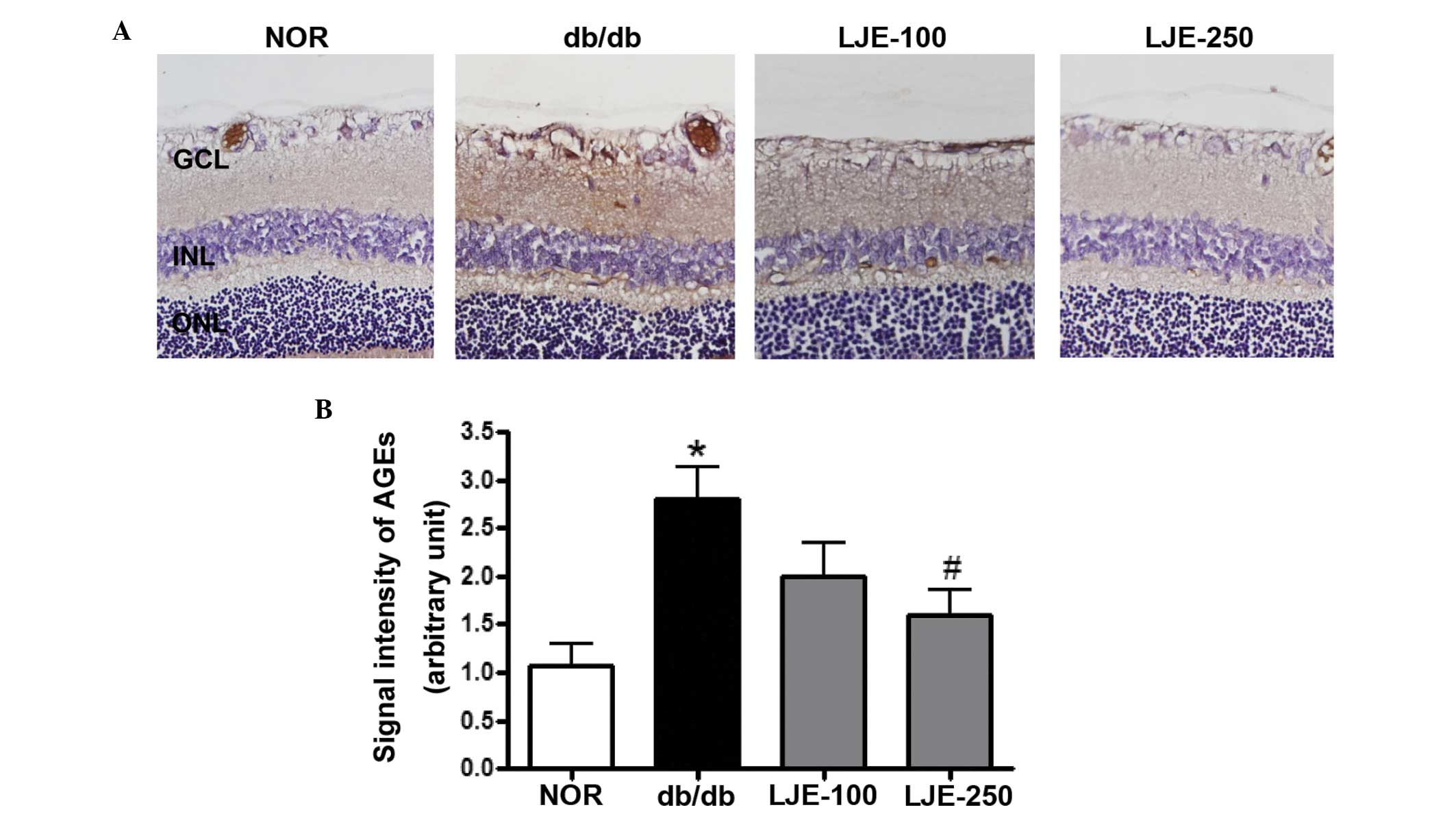 | Figure 1Accumulation of AGEs. (A)
Representative immunostaining of AGEs in the retina (magnification,
×200). db/db, diabetic db/db mice; LJE-100, db/db mice treated with
LJE (100 mg/kg); LJE-250, db/db mice treated with LJE (250 mg/kg).
(B) Quantitative analysis of AGE signal intensity. Values are
expressed as the mean ± standard error of the mean, n=8.
*P<0.01, vs. NOR mice, #P<0.01, vs.
vehicle-treated db/db mice. GCL, ganglion cell layer; INL, inner
nuclear layer; ONL, outer nuclear layer; AGEs. advanced glycation
end products; NOR. normal; LJE, Litsea japonica extract. |
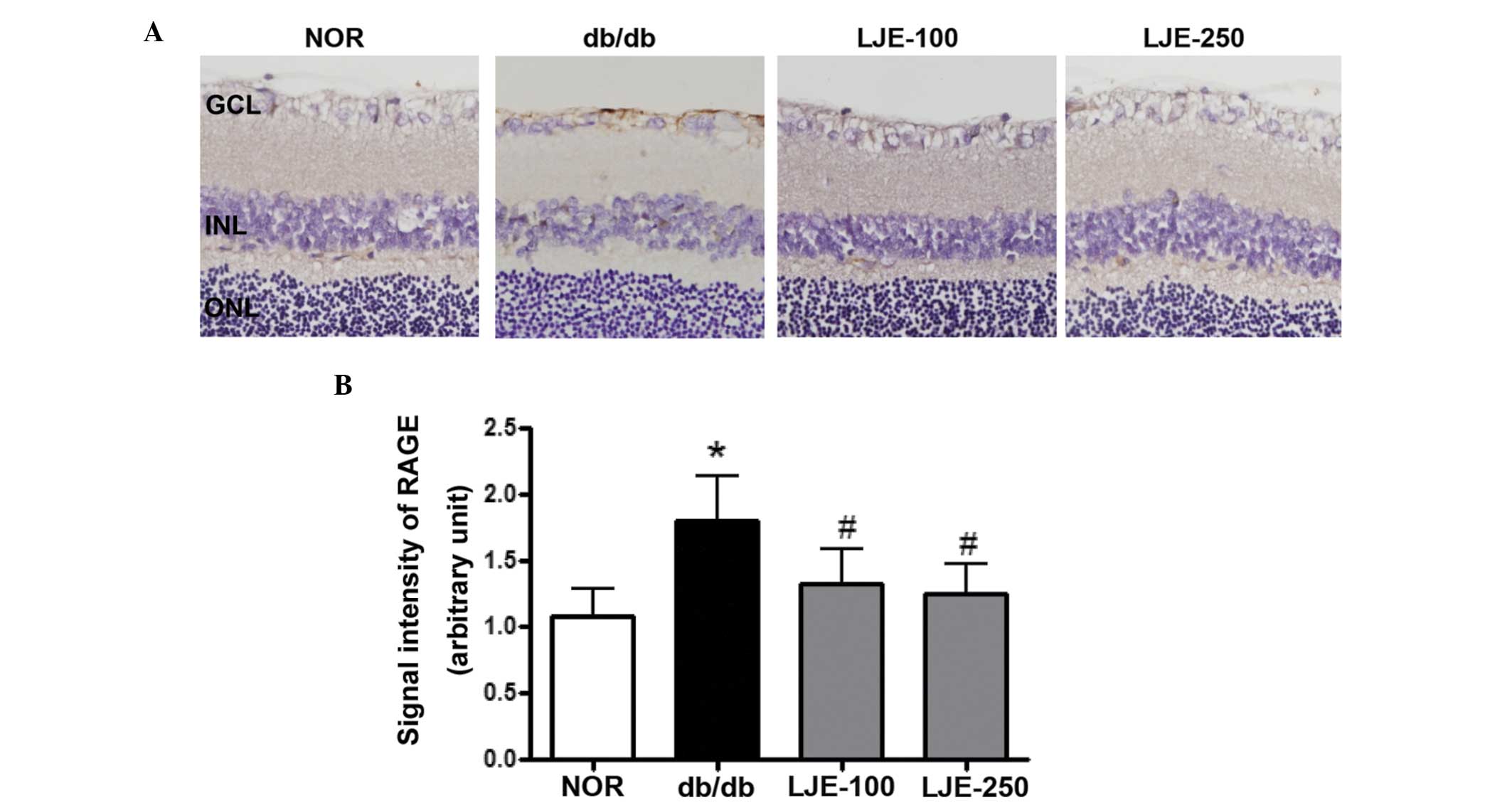 | Figure 2Expression of RAGE. (A) Representative
immunostaining of RAGE in the retina (magnification, ×200). db/db,
diabetic db/db mice; LJE-100, db/db mice treated with LJE (100
mg/kg); LJE-250, db/db mice treated with LJE (250 mg/kg). (B)
Quantitative analysis of RAGE signal intensity. Values are
expressed as the mean ± standard error of the mean, n=8.
*P<0.01, vs. NOR mice; #P<0.01, vs.
vehicle-treated db/db mice. GCL, ganglion cell layer; INL, inner
nuclear layer; ONL, outer nuclear layer; RAGE, receptor for
advanced glycation end products; NOR. normal; LJE, Litsea
japonica extract. |
Apoptosis in retinal neuronal cells
To characterize the death of neurons in the ganglion
cell layer (GCL) of the vehicle-treated db/db mice, TUNEL staining
was performed. A significant increase in TUNEL-positive cells were
observed in the GCLs of the vehicle-treated db/db mice compared
with the normal mice (Fig. 3). The
presence of TUNEL-positive cells was not limited to the GCL. The
inner nuclear layer demonstrated occasional positive cells, as did
the outer nuclear layer of the photoreceptor cell nuclei. However,
treatment of the db/db mice with LJE prevented an increase in
positive cells, similar to that observed in the normal mice.
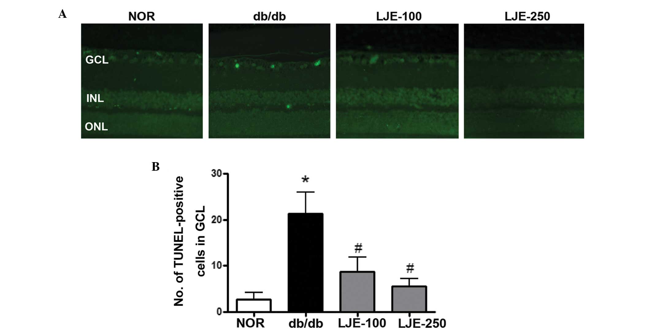 | Figure 3Apoptosis of retinal ganglion cells.
(A) Retinal sections were stained with TUNEL (green). Apoptotic
ganglion cells were observed in the vehicle-treated db/db mice
(magnification, ×200). db/db, diabetic db/db mice; LJE-100, db/db
mice treated with LJE (100 mg/kg); LJE-250, db/db mice treated with
LJE (250 mg/kg). (B) Quantitative analysis of TUNEL-positive cells
in GCL. Values are expressed as the mean ± standard error of the
mean, n=8. *P<0.01, vs. NOR mice,
#P<0.01, vs. vehicle-treated db/db mice. TUNEL,
terminal deoxynucleotidyl transferase dUTP nick end labeling; GCL,
ganglion cell layer; INL, inner nuclear layer; ONL, outer nuclear
layer; NOR, normal; LJE, Litsea japonica extract. |
LJE inhibits the activation of NF-κB in
the retina
NF-κB is a common downstream signal of AGEs
(24). Since inhibition of NF-κB
activity is considered one of the mechanisms promoting apoptosis
(25,26), the present study examined whether
LJE inhibited the activation of NF-κB. EMSA analysis of the nuclear
protein revealed consistently increased DNA binding activity of
NF-κB in the vehicle-treated db/db mice compared with the normal
mice, with a 3.1-fold increase (P<0.01; Fig. 4). LJE significantly inhibited the
DNA binding activity of NF-κB. In addition, southwestern
histochemistry was performed to determine the activity of NF-κB the
retinal tissue. This technique allows the localization of activated
nuclear factor in the cellular nucleus and, using this novel
method, marked NF-κB activity was found predominantly in the nuclei
in the GCL and in the inner nuclear layer in the vehicle-treated
db/db mice (Fig. 5A). Morphometric
analysis revealed that the expression of activated NF-κB in the
vehicle-treated db/db mice was significantly increased compared
with the normal mice, whereas LJE significantly inhibited the
activated NF-κB (Fig. 5B).
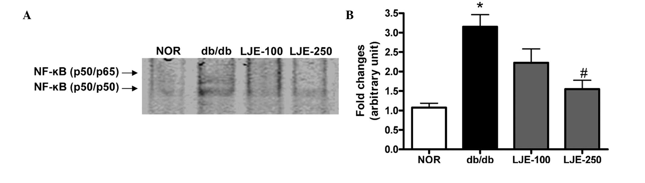 | Figure 4Activation of NF-κB. (A) The NF-κB
DNA-binding activity was measured by EMSA. db/db, diabetic db/db
mice; LJE-100, db/db mice treated with LJE (100 mg/kg); LJE-250,
db/db mice treated with LJE (250 mg/kg) (B) Data are expressed as
the mean ± standard error of the mean, n=8. *P<0.01,
vs. NOR mice, #P<0.01, vs. vehicle-treated db/db
mice. NF-κB, nuclear factor-κB.. NOR, normal; LJE, Litsea
japonica extract. |
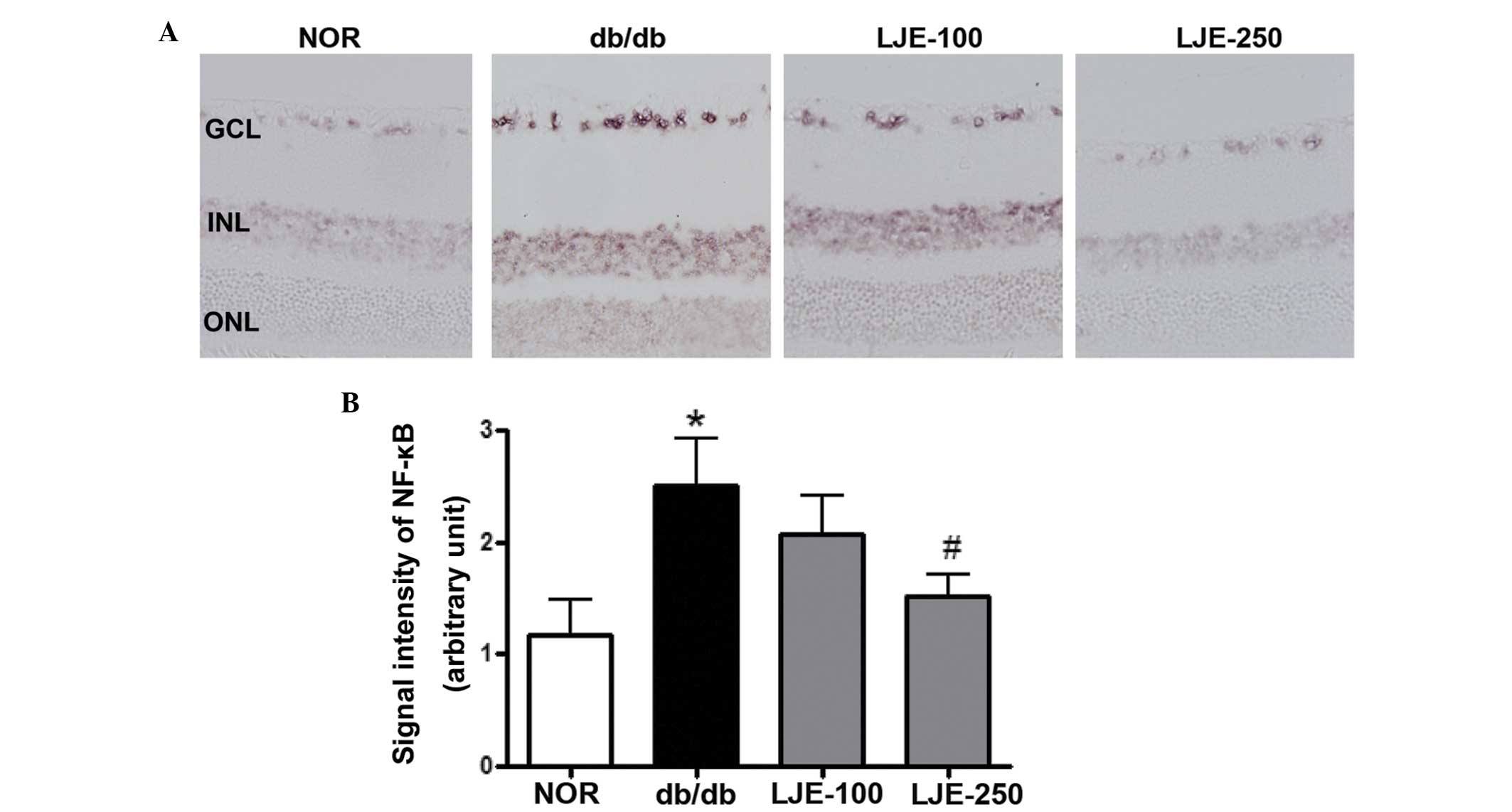 | Figure 5SWH for in situ detection of
active NF-κB. (A) Positive signals for activated NF-κB were
predominantly detected in the nuclei of the retinal ganglion cells
and inner nuclear cells in the vehicle-treated db/db mice.
(magnification, ×200). db/db, diabetic db/db mice; LJE-100, db/db
mice treated with LJE (100 mg/kg); LJE-250, db/db mice treated with
LJE (250 mg/kg). (B) Quantitative analysis of SWH-positive signal
intensity. Data are expressed as the mean ± standard error of the
mean, n=8. *P<0.01, vs. NOR mice,
#P<0.01, vs. vehicle-treated db/db mice. SWH.
southwestern histochemistry; NF-κB, nuclear factor-κB; GCL,
ganglion cell layer; INL, inner nuclear layer; ONL, outer nuclear
layer; NOR. normal; LJE, Litsea japonica extract. |
Discussion
In streptozotocin-induced diabetic rats and
postmortem human retinas, TUNEL assays have revealed that diabetes
increases apoptosis in neurons, particularly in the inner retina,
where retinal ganglion cells are located (27,28).
The rate of neural apoptosis remains constant throughout the
duration of diabetes (3). As
neurons are unable to proliferate, the apoptosis of these cells
leads to chronic neurodegeneration. In the present study, the
preventive effect of LJE on diabetes-induced injury of retinal
ganglion cells was evaluated. LJE, a herbal AGE inhibitor, reduced
diabetes-induced apoptosis of the retinal ganglion cells. In
addition, LJE prevented AGE accumulation in the neural retina and
decreased the expression of RAGE in the retinal tissues. LJE
marginally decreased the levels of blood glucose, but caused no
reduction in the levels of HbA1c in the db/db mice. Therefore, LJE
had anti-apoptotic effects in the diabetic neural retinas without a
substantial reduction in blood glucose. These results suggested
that, even in hyperglycemia, diabetes-induced retinal
neurodegeneration was attenuated by LJE.
The potential mechanisms of diabetic retinopathy are
numerous. The accumulation of AGEs during the Maillard reaction is
associated with the risk of diabetic retinopathy and levels of
serum AGEs correlate with the degree of diabetic retinopathy
(29,30). In addition, retinal ganglion cells
are the most vulnerable cell population in the retina (31). AGE-RAGE interaction elicits the
induction of apoptosis in various types of cell (12,32,33)
and the inhibition of AGE formation improves diabetic retinopathy
(32–34). In the present study, LJE exhibited
the properties of an AGE inhibitor in retinal tissue, and treatment
with LJE resulted in a decline in the cellular damage mediated by
AGEs. In the present study, three flavonoids (epicatechin,
quercitrin and afzelin) were identified in LJE. These flavonoids
exhibit significant inhibitory activities on the formation of AGEs,
with previously reported half maximal inhibitory concentrations of
144, 77.8 and 58.9 μM, respectively (35–37).
Therefore, the ability of LJE to protect against retinal
neurodegeneration may be due to the effect of this compound.
Notably, NF-κB has been previously implicated in the
development of diabetic retinopathy (25). AGEs interact with RAGE, inducing
the subsequent activation of NF-κB and NF-κB-controlled
pro-apoptotic molecules (38).
Apoptosis of retinal pericytes and the retinal neuronal cells is
also associated with NF-κB (25,26).
Although the activation of NF-κB in the retina may be involved in
retinal cell death or survival (39,40),
the activation of NF-κB due to hyperglycemia induces the
accelerated loss of retinal pericyts (25) and retinal capillary cell death
(26). In the present study,
southwestern histochemistry demonstrated that NF-κB was markedly
activated in the ganglion cell layer of the db/db mice. This result
suggested that the activation of NF-κB activation was responsible
for the loss of ganglion cells. Treatment with LJE almost
completely inhibited this activation of NF-κB.
In conclusion, LJE suppressed the accumulation of
AGEs in neural retinas. In addition, the expression of RAGE, which
is important in the pro-apoptotic signaling pathway, was restored.
Although the pathogenesis of diabetic nephropathy is
multifactorial, AGE/RAGE signaling is a common pathway for the
progression of diabetic retinopathy. Therefore, the neural
protective effect of LJE may be, at least partly, attributed to its
effect on AGEs, and LJE may be a beneficial agent in protecting
against diabetes-induced retinal neurodegeneration.
Acknowledgments
This study was supported by a grant (no. K14040)
from the Korean Institute of Oriental Medicine.
References
|
1
|
Mizutani M, Kern TS and Lorenzi M:
Accelerated death of retinal microvascular cells in human and
experimental diabetic retinopathy. J Clin Invest. 97:2883–2890.
1996. View Article : Google Scholar : PubMed/NCBI
|
|
2
|
Asnaghi V, Gerhardinger C, Hoehn T,
Adeboje A and Lorenzi M: A role for the polyol pathway in the early
neuroretinal apoptosis and glial changes induced by diabetes in the
rat. Diabetes. 52:506–511. 2003. View Article : Google Scholar : PubMed/NCBI
|
|
3
|
Barber AJ, Lieth E, Khin SA, Antonetti DA,
Buchanan AG and Gardner TW: Neural apoptosis in the retina during
experimental and human diabetes. Early onset and effect of insulin.
J Clin Invest. 102:783–791. 1998. View
Article : Google Scholar : PubMed/NCBI
|
|
4
|
Kern TS and Barber AJ: Retinal ganglion
cells in diabetes. J Physiol. 586:4401–4408. 2008. View Article : Google Scholar : PubMed/NCBI
|
|
5
|
Brownlee M: Advanced protein glycosylation
in diabetes and aging. Annu Rev Med. 46:223–234. 1995. View Article : Google Scholar : PubMed/NCBI
|
|
6
|
Koga K, Yamagishi S, Okamoto T, et al:
Serum levels of glucose-derived advanced glycation end products are
associated with the severity of diabetic retinopathy in type 2
diabetic patients without renal dysfunction. Int J Clin Pharmacol
Res. 22:13–17. 2002.PubMed/NCBI
|
|
7
|
Miura J, Yamagishi S, Uchigata Y, et al:
Serum levels of non-carboxymethyllysine advanced glycation
endproducts are correlated to severity of microvascular
complications in patients with Type 1 diabetes. J Diabetes
Complications. 17:16–21. 2003. View Article : Google Scholar
|
|
8
|
Hammes HP, Alt A, Niwa T, et al:
Differential accumulation of advanced glycation end products in the
course of diabetic retinopathy. Diabetologia. 42:728–736. 1999.
View Article : Google Scholar : PubMed/NCBI
|
|
9
|
Lecleire-Collet A, Tessier LH, Massin P,
et al: Advanced glycation end products can induce glial reaction
and neuronal degeneration in retinal explants. Br J Ophthalmol.
89:1631–1633. 2005. View Article : Google Scholar : PubMed/NCBI
|
|
10
|
Kasper M, Roehlecke C, Witt M, et al:
Induction of apoptosis by glyoxal in human embryonic lung
epithelial cell line L132. Am J Respir Cell Mol Biol. 23:485–491.
2000. View Article : Google Scholar : PubMed/NCBI
|
|
11
|
Kaji Y, Amano S, Usui T, et al: Expression
and function of receptors for advanced glycation end products in
bovine corneal endothelial cells. Invest Ophthalmol Vis Sci.
44:521–528. 2003. View Article : Google Scholar : PubMed/NCBI
|
|
12
|
Yamagishi S, Inagaki Y, Amano S, Okamoto
T, Takeuchi M and Makita Z: Pigment epithelium-derived factor
protects cultured retinal pericytes from advanced glycation end
product-induced injury through its antioxidative properties.
Biochem Biophys Res Commun. 296:877–882. 2002. View Article : Google Scholar : PubMed/NCBI
|
|
13
|
Deane R, Du Yan S, Submamaryan RK, et al:
RAGE mediates amyloid-beta peptide transport across the blood-brain
barrier and accumulation in brain. Nat Med. 9:907–913. 2003.
View Article : Google Scholar : PubMed/NCBI
|
|
14
|
Hammes HP, Martin S, Federlin K, Geisen K
and Brownlee M: Aminoguanidine treatment inhibits the development
of experimental diabetic retinopathy. Proc Natl Acad Sci USA.
88:11555–11558. 1991. View Article : Google Scholar : PubMed/NCBI
|
|
15
|
Kern TS and Engerman RL: Pharmacological
inhibition of diabetic retinopathy: aminoguanidine and aspirin.
Diabetes. 50:1636–1642. 2001. View Article : Google Scholar : PubMed/NCBI
|
|
16
|
Kern TS, Tang J, Mizutani M, et al:
Response of capillary cell death to aminoguanidine predicts the
development of retinopathy: comparison of diabetes and
galactosemia. Invest Ophthalmol Vis Sci. 41:3972–3978.
2000.PubMed/NCBI
|
|
17
|
Vasan S, Foiles PG and Founds HW:
Therapeutic potential of AGE inhibitors and breakers of AGE protein
cross-links. Expert Opin Investig Drugs. 10:1977–1987. 2001.
View Article : Google Scholar
|
|
18
|
Harris CS, Beaulieu LP, Fraser MH, et al:
Inhibition of advanced glycation end product formation by medicinal
plant extracts correlates with phenolic metabolites and antioxidant
activity. Planta Med. 77:196–204. 2011. View Article : Google Scholar
|
|
19
|
Lee SY, Min BS, Kim JH, et al: Flavonoids
from the leaves of Litsea japonica and their anti-complement
activity. Phytother Res. 19:273–276. 2005. View Article : Google Scholar : PubMed/NCBI
|
|
20
|
Min BS, Lee SY, Kim JH, et al: Lactones
from the leaves of Litsea japonica and their anti-complement
activity. J Nat Prod. 66:1388–1390. 2003. View Article : Google Scholar : PubMed/NCBI
|
|
21
|
Sohn EJ, Kim J, Kim CS, et al: The extract
of Litsea japonica reduced the development of diabetic nephropathy
via the inhibition of advanced Glycation end products accumulation
in db/db mice. Evid-Based Compl Alt Med. 2013:7694162013.
|
|
22
|
Sohn EJ, Kim CS, Kim YS, et al: Effects of
magnolol (5,5′-diallyl-2,2′-dihydroxybiphenyl) on diabetic
nephropathy in type 2 diabetic Goto-Kakizaki rats. Life Sci.
80:468–475. 2007. View Article : Google Scholar
|
|
23
|
Hernández-Presa MA, Gómez-Guerrero C and
Egido J: In situ non-radioactive detection of nuclear factors in
paraffin sections by Southwestern histochemistry. Kidney Int.
55:209–214. 1999. View Article : Google Scholar : PubMed/NCBI
|
|
24
|
Yamagishi S, Takeuchi M, Matsui T,
Nakamura K, Imaizumi T and Inoue H: Angiotensin II augments
advanced glycation end product-induced pericyte apoptosis through
RAGE overexpression. FEBS Lett. 579:4265–4270. 2005. View Article : Google Scholar : PubMed/NCBI
|
|
25
|
Romeo G, Liu WH, Asnaghi V, Kern TS and
Lorenzi M: Activation of nuclear factor-kappaB induced by diabetes
and high glucose regulates a proapoptotic program in retinal
pericytes. Diabetes. 51:2241–2248. 2002. View Article : Google Scholar : PubMed/NCBI
|
|
26
|
Kowluru RA, Koppolu P, Chakrabarti S and
Chen S: Diabetes-induced activation of nuclear transcriptional
factor in the retina and its inhibition by antioxidants. Free Radic
Res. 37:1169–1180. 2003. View Article : Google Scholar
|
|
27
|
Hammes HP, Federoff HJ and Brownlee M:
Nerve growth factor prevents both neuroretinal programmed cell
death and capillary pathology in experimental diabetes. Mol Med.
1:527–534. 1995.PubMed/NCBI
|
|
28
|
Kerrigan LA, Zack DJ, Quigley HA, Smith SD
and Pease ME: TUNEL-positive ganglion cells in human primary
open-angle glaucoma. Arch Ophthalmol. 115:1031–1035. 1997.
View Article : Google Scholar : PubMed/NCBI
|
|
29
|
Chiarelli F, Catino M, Tumini S, et al:
Advanced glycation end products in adolescents and young adults
with diabetic angiopathy. Pediatr Nephrol. 14:841–846. 2000.
View Article : Google Scholar : PubMed/NCBI
|
|
30
|
Ono Y, Aoki S, Ohnishi K, Yasuda T, Kawano
K and Tsukada Y: Increased serum levels of advanced glycation
end-products and diabetic complications. Diabetes Res Clin Pract.
41:131–137. 1998. View Article : Google Scholar : PubMed/NCBI
|
|
31
|
Funk RH and Schmidt KG: Characteristic
features of optic nerve ganglion cells and approaches for
neuroprotection. From intracellular to capillary processes and
therapeutic considerations. Ophthalmologe. 101:1062–1070. 2004.In
German. View Article : Google Scholar : PubMed/NCBI
|
|
32
|
Yamagishi S, Hsu CC, Taniguchi M, et al:
Receptor-mediated toxicity to pericytes of advanced glycosylation
end products: a possible mechanism of pericyte loss in diabetic
microangiopathy. Biochem Biophys Res Commun. 213:681–687. 1995.
View Article : Google Scholar : PubMed/NCBI
|
|
33
|
Yamagishi S, Amano S, Inagaki Y, et al:
Advanced glycation end products-induced apoptosis and
overexpression of vascular endothelial growth factor in bovine
retinal pericytes. Biochem Biophys Res Commun. 290:973–978. 2002.
View Article : Google Scholar : PubMed/NCBI
|
|
34
|
Yamagishi S, Nakamura K and Matsui T:
Advanced glycation end products (AGEs) and their receptor (RAGE)
system in diabetic retinopathy. Curr Drug Discov Technol. 3:83–88.
2006. View Article : Google Scholar : PubMed/NCBI
|
|
35
|
Lee EH, Song DG, Lee JY, Pan CH, Um BH and
Jung SH: Flavonoids from the leaves of Thuja orientalis inhibit the
aldose reductase and the formation of advanced glycation
endproducts. J Korean Soc Appl Biol Chem. 52:448–455. 2009.
View Article : Google Scholar
|
|
36
|
Jang DS, Kim JM, Lee YM, et al: Flavonols
from Houttuynia cordata with protein Glycation and Aldose reductase
inhibitory activity. Nat Prod Sci. 12:210–213. 2006.
|
|
37
|
Matsuda H, Wang T, Managi H and Yoshikawa
M: Structural requirements of flavonoids for inhibition of protein
glycation and radical scavenging activities. Bioorg Med Chem.
11:5317–5323. 2003. View Article : Google Scholar : PubMed/NCBI
|
|
38
|
Kim J, Kim KM, Kim CS, et al: Puerarin
inhibits the retinal pericyte apoptosis induced by advanced
glycation end products in vitro and in vivo by inhibiting NADPH
oxidase-related oxidative stress. Free Radic Biol Med. 53:357–365.
2012. View Article : Google Scholar : PubMed/NCBI
|
|
39
|
Wu T, Chiang SK, Chau FY and Tso MO:
Light-induced photoreceptor degeneration may involve the NF
kappaB/caspase-1 pathway in vivo. Brain Res. 967:19–26. 2003.
View Article : Google Scholar : PubMed/NCBI
|
|
40
|
Choi JS, Kim JA, Kim DH, et al: Failure to
activate NF-kappaB promotes apoptosis of retinal ganglion cells
following optic nerve transection. Brain Res. 883:60–68. 2000.
View Article : Google Scholar : PubMed/NCBI
|


