Introduction
Lung cancer is the leading cause of
cancer-associated mortality, with an increasing incidence worldwide
(1). Despite improvements in
diagnostic imaging, surgery, radiotherapy and chemotherapy, the
overall survival rate of patients with lung cancer remains poor,
with only 14% of patients surviving 5 years from the date of
diagnosis (1). As non-small cell
lung cancer (NSCLC) is the most common type of lung cancer, the
development of effective therapeutic targets for NSCLC is urgently
required (1).
The development and progression of NSCLC are
associated with the dysregulation of oncogenes or tumor
suppressors, including microRNAs (miRNAs) (2). miRNAs are 18–25 nucleotide,
non-coding RNAs, which can result in the inhibition of gene
expression at the post-transcriptional level, through direct
binding to the 3′-untranslational region (UTR) of mRNAs (3). In addition, the dysregulation of
miRNAs has been reported to be associated with the development and
progression of NSCLC, including miRNA (miR)-7 (4,5).
miR-7 has been demonstrated to act as a tumor suppressor in NSCLC,
through targeting B-cell lymphoma (BCL)-2 and PA28γ (6,7). As
one miRNA can directly bind to several target mRNAs, whether other
target genes of miR-7 exist in NSCLC remains to be fully
elucidate.
Paired box 6 (Pax6) has been demonstrated as a
highly conserved transcription factor during embryogenesis, and is
involved in the development and function of the central nervous
system, endocrine glands, eyes and pancreas (8–12).
The role of Pax6 in the development and progression of cancer been
gradually revealed (13,14). Zhao et al reported that
shRNA-induced PAX6 downregulation notably suppressed proliferation
and cell cycle progression in NCSLC cells. They further
demonstrated that the extracellular signal-regulated kinase (ERK)
and p38 mitogen-activated protein kinase (MAPK) signaling pathways
are involved in Pax6-mediated cell cycle progression in NCSLC cells
(15).
The present study aimed to examine the role of miR-7
in the regulation of NSCLC in vitro. In addition, as a
previous study demonstrated a target association between miR-7 and
Pax6 in colon cancer (16), the
present study also investigated whether Pax6 is involved in the
regulatory effect of miR-7 on NSCLC cells.
Materials and methods
Agents
TRIzol reagent, fetal bovine serum (FBS)
Lipofectamine 2000 and the mirVana™ qRT-PCR miRNA Detection kit
were purchased from Invitrogen Life Technologies (Carlsbad, CA,
USA). Mouse anti-Pax6, -total ERK, -phosphorylated (p)-ERK, -p38
MAPK and -GAPDH primary antibodies, and rabbit anti-mouse secondary
antibody were purchased from Abcam (Cambridge, UK). Bioinformatics
analysis was conducted to predicate the putative target genes of
miR-7 using the Targetscan online software (http://www.targetscan.org/). The Quick-Change
Site-Directed Mutagenesis kit was purchased from Stratagene (La
Jolla, CA, USA). The PsiCHECK™2 vector was purchased from Promega
Corporation (Madison, WI, USA). The enhanced chemiluminescence
(ECL) kit was purchased from Pierce Biotechnology, Inc. (Rockford,
IL, USA).
Cell lines and cell culture
The A549, H460, SK-MES-1 and SPC-A1 human NSCLC cell
lines, and normal BEAS-2B human lung epithelial cell line were
purchased from the Cell bank of Central South University (Changsha,
China). All cells were cultured in Dulbecco's modified Eagle's
medium (DMEM; Invitrogen Life Technologies) supplemented with 10%
FBS at 37°C with 5% CO2.
RNA extraction and reverse
transcription-quantitative polymerase chain reaction (RT-qPCR)
Total RNA was extracted from the cells using TRIzol
reagent, according to the manufacturer's instructions. The relative
expression level of miR-7 was determined by RT-qPCR using a
mirVana™ qRT-PCR miRNA Detection kit, according to the
manufacturer's instruction. Specific primer sets for miR-7 and U6
(internal reference) were obtained from GeneCopoeia, Inc. (Maryland
Rockville, MD, USA). The PCR cycling conditions were as follows:
95°C for 10 min, denaturation at 95°C for 15 sec and an
annealing/elongation step at 60°C for 1 min for 40 cycles. The data
were analyzed with SDS relative quantification software version
2.2.2 (Applied Biosystems, Foster City, CA, USA). The 2-ΔΔCt method
was used for the relative quantification of the differences in
expression levels of each target.
Western blotting
The cells (107) were solubilized in cold
radioimmunoprecipitation assay lysis buffer (Beyotime Institute of
Biotechnology, Shanghai, China). The proteins (60 µg) were
separated with 12% SDS-PAGE (Beijing Biolab Science and Technology
Co., Ltd., Beijing, China), and transferred onto a polyvinylidene
difluoride (PVDF) membrane (Invitrogen Life Technologies), which
was then incubated with Tris-buffered saline with Tween 20 (TBST;
Fanke Biotech Co., Ltd., Shanghai, China) containing 5% milk at
room temperature for 3 h. The PVDF membrane was then incubated with
primary antibodies at room temperature for 3 h. The primary
antibodies were as follows: Monoclonal mouse anti-human PAX6
(1:100, cat. no. ab78545), monoclonal mouse anti-human ERK (1:100,
cat. no. ab119933), polyclonal rabbit anti-human p-ERK (1:200, cat.
no. ab131438), monoclonal mouse anti-human p38 MAPK, (1:100, cat.
no. ab31828) and monoclonal rabbit anti-human p-p38 MAPK, (1:100,
cat. no. EPR16587). This was followed by incubation with rabbit
anti-mouse IgG, (cat. no. ab46540) and mouse anti-rabbit IgG (cat.
no. ab99700) secondary antibodies at room temperature for 40 min.
All antibodies were purchased from Abcam. Chemiluminescent
detection was performed using an ECL kit. The relative protein
expression was analyzed using Image-Pro plus software 6.0 (Media
Cybernetics, Inc., Rockville, MD, USA), and presented as the
density ratio, vs. GAPDH.
Transfection
The plasmid of Pax6, scramble miRNA mimics, miR-7
mimics and miR-7 inhibitor were constructed by Nlunbio (Changsha,
China). Lipofectamine 2000 was used to perform transfection,
according to the manufacture's instruction. Briefly, the plasmid or
miRNA mimics and Lipofectamine 2000 were diluted with 100 nM
serum-free medium, respectively. The diluted Lipofectamine 2000 was
added into the diluted plasmid or miRNA mimics, respectively, and
incubated for 20 min at room temperature, following which they were
added to the cell suspension. The cells (105 cells/ml),
which were suspended in DMEM were then incubated at 37°C, 5%
CO2 for 6 h. Following incubation, the medium in each
well was replaced with normal serum-containing medium, and the
cells were cultured for 24 h prior to performing the subsequent
assays. The successful transfection was confirmed by detecting the
expression levels of PAX6 or miR-7, respectively.
Dual luciferase reporter assays
A Quick-Change Site-Directed Mutagenesis kit was
used to generate a mutant 3′-UTR of Pax6, according to the
manufacturer's instructions. The wild-type or mutant 3′-UTR of Pax6
were inserted into the psiCHECK™2 vector, respectively. A549 cells
(105 cells/well), which were cultured to ~70%
confluence, were the transfected with either the
psiCHECK™2-Pax6-3′-UTR or psiCHECK™2-mutant Pax6 -3′-UTR vector,
with or without 100 nM miR-7 mimic, respectively. Following
transfection for 48 h, the luciferase activities were determined
using an LD400 luminometer (Beckman Coulter, Fullerton, CA, USA).
The activity of Renilla luciferase was normalized to that of
firefly luciferase.
Cell proliferation assay
An MTT assay was performed to measure cell
proliferation. Cells in the exponential growth were plated, at a
final concentration of 2,000 cells per well, into 96-well plates.
The viability of the cells were evaluated using an MTT assay (20
µl MTT) 24, 48, 72 and 96 h after seeding. The optical
density (OD) at 570 nm of each well was measured using an ELISA
reader (ELX-800; BioTek Instruments, Inc., Winooski, VT, USA).
Cell invasion assay
The invasive abilities of the A549 cells were
determined using 24-well Transwell chambers (Chemicon, Temecula,
CA, USA), which contained a layer of Matrigel. For each group, the
cell suspension was added to the upper chamber, and DMEM,
containing 10% FBS, was added to the lower chamber. Following
incubation for 24 h at 37°C, the non-invading cells and the matrix
gel on the interior of the inserts were removed using a
cotton-tipped swab. The invasive cells on the lower surface of the
membrane were stained with gentian violet, followed by rinsing with
water and air drying. Subsequently, the number of cells were
counted in five randomly-selected fields under an inverted
microscope (IX71; Olympus, Tokyo, Japan).
Statistical analysis
The results are expressed as the mean ± standard
deviation of at least three independent experiments. Statistical
analysis was performed using SPSS 17 software (SPSS, Inc., Chicago,
IL, USA). Statistical analysis of differences was performed using
one-way analysis of variance. P<0.01 was considered to indicate
a statistically significant difference.
Results
miR-7 is downregulated, while Pax6 is
upregulated in NSCLC cell lines
To determine the role of miR-7 in NSCLC, the present
study first examined the expression level of miR-7 in the A549,
H460, SK-MES-1 and SPC-A1 human NSCLC cell lines, and BEAS-2B
normal human lung epithelial cell line. As shown in Fig. 1A, the expression level of miR-133a
was significantly reduced in the NSCLC cell lines, compared with
the BEAS-2B normal human lung epithelial cells. Subsequently, the
expression level of Pax6 was determined by performing RT-qPCR. As
shown in Fig. 1B, the mRNA level
of Pax6 was upregulated in the NSCLC cell lines, compared with the
BEAS-2B normal human lung epithelial cells. Accordingly, these data
demonstrated that miR-7 was downregulated, while Pax6 was
upregulated in the NSCLC cell lines.
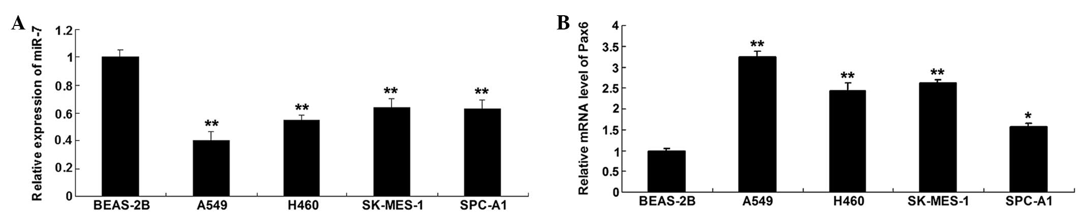 | Figure 1(A) Expression levels of miR-7 were
determined using RT-qPCR in the A549, H460, SK-MES-1 and SPC-A1
human NSCLC cell lines, and BEAS-2B normal human lung epithelial
cells. **P<0.01, vs. BEAS-2B. (B) mRNA expression
levels of Pax6 were examined using RT-qPCR in the A549, H460,
SK-MES-1 and SPC-A1 human NSCLC cell lines, and BEAS-2B normal
human lung epithelial cells. *P<0.05, vs. BEAS-2B;
**P<0.01 vs. BEAS-2B. GAPDH was used as an internal
control. Data are expressed as the mean ± standard deviation.
NSCLC, non-small cell lung cancer; RT-qPCR, reverse
transcription-quantitative polymerase chain reaction. miR,
microRNA. |
As the A549 cells exhibited the most marked changes
in expression levels of miR-7 and Pax6 among the four NSCLC cell
lines, when compared with the BEAS-2B normal human lung epithelial
cells (Fig. 1B), the A549 cell
line was selected for examination in the subsequent
experiments.
miR-7 negatively regulates the protein
expression of its target, Pax6, in A549 NSCLC cells
Based on the above data, the regulatory association
between miR-7 and Pax6 in A549 NSCLC cells were further
investigated. Following transfection with miR-7 mimics or
inhibitor, the level of miR-7 in the A549 NSCLC cells was examined,
and the data revealed that the transfection had been successful
(Fig. 2A). The protein level of
Pax6 in each group was then determined by performing western
blotting, which demonstrated that, in the A549 NSCLC cells
transfected with miR-7 mimics, the protein level of Pax6 was
reduced (Fig. 2B). By contrast,
inhibition of miR-7 led to an increase in the protein expression of
Pax6 in the A549 NSCLC cells (Fig.
2B). Based on the bioinformatical prediction that the putative
seed sequences for miR-7 at the 3′UTR of Pax6 is conserved
(Fig. 2C), the present study
performed a luciferase reporter assay to determine whether Pax6 was
a target of miR-7. The resulting data indicated that the luciferase
activity was reduced only in the A549 NSCLC cells that were
co-transfected with the miR-7 mimics and wild-type Pax6 3′UTR. In
the remaining groups, the luciferase activity was unchanged
(Fig. 2D). These findings
indicated that Pax6 was a direct target of miR-7 in the A549 NSCLC
cells.
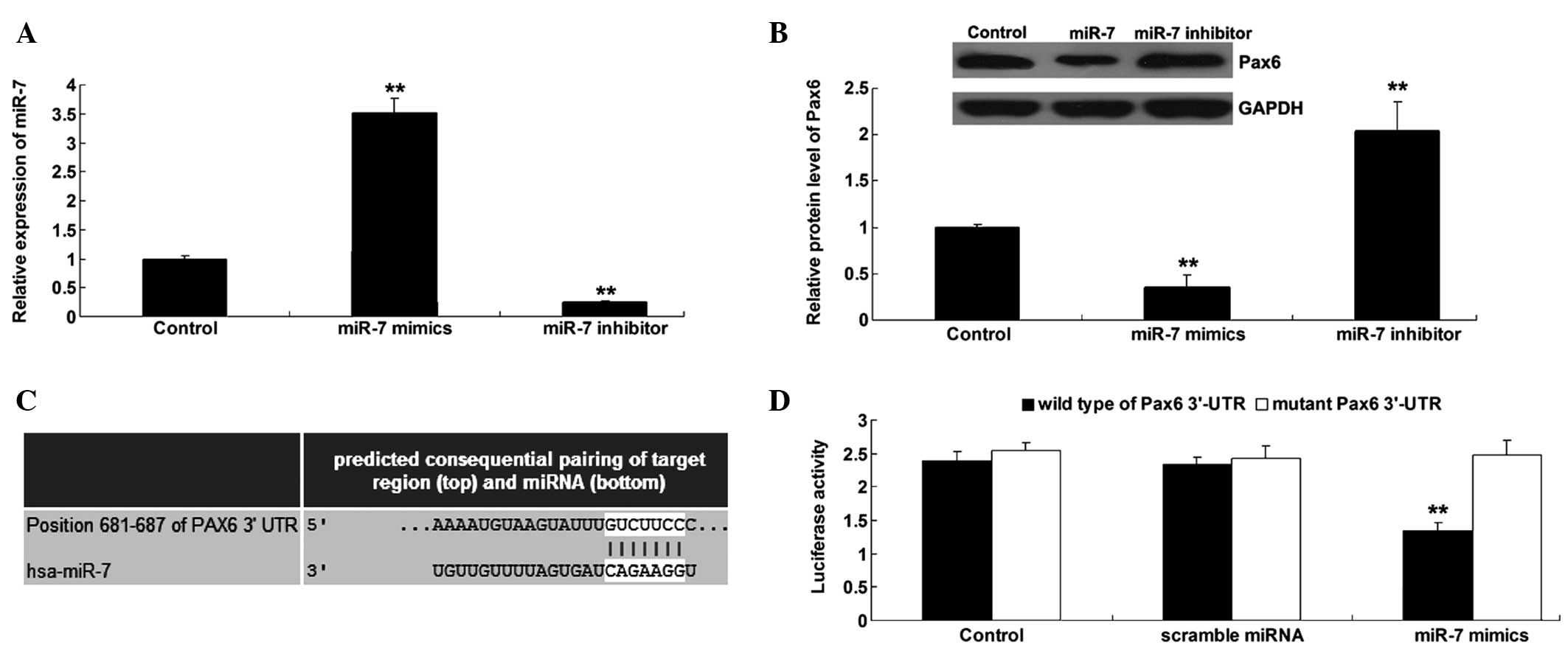 | Figure 2(A) Expression levels of miR-7 were
examined using RT-qPCR in A549 NSCLC cells transfected with miR-7
mimics or inhibitor, respectively. Untreated A549 cells were used
as a control (**P<0.01, vs. control). (B) Protein
levels of Pax6 were determined using western blotting in A549 NSCLC
cells transfected with miR-7 mimics or inhibitor, respectively.
Untreated A549 cells were used as a control
(**P<0.01, vs. control). (C) Putative seed sequences
of miR-7 in the 3′-UTR of Pax6 mRNA were indicated from online
bioinformatics analysis software (Targetscan; http://www.targetscan.org). (D) Data of the luciferase
reporter assay demonstrated that the luciferase activity was
reduced only in the A549 NSCLC cells co-transfected with the miR-7
mimics and wild-type Pax6 3′UTR, and were unchanged in the
remaining groups. A549 cells transfected only with wild-type or
mutant type Pax6 3′-UTR were used as controls
(**P<0.01, vs. control). Data are expressed as the
mean ± standard deviation. NSCLC, non-small cell lung cancer;
RT-qPCR, reverse transcription-quantitative polymerase chain
reaction. miR, microRNA; UTR, untranslated region. |
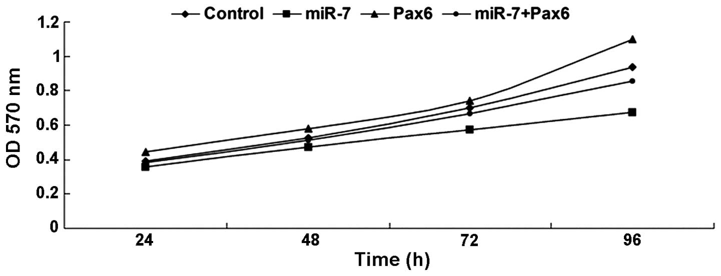
Overexpression of miR-7 inhibits NSCLC
A549 cell proliferation by targeting Pax6
To further investigate the effect of Pax6 and miR-7
on NSCLC cell proliferation, an MTT assay was performed. As shown
in Fig. 3, in the
miR-7-overexpressed A549 cells, the cell proliferation was
downregulated, whereas in the Pax6-overexpressed A549 cells, the
cell proliferation rate was upregulated. In addition, upregulation
of Pax6 attenuated the inhibitory effect of miR-7 overexpression on
A549 cell proliferation. These data suggested that overexpression
of miR-7 inhibited A549 cell proliferation by targeting Pax6.
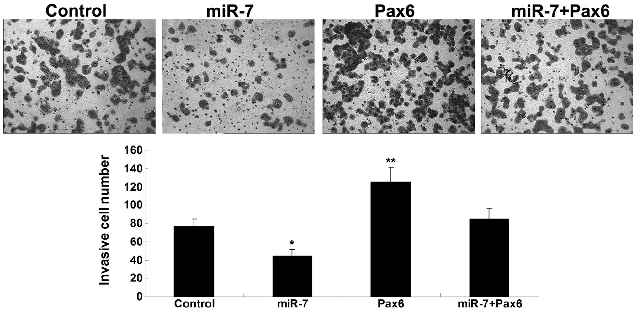
Overexpression of miR-7 inhibits NSCLC
A549 cell invasion through inhibition of Pax6
The present study subsequently further investigated
the roles of miR-7 and Pax6 in the regulation of A549 NSCLC cell
invasion. The findings demonstrated that upregulation of miR-7
inhibited A549 NSCLC cell invasion, whereas in the
Pax6-overexpressed A549 NSCLC cells, cell invasion was upregulated.
Furthermore, restoration of the expression of Pax6 reversed the
suppressive effect of miR-7 overexpression on NSCLC A549 cell
proliferation. These data suggested that overexpression of miR-7
inhibited NSCLC cell invasion through the inhibition of Pax6
(Fig. 4).
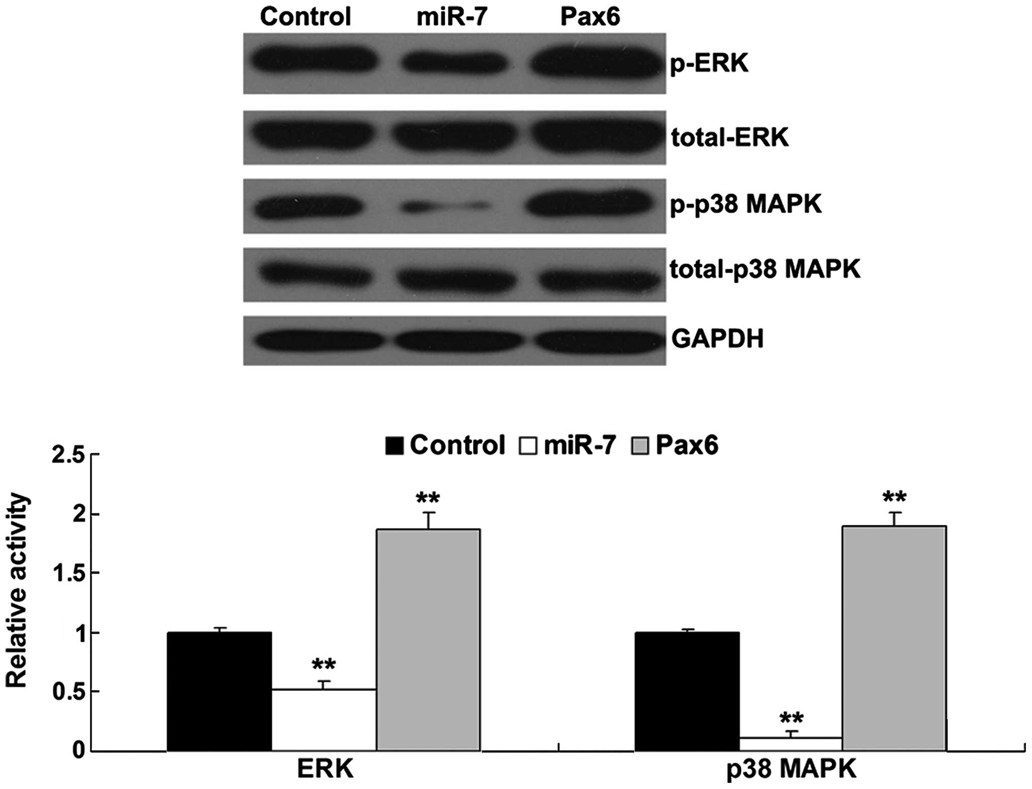
Activities of the ERK and p38 MAPK
pathways are mediated by miR-7 and Pax6 in A549 NSCLC cells
The activities of the ERK and p38 MAPK signaling
pathways were determined following the overexpression of Pax6 or
miR-7 in the A549 human NSCLC cells. As shown in Fig. 5, the phosphorylated protein levels
of ERK and p38 MAPK were suppressed in the miR-7-overexpressed A549
cells, but were increased in the Pax6-overexpressed A549 cells.
These data indicated that the activities of the ERK and p38 MAPK
signalingpathways were downregulated by the overexpression of
miR-7, but upregulated by the overexpression of Pax6 in A549 NSCLC
cells.
Discussion
In the present study, the roles of miR-7 and Pax6,
as well as their associations, were investigated in NSCLC cells.
The results demonstrated that miR-7 was downregulated, while Pax6
upregulated in the NSCLC cell lines. Further investigation
identified Pax6 as a target of miR-7 in the A549 NSCLC cells, and
the protein expression of Pax6 was negatively mediated by miR-7.
Overexpression of miR-7 significantly inhibited A549 cell
proliferation and invasion, which was reversed by the upregulation
of Pax6. In addition, the ERK and p38 MAPK signaling pathways were
downregulated in miR-7-overexpressed A549 cells, but activated in
Pax6-overexpressed A549 cells.
It has been demonstrated that miR-7 acts as a tumor
suppressor in various types of cancer. Reduced levels of miR-7 have
been linked to the development of cancer and metastasis (17). Zhou et al reported that
miR-7 inhibits tumor metastasis and reverses epithelial-mesenchymal
transition through inhibition of AKT and ERK signaling in
epithelial ovarian cancer (18).
Xie et al demonstrated that miR-7 inhibits gastric cancer
cell invasion and metastasis by suppressing the expression of
epidermal growth factor receptor (19). In adddition, miR-7 has been
reported to inhibit colorectal cancer cell proliferation and induce
apoptosis (20). The role of miR-7
in NSCLC has also been reported. Xiong et al revealed that
miR-7 has a suppressive effect in NSCLC by targeting PA28γ
(6). In addition miR-7 has been
observed to inhibit the growth of A549 NSCLC cells through
targeting BCL-2 (7). As a single
miRNA has multiple targets, and one gene can be regulated by
various miRNAs (21), the presents
study aimed to identify other targets of miR-7 in NSCLC cells, and
demonstrated that the inhibition of Pax6 was involved in
miR-7-overexpression-induced downregulation of A549 NSCLC cell
proliferation and invasion.
As a transcription factor, Pax6 mediates the
expression of various genes. In addition, the expression of Pax6
has been reported to be upregulated in pancreatic cancer and
retinoblastoma, but downregulated in glioma and prostate cancer,
suggesting that Pax6 may have different roles in different types of
cancer (13,22–24).
In the present study, the results suggested that, as a target of
miR-7, Pax6 acts as an oncogene in NSCLC through promoting NSCLC
cell proliferation and invasion. Previously, Zhao et al
investigated the role of Pax6 in NSCLC cell proliferation and
reported that inhibition of the expression of Pax6 suppresses cell
growth and colony formation in A549 and H1299 NSCLC cells (15). In addition, the percentage of NSCLC
cells in the G1-phase is increased following a reduction in the
expression of Pax6, indicating that the inhibition of Pax6 induces
cell cycle arrest at the G1 phase (15). It was also demonstrated that the
activitites of the ERK and p38 MAPK signaling pathways were
suppressed in Pax6 knockdown cells (15), consistent with the data obtained
the present study, that overexpression of Pax6 induced upregulation
in the activity of ERK and p38 MAPK signaling. It has been well
established that the ERK and p38 MAPK signaling pathways are
involved in the development and progression of various types of
cancer, including NSCLC, and targeting components of the ERK and
MAPK signaling pathways offers an attractive strategy in the
development of novel therapeutic approaches to treat NSCLC
(25,26).
In addition, the present study demonstrated for the
first time, to the best of our knowledge, that miR-7/Pax6 is
involved in the regulation of NSCLC cell invasion. The role of Pax6
in cancer cell invasion has also been demonstrated in several other
types of cancers, including glioma and colon cancer (27). Cheng et al reported that
Pax6 inhibits cell proliferation and invasion in glioma cells
(27), and, Liu et al
suggested that brain fatty acid-binding protein 7 is a target gene
of Pax6, and is involved in the regulation of glioma cell invasion
(28). In glioblastoma cells, Pax6
has been reported to suppress cell invasion, at least in part,
through inhibiting the protein expression of matrix
metalloproteinase (MMP) 2 (29).
Huang et al also demonstrated that MMP2 and MMP9 are
downstream effectors of Pax7 in the regulation of glioblastoma cell
invasion (13).
In conclusion, the present study demonstrated that
miR-7 inhibited NSCLC cell proliferation and invasion through the
direct targeting of Pax6, suggesting that Pax6 and miR-7 may serve
as potential therapeutic targets in the treatment of NSCLC.
References
|
1
|
Greenlee RT, Murray T, Bolden S and Wingo
PA: Cancer statistics, 2000. CA Cancer J Clin. 50:7–33. 2000.
View Article : Google Scholar : PubMed/NCBI
|
|
2
|
Kang SM and Lee HJ: MicroRNAs in human
lung cancer. Exp Biol Med (Maywood). 239:1505–1513. 2014.
View Article : Google Scholar
|
|
3
|
Huang X, Liang M, Dittmar R and Wang L:
Extracellular microRNAs in urologic malignancies: Chances and
challenges. Int J Mol Sci. 14:14785–14799. 2013. View Article : Google Scholar : PubMed/NCBI
|
|
4
|
Zagryazhskaya A and Zhivotovsky B: miRNAs
in lung cancer: A link to aging. Ageing Res Rev. 17:54–67. 2014.
View Article : Google Scholar : PubMed/NCBI
|
|
5
|
Giles KM, Barker A, Zhang PM, Epis MR and
Leedman PJ: MicroRNA regulation of growth factor receptor signaling
in human cancer cells. Methods Mol Biol. 676:147–163. 2011.
View Article : Google Scholar
|
|
6
|
Xiong S, Zheng Y, Jiang P, Liu R, Liu X,
Qian J, Gu J, Chang L, Ge D and Chu Y: PA28gamma emerges as a novel
functional target of tumour suppressor microRNA-7 in non-small-cell
lung cancer. Br J Cancer. 110:353–362. 2014. View Article : Google Scholar :
|
|
7
|
Xiong S, Zheng Y, Jiang P, Liu R, Liu X
and Chu Y: MicroRNA-7 inhibits the growth of human non-small cell
lung cancer A549 cells through targeting BCL-2. Int J Biol Sci.
7:805–814. 2011. View Article : Google Scholar : PubMed/NCBI
|
|
8
|
Shaham O, Menuchin Y, Farhy C and
Ashery-Padan R: Pax6: A multi-level regulator of ocular
development. Prog Retin Eye Res. 31:351–376. 2012. View Article : Google Scholar : PubMed/NCBI
|
|
9
|
Georgala PA, Carr CB and Price DJ: The
role of Pax6 in forebrain development. Dev Neurobiol. 71:690–709.
2011. View Article : Google Scholar : PubMed/NCBI
|
|
10
|
Matsumoto Y and Osumi N: Role of Pax6 in
the developing central nervous system. Brain Nerve. 60:365–374.
2008.In Japanese. PubMed/NCBI
|
|
11
|
Simpson TI and Price DJ: Pax6; a
pleiotropic player in development. BioEssays. 24:1041–1051. 2002.
View Article : Google Scholar : PubMed/NCBI
|
|
12
|
Gosmain Y, Cheyssac C, Heddad Masson M,
Dibner C and Philippe J: Glucagon gene expression in the endocrine
pancreas: The role of the transcription factor Pax6 in α-cell
differentiation, glucagon biosynthesis and secretion. Diabetes Obes
Metab. 13(Suppl 1): 31–38. 2011. View Article : Google Scholar
|
|
13
|
Huang BS, Luo QZ, Han Y, Li XB, Cao LJ and
Wu LX: microRNA-223 promotes the growth and invasion of
glioblastoma cells by targeting tumor suppressor PAX6. Oncol Rep.
30:2263–2269. 2013.PubMed/NCBI
|
|
14
|
Bai SW, Li B, Zhang H, Jonas JB, Zhao BW,
Shen L and Wang YC: Pax6 regulates proliferation and apoptosis of
human retinoblastoma cells. Invest Ophthalmol Vis Sci.
52:4560–4570. 2011. View Article : Google Scholar
|
|
15
|
Zhao X, Yue W, Zhang L, Ma L, Jia W, Qian
Z, Zhang C and Wang Y: Downregulation of PAX6 by shRNA inhibits
proliferation and cell cycle progression of human non-small cell
lung cancer cell lines. PLoS One. 9:e857382014. View Article : Google Scholar : PubMed/NCBI
|
|
16
|
Li Y, Li Y, Liu Y, Xie P, Li F and Li G:
PAX6, a novel target of microRNA-7, promotes cellular proliferation
and invasion in human colorectal cancer cells. Dig Dis Sci.
59:598–606. 2014. View Article : Google Scholar
|
|
17
|
Kalinowski FC, Brown RA, Ganda C, Giles
KM, Epis MR, Horsham J and Leedman PJ: microRNA-7: A tumor
suppressor miRNA with therapeutic potential. Int J Biochem Cell
Biol. 54:312–317. 2014. View Article : Google Scholar : PubMed/NCBI
|
|
18
|
Zhou X, Hu Y, Dai L, Wang Y, Zhou J, Wang
W, Di W and Qiu L: MicroRNA-7 inhibits tumor metastasis and
reverses epithelial-mesenchymal transition through AKT/ERK1/2
inactivation by targeting EGFR in epithelial ovarian cancer. PLoS
One. 9:e967182014. View Article : Google Scholar : PubMed/NCBI
|
|
19
|
Xie J, Chen M, Zhou J, Mo MS, Zhu LH, Liu
YP, Gui QJ, Zhang L and Li GQ: miR-7 inhibits the invasion and
metastasis of gastric cancer cells by suppressing epidermal growth
factor receptor expression. Oncol Rep. 31:1715–1722.
2014.PubMed/NCBI
|
|
20
|
Xu K, Chen Z, Qin C and Song X: miR-7
inhibits colorectal cancer cell proliferation and induces apoptosis
by targeting XRCC2. Onco Targets Ther. 7:325–332. 2014. View Article : Google Scholar : PubMed/NCBI
|
|
21
|
Ambros V: The functions of animal
microRNAs. Nature. 431:350–355. 2004. View Article : Google Scholar : PubMed/NCBI
|
|
22
|
Mascarenhas JB, Young KP, Littlejohn EL,
Yoo BK, Salgia R and Lang D: PAX6 is expressed in pancreatic cancer
and actively participates in cancer progression through activation
of the MET tyrosine kinase receptor gene. J Biol Chem.
284:27524–27532. 2009. View Article : Google Scholar : PubMed/NCBI
|
|
23
|
Wang J, Wang X, Wu G, Hou D and Hu Q:
MiR-365b-3p, down-regulated in retinoblastoma, regulates cell cycle
progression and apoptosis of human retinoblastoma cells by
targeting PAX6. FEBS Lett. 587:1779–1786. 2013. View Article : Google Scholar : PubMed/NCBI
|
|
24
|
Shyr CR, Tsai MY, Yeh S, Kang HY, Chang
YC, Wong PL, Huang CC, Huang KE and Chang C: Tumor suppressor PAX6
functions as androgen receptor co-repressor to inhibit prostate
cancer growth. Prostate. 70:190–199. 2010.
|
|
25
|
Ciuffreda L, Incani UC, Steelman LS,
Abrams SL, Falcone I, Curatolo AD, Chappell WH, Franklin RA, Vari
S, Cognetti F, et al: Signaling Intermediates (MAPK and PI3K) as
Therapeutic Targets in NSCLC. Curr Pharm Des. 20:3944–3957. 2014.
View Article : Google Scholar
|
|
26
|
Chen Y, Nowak I, Huang J, Keng PC, Sun H,
Xu H, Wei G and Lee SO: Erk/MAP kinase signaling pathway and
neuroendocrine differentiation of non-small-cell lung cancer. J
Thorac Oncol. 9:50–58. 2014. View Article : Google Scholar
|
|
27
|
Cheng Q, Cao H, Chen Z, Ma Z, Wan X, Peng
R and Jiang B: PAX6, a novel target of miR-335, inhibits cell
proliferation and invasion in glioma cells. Mol Med Rep.
10:399–404. 2014.PubMed/NCBI
|
|
28
|
Liu RZ, Monckton EA and Godbout R:
Regulation of the FABP7 gene by PAX6 in malignant glioma cells.
Biochem Biophys Res Commun. 422:482–487. 2012. View Article : Google Scholar : PubMed/NCBI
|
|
29
|
Mayes DA, Hu Y, Teng Y, Siegel E, Wu X,
Panda K, Tan F, Yung WK and Zhou YH: PAX6 suppresses the
invasiveness of glioblastoma cells and the expression of the matrix
metalloproteinase-2 gene. Cancer Res. 66:9809–9817. 2006.
View Article : Google Scholar : PubMed/NCBI
|