Introduction
Currently, total knee and hip replacements are
commonly used for the reconstruction of injured or degenerated
joints. The tight fixation of the implant to the surrounding tissue
is mostly responsible for stable integration into the human body
and should preclude micromovements that may lead to the failure of
the implant. Implant fixation can be realized via cementless means
or with bone cement. The type of implant fixation and the
integration into the body by bone ingrowth highly influences the
stability and appropriate use of artificial joints. Due to the
standard use of artificial joints by the patient, the friction
between sliding parts of the implant and the physiological
environment, wear particles are produced from various components of
the implant or from the bone-implant interface as well as between
the bone and cement mantle (1–3). The
aseptic loosening of implants due to these particles and
tribological products is one of the most common problems occurring
in the field of joint endoprosthetics (4). These wear particles can cause
osteolytic processes as a result of osteoblastic inhibition and
osteoclastic activation (4). Other
cell types, including macrophages, fibroblasts, mesenchymal stem
cells and lymphocytes are also involved in the osteolytic cascade
(5). Wear debris from all
orthopedic materials may induce a lot of cellular responses with
less specific characteristics of the respective metal and certain
types of debris can be more cytotoxic than others (3,6).
However, the pathophysiological background has not been fully
elucidated and effective therapies are not currently available.
Nevertheless, it is well known that various proteins and signaling
molecules are involved in the osteolytic process. This process
includes osteoblast- and macrophage-secreted matrix
metalloproteinases (MMPs), of which MMPs 1, 2, 9 and 13 are of
particular importance for aseptic implant loosening (7,8).
These MMPs are responsible for the degradation of all components of
the extracellular matrix resulting in a loss of bone matrix.
Additionally, bone resorption, as a response to wear debris, is
mediated by cytokines, including tumor necrosis factor (TNF)α and
interleukin (IL)-1β. Together, they can induce the expression of
the receptor activator of nuclear factor-κB (RANK) ligand (RANKL),
which is the general regulator of osteoclast generation as well as
activation (9). As a result, these
bone-resorbing osteoclasts enhance the matrix degrading effect of
MMPs. A number of cytokines and growth factors are involved in the
signaling pathway of MMP expression (10,11).
As mentioned above, the RANK/RANKL/osteoprotegerin
(OPG) system is important in the differentiation of osteoclasts
(12). Osteoblasts synthesize the
effector protein RANKL, which can bind to the RANK receptor on
osteoclastic progenitor cells. OPG, also expressed by osteoblasts,
is able to bind RANKL inhibiting its binding to RANK (13). Therefore, the RANK/RANKL/OPG system
is another effector system to sustain the periprosthetic balance
between bone resorption and formation (14).
Wear debris also affects osteoblasts, which are
negatively influenced by decreasing the synthetic capacity of
collagen type 1, the major component of bone extracellular matrix
(4,15–17).
Additionally, the apoptosis rate of osteoblasts is clearly
increased (4,17).
As extracellular matrix degradation and remodeling
processes around implants have been considered the main biological
events during periprosthetic loosening (5), the aim of the present study was to
analyze the influence of different wear particles on the expression
of matrix degrading MMPs and proinflammatory cytokines in human
osteoblasts and macrophages. Monocytes/macrophages are the
predominant cells involved in inflammatory particle-induced
osteolysis due to their phagocytic role and cytokine release
(6,18,19).
In the current study, it was determined which of these two cell
types was more responsible for increased cytokine and MMP
expression, and thus for the stimulation of matrix degradation.
Furthermore, it was determined which type of MMP was induced by
wear particles in the two cell types.
In contrast to other studies conducted using
commercially available particles, particles generated from various
implant materials (titanium and cobalt chromium alloys) were used.
Following exposure of human osteoblasts and monocytes/macrophages
to these particles, the release of different MMPs and cytokines was
analyzed. Additionally, the release of tissue inhibitors of MMPs
(TIMPs) and their impact on MMP inhibition was determined.
Materials and methods
Generation and preparation of abrasive
wear particles
Particles were generated by simulating the
interfacial wear between total hip stems and bone cement using a
special wear simulator (1). For
particle generation, standard total hip stems made of either a
titanium alloy (Ti-6Al-7Nb) or a Cobalt-Chromium alloy
(Co-28Cr-6Mo) were used (20). The
implants were straight stems characterized by a defined surface
roughness (Rz) value of ~7 µm (smooth surface, Co-28Cr-6Mo)
and 20 µm (rough surface, Ti-6Al-7Nb) were used. The total
hip stems were cemented with commercially available
polymethylmethacrylate (PMMA) bone cement (Palacos® R,
Hereaus Kulzer GmbH, Wehrheim, Germany), containing 15%
zirconiumoxide (ZrO2) particles as a radio-opaque
additive.
Prior to cell biological experiments, the quantities
of metallic particles from the generated wear debris (a
conglomerate of metallic and bone cement particles) were determined
by atomic absorption spectroscopy and were shown to be 424±93
µg Ti for Ti-6Al-7Nb stems and 376±83 µg Co for
Co-28Cr-6Mo stems (21).
Additionally, >99% of the total particles were non-metallic
(21). The mean particle size was
determined by size distribution measurements and ranged from 2.8
µm (Co-28Cr-6Mo) to 4.4 µm (Ti-6Al-7Nb) whereby 90%
of the particles were <15 µm (21).
Moreover, commercially pure titanium powder with an
average particle size of 3 µm (cp-Ti; Grade E, dry;
Chemetall GmbH, Frankfurt, Germany), pure polycrystalline
ZrO2 particles with mean size of 1.75 µm
(Selectipur, Hereaus Kulzer GmbH), as well as particles from PMMA
(Hereaus Kulzer GmbH) and titanium dioxide (TiO2) with a
mean size of 1.8 µm (BioCer Entwicklungs-GmbH, Bayreuth,
Germany) were used as references.
Generated wear particles and commercially available
reference particles (5 mg of each) were sterilized using gamma
radiation. Subsequently, a stock solution (10 mg/ml) was prepared
by suspending the particles in 0.5 ml sterile phosphate-buffered
saline (PAA Laboratories GmbH, Coelbe, Germany). Further dilutions
with cell culture medium created the final particle concentrations
of 0.1 and 0.01 mg/ml.
Isolation and cultivation of human
primary osteoblasts
Human primary osteoblasts were isolated under
sterile conditions from bone marrow-derived femoral heads of
patients undergoing primary total hip replacement. All samples were
collected after participants had signed written informed consent
forms and approval by the Local Ethical Committee of the University
of Rostock (Rostock, Germany; no. AZ: 2010–10) had been
obtained.
Cancellous bone was extracted from femoral heads,
suspended in sterile phosphate-buffered saline (PBS) and washed
twice. Osteoblasts were obtained using the protocol of Lochner
et al (4).
Freshly isolated osteoblast-like cells were plated
in 25 cm2 culture flasks in 8 ml osteogenic medium
(Dulbecco's modified Eagle's medium; Biochrom AG Biotechnologie,
Berlin, Germany), supplemented with 10% fetal calf serum, 1%
penicillin/streptomycin, 1% amphotericin B, 1% HEPES, 50
µg/ml ascorbic acide, 10 mm β-glycerophosphate and 100 nM
dexamethasone, and were incubated at 37°C in a humidified
atmosphere of 5% CO2 and 95% air. The culture medium was
changed every other day and non-adherent cells were aspirated. The
progress of proliferation was determined by microscopy. When a
confluency of 90% was reached, the cells were subcultured at a
ratio of 1:6. The cultivated cells were used in experiments at
passage three. To verify the osteogenic character of isolated
cells, alkaline phosphatase staining with fuchsin and substrate
chromogen (Dako, Hamburg, Germany) was performed, according to the
manufacturer's instructions. To exclude the presence of
monocytes/macrophages in the osteoblastic culture, flow cytometry
was performed on representative samples from two donors (4). Since in the primary cell cultures, no
CD68 positive cells (selective for monocytes) and only 2.4% CD14
positive cells (selective for monocytes and macrophages) were
detected, a contamination of human primary osteoblasts with
monocytes/macrophages could be excluded (4).
For in vitro experiments, osteoblasts were
transferred to 96-well plates with 3,000 cells per well in 200
µl complete medium. The human osteoblasts were allowed to
adhere for 24 h at which time the particle-free medium was replaced
with particle-containing medium for 48 h. Osteoblasts incubated
with normal culture medium served as test controls.
Supernatants from osteoblasts incubated with
particle-containing medium were investigated for the expression of
cytokines IL-6, IL-8, monocyte chemoattractant protein-1 (MCP1) and
vascular endothelial growth factor (VEGF), as well as for the
synthesis of MMPs 1, 3, 8 and 10, and their inhibitors, TIMPs 1–4.
These data were collected to assess whether osteoblasts, being
bone-forming cells, contribute with their induction of osteoclast
differentiation and matrix resorption to osteolysis following
contact with abrasive wear particles.
Cultivation of human
monocytes/macrophages
The THP-1 monocytic cell line is an ideal cell line
for differentiation experiments of macrophages as it is similar in
morphology and metabolism to macrophages. Therefore, differentiated
THP-1 macrophages have already been used in several in vitro
studies concerning macrophage involvement in inflammatory processes
(22). To induce differentiation
within 48 h of incubation, phorbol 12-myristate 13-acetate (PMA)
can be used (22).
In the present experiments, THP-1 cells (American
Type Culture Collection, Rockville, MD, USA) were maintained in
continuous culture in RPMI-1640 medium (Biochrom AG Biotechnologie)
supplemented with 10% fetal calf serum (FCS), 100 U/ml L-glutamine,
200 mU/ml penicillin and 0.2 mg/ml streptomycin. Cells were
incubated at 37°C in a humidified atmosphere of 5% CO2
and 95% air. For differentiation into macrophage-like adherent
cells, THP-1 cells were transferred into 6-well plates with
2×10⁶ cells per well in 3 ml differentiation medium.
Differentiation medium was composed of RPMI-1640 and supplements,
as well as 25 nM PMA for the maturation of monocytes into
macrophage-like adherent cells. All medium contents were purchased
from PAA Laboratories GmbH (Linz, Austria).
Following differentiation into macrophage-like cells
within 48 h, the particle-free differentiation medium was replaced
with the respective particle-containing medium (stem-derived wear
particles and commercially available reference particles) for a
further 48 h. Macrophages incubated with normal culture medium
served as test controls.
Supernatants from the macrophage culture were
investigated for the expression of cytokines (IL-6, IL-8, MCP1 and
VEGF) as well as for the synthesis of MMPs 1, 3, 8, 10 and 13, and
their inhibitors, TIMPs 1–4. These data were collected to assess
the extent to which macrophages contribute to matrix resorption and
osteolysis after contact with abrasive wear particles.
Protein detection using multiplex
technology
The effects of particle exposition on the release of
cytokines (IL-6, IL-8, MCP1 and VEGF), ECM-degrading MMPs (MMPs 1,
3, 8, 9 and 10) and endogenous TIMPs (TIMP 1, 2, 3 and 4) were
analyzed by Multiplex enzyme-linked immunosorbent assays.
Therefore, the supernatants of osteoblasts and macrophage-like
cells were quantified after 48 h of incubation using the Multiplex
Cytokine Assay (Bio-Plex Pro Human Cytokines Group I 5-plex,
Bio-Rad Laboratories Inc., Munich, Germany), the Multiplex MMP
Assay (WideScreen Human MMP Panel, Novagen, Merck Chemicals Ltd.,
Nottingham, UK) and the Fluorokine MAP Human TIMP Multiplex kit
(R&D Systems, Abingdon, UK). All assays are based on
simultaneous detection of different mediators in a single well of a
96-well microplate involving fluorochrome-labeled microsphere
beads. For the assays, osteoblast or macrophage supernatants,
standard solutions, and blanks were dispensed into the
corresponding wells of a 96-well filter plate already containing
anti-cytokine/MMP/TIMP-conjugated beads. The assays were performed
according to the manufacturer's instructions. The concentration of
cytokines, MMPs and TIMPs were finally detected by the Bio-Plex 200
system using the Bio-Plex Manager software 4.1.1 (both from Bio-Rad
Laboratories Inc., Munich, Germany). Protein concentrations were
calculated using a standard curve derived from a recombinant
standard supported by the multiplex assays.
Data illustration and statistical
analysis
Data are presented as box plots. Boxes denote
interquartile ranges, horizontal lines within the boxes denote
medians, and whiskers denote minimum and maximum values. For all
analyses, cultures of human osteoblasts from a minimum of three
independent donors and of the THP-1 cell line from a minimum of
three independent experiments were used (in duplicates). Since the
data obtained were not normally distributed, the statistical
significance between two datasets was calculated with the
Mann-Whitney U test using SPSS 20 (IBM Deutschland, Ehningen,
Germany). P≤0.05 was considered to indicate a statistically
significant difference.
Results
MMP expression by human osteoblasts and
macrophages
MMPs are zinc-dependent proteases that degrade,
among other proteins, collagens of the extracellular matrix. After
48 h of particle exposure, the levels of MMPs were determined by a
multiplex ELISA.
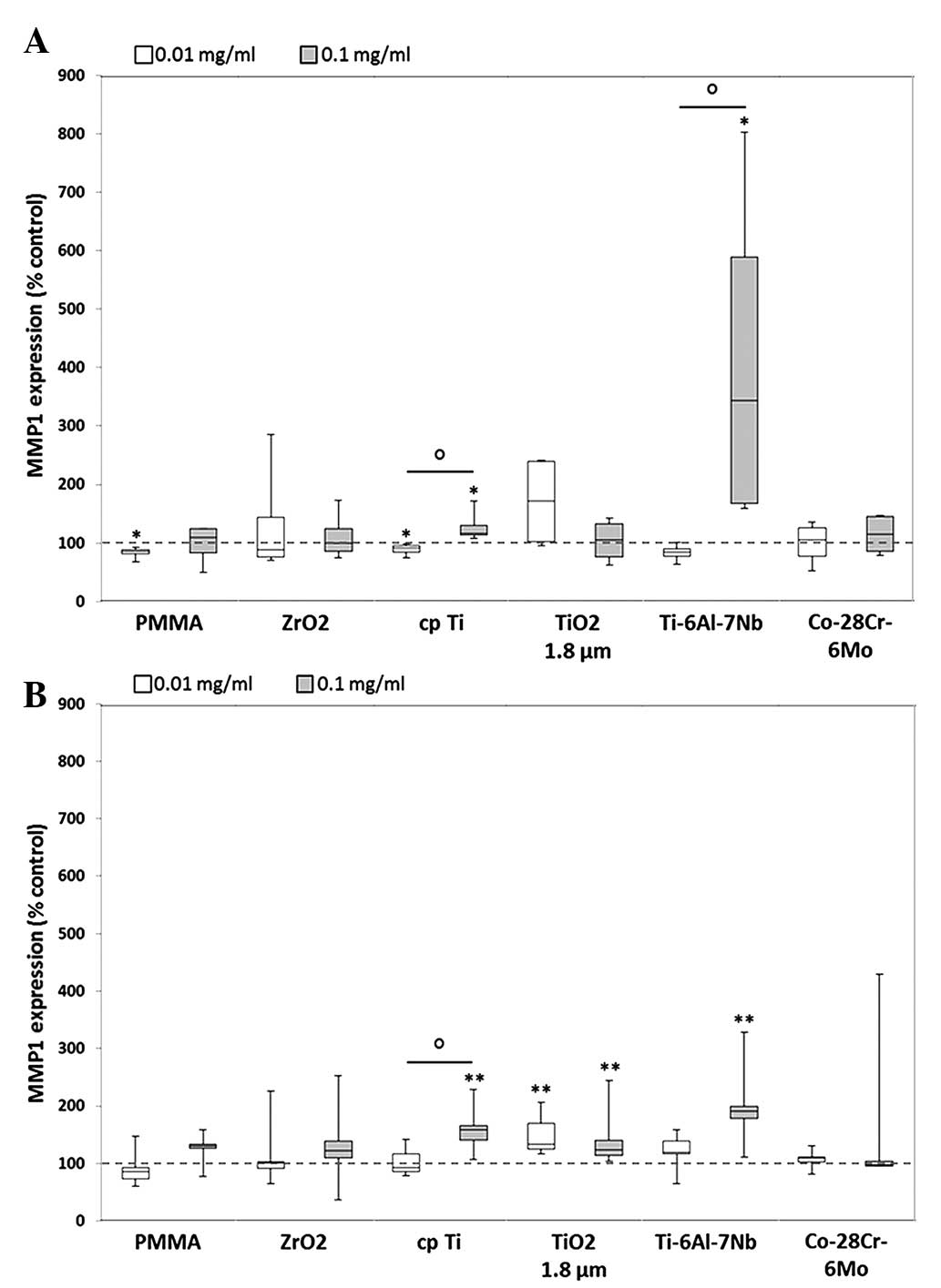
Compared with unstimulated cells, MMP1 expression
was significantly induced following exposure to a higher
concentration (0.1 mg/ml) of the cpTi reference particles
(osteoblasts: 1.2-fold increase, P=0.014; macrophages: 1.6-fold
increase, P=0.005) and TiO2 (macrophages: 1.2-fold
increase, P=0.005) as well as from hip stem-derived particles of
Ti-6Al-7Nb (osteoblasts: 3.4-fold increase, P=0.014 macrophages:
1.9-fold increase, P= 0.005) in the two cell types. Furthermore,
significant differences between particle concentrations were
observed following exposure to particles from cpTi (P=0.021) and
Ti-6Al-7Nb (P=0.021) in osteoblasts as well as for cpTi particles
(P=0.028) in macrophages. By contrast, particles from Co-28Cr-6Mo
stems as well as reference particles from PMMA and ZrO2
did not induce MMP1 expression in human osteoblasts and macrophages
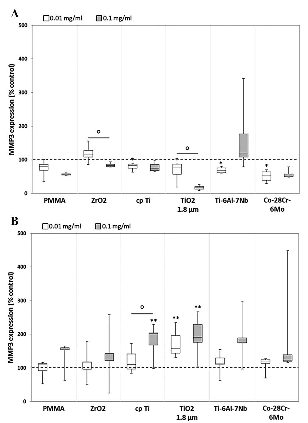
(Fig. 1). MMP3 protein was mostly
downregulated in human osteoblasts following exposure to reference
and hip stem-derived particles. Significant differences compared
with unstimulated cells were shown for the lower particle
concentration of cpTi (0.8-fold decrease, P=0.014), TiO2
(0.8-fold decrease, P=0.014), Ti-6Al-7Nb (0.7-fold decrease,
P=0.014) and Co-28Cr-6Mo (0.5-fold decrease, P=0.014).
Additionally, concentration-dependent differences were visible for
ZrO2 [1.2-fold increase (0.01 mg/ml) vs. 0.8-fold
decrease (0.1 mg/ml), P= 0.043] and TiO2 [0.8-fold
decrease (0.01 mg/ml) vs. 0.2-fold increase (0.1 mg/ml), P=0.043]
particles (Fig. 2A). In human
macrophages, particle exposure resulted in an increase in MMP3
protein (Fig. 2B); a greater
higher particle concentration generally resulted in increased MMP3
levels, particularly with titanium-based particles [cpTi: 1.1-fold
increase (0.01 mg/ml), 2-fold increase, P=0.005 (0.1 mg/ml); and
TiO2: 1.6-fold increase (0.01 mg/ml), 1.9-fold increase
(0.1 mg/ml); both P=0.005].
Compared with unstimulated cells, MMP8 protein
expression was induced in human osteoblasts following exposure to
all particles, with the exception of cpTi (0.01 mg/ml: 0.9-fold
decrease) and TiO2 (0.1 mg/ml: 0.9-fold decrease). A
significant upregulation was determined for particles from
ZrO2 (0.1 mg/ml: 1.2-fold, P=0.029) and Ti-6Al-7Nb (0.1
mg/ml: 1.5-fold, P=0.029) following exposure to the higher particle
concentration. In human macrophages, a concentration-dependent
upregulation of MMP8 protein was also determined following
treatment with reference and Ti-6Al-7Nb particles, with higher
protein levels following exposure to the higher particle
concentration [PMMA: 0.8-fold decrease, P=0.008 (0.01 mg/ml) vs.
1.2-fold increase (0.1 mg/ml); ZrO2: 1.0-fold (0.01
mg/ml) vs. 1.3-fold increase (0.1 mg/ml); cpTi: 0.9-fold decrease
(0.01 mg/ml) vs. 1.3-fold increase, P=0.008 (0.1 mg/ml), P=0.032
(0.01 mg/ml vs. 0.1 mg/ml); Ti-6Al-7Nb: 1.1-fold increase (0.01
mg/ml) vs. 1.4-fold increase (0.1 mg/ml)] (data not shown).
Furthermore, synthesis of MMP10 protein was
down-regulated in human osteoblasts for all particles except for
ZrO2 (0.01 mg/ml: 1.5-fold increase) and Ti-6Al-7Nb (0.1
mg/ml: 11-fold increase) particles, which resulted in increased
MMP10 expression following exposure. In human macrophages, MMP10
protein content was increased following exposure to all particles
with a tendency for higher protein amounts following stimulation
with 0.1 mg/ml particle suspension compared with 0.01 mg/ml [PMMA:
1.0-fold vs. 1.3-fold, ZrO2: 1.0-fold vs. 1.3-fold,
cpTi: 1.0-fold vs. 1.5-fold, Ti-6Al-7Nb: 1.2-fold vs. 1.6-fold,
Co-28Cr-6Mo: 1.1-fold vs. 1.2-fold, all 0.01 mg/ml vs. 0.1 mg/ml,
respectively] (data not shown).
Finally, induction of MMP13 protein expression was
only observed in human macrophages following exposure to
TiO2 (0.01 mg/ml: 1.2-fold; 0.1 mg/ml: 1.5-fold) and
Ti-6Al-7Nb (0.01 mg/ml: 1.2-fold) particles; however, this was not
identified to be significantly different compared with the
unstimulated control (data not shown).
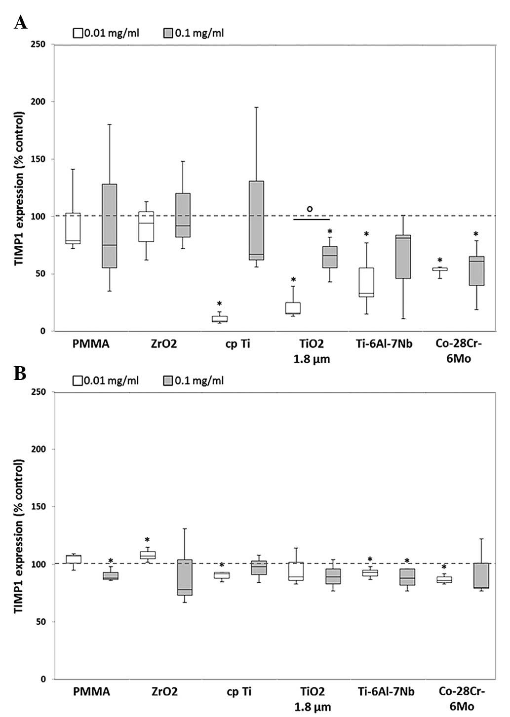
Expression of TIMPs
The effects of MMPs are naturally inhibited by
endogenous TIMPs, which comprise a family of four specific members.
Thus, the activity of MMPs is regulated by the binding of TIMPs
(23).
Expression of TIMP1 and TIMP2 was decreased in the
two cell types compared with unstimulated cells. After exposure to
the lower particle concentration, a significant decrease in TIMP1
content in human osteoblasts was shown for cpTi (0.09-fold),
TiO2 (0.16-fold), Ti-6Al-7Nb (0.33-fold) and Co-28Cr-6Mo
(0.55-fold) (all P=0.014; Fig.
3A). In human macrophages, a significant reduction of TIMP1
could be shown for all particles except for ZrO2. Here,
marginally increased levels of ZrO2 (0.01 mg/ml:
1.1-fold) were determined following exposure to lower particle
concentrations (Fig. 3B).
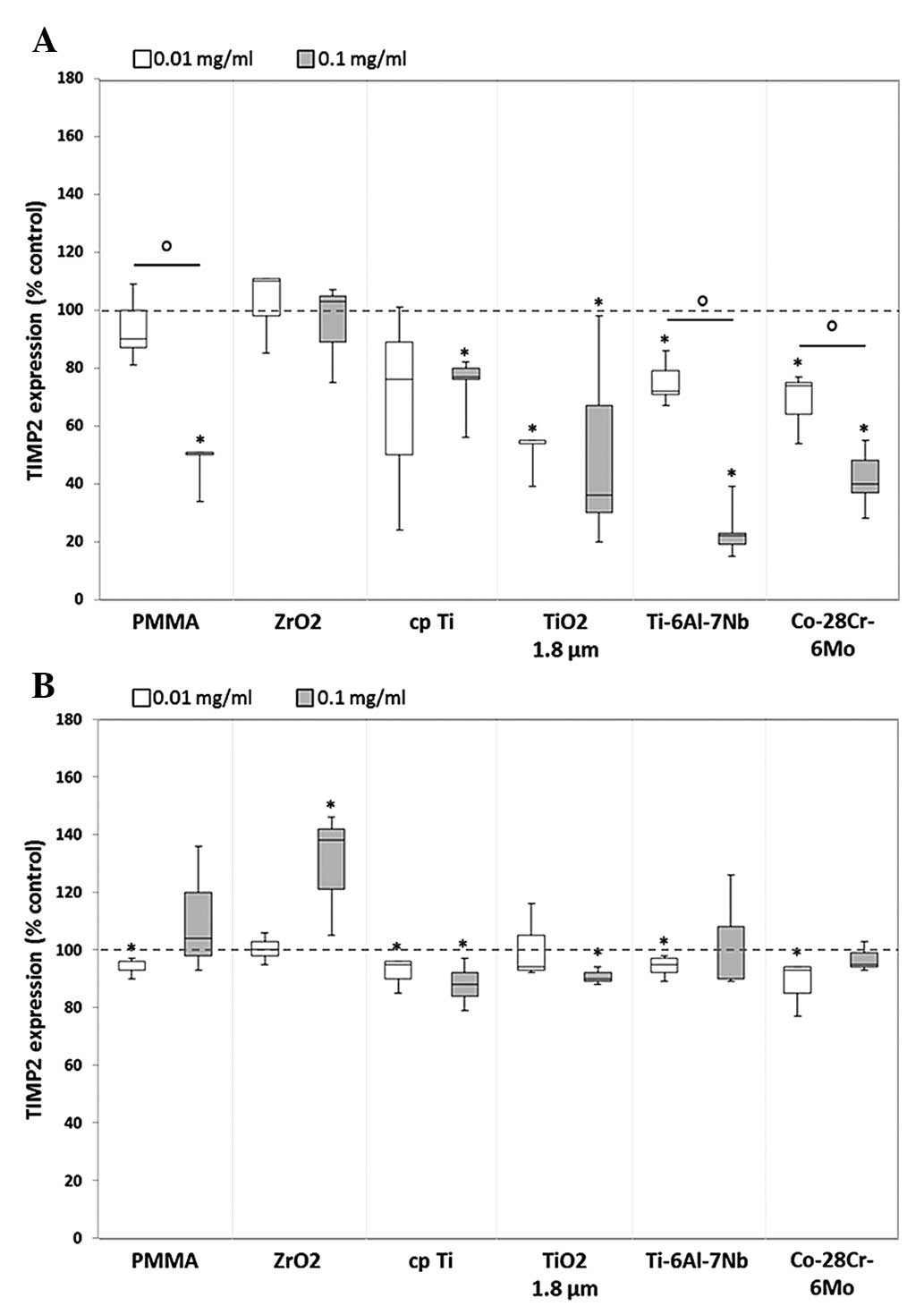
The TIMP2 expression level in human osteoblasts
showed a greater decrease when exposed to the higher dose of each
particle (Fig. 4A). Significant
differences between the two particle concentrations could be shown
following exposure to particles from PMMA [0.9-fold decrease (0.01
mg/ml) vs. 0.5-fold decrease (0.1 mg/ml); P=0.029), Ti-6Al-7Nb
[0.7-fold decrease (0.01 mg/ml) vs. 0.2-fold decrease (0.1 mg/ml);
P=0.021) and Co-28Cr-6Mo (0.7-fold decrease (0.01 mg/ml) vs.
0.4-fold decrease (0.1 mg/ml); P=0.043]. In human macrophages, a
similar expression profile to that of TIMP1 was visible for TIMP2.
Except for ZrO2 (0.1 mg/ml: 1.4-fold increase, P=0.037),
slightly decreased or constant protein amounts (compared with the
unstimulated control) were determined following particle exposure
without dose-dependent differences (Fig. 4B). For TIMP3 and TIMP4, no protein
expression was identified in human osteoblasts and macrophages
following particle exposure.
Release of cytokines following particle
exposure
Particle-induced communication among cells, such as
osteoblasts and macrophages, resulted in the production of
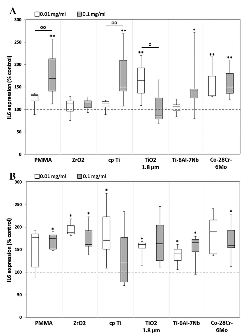
proinflammatory cytokines, including IL-6, IL-8 and MCP1 (24). Regarding the IL-6 release from
human osteoblasts, a significant upregulation in the protein level
was determined following particle exposure, except in the case of
ZrO2. Compared with unstimulated cells, higher
concentrations (0.1 mg/ml) of hip stem-derived particles resulted
in a higher IL-6 synthesis rate (Ti-6Al-7Nb: 1.4-fold, P=0.017;
Co-28Cr-6Mo: 1.5-fold) in human osteoblasts (Fig. 5A). Additionally, significant
dose-dependent differences were observed following exposure to the
reference particles PMMA [1.3-fold increase (0.01 mg/ml) vs.
1.7-fold increase (0.1 mg/ml), P=0.009], cpTi [1.1-fold increase
(0.01 mg/ml) vs. 1.5-fold increase (0.1 mg/ml), P=0.009] and
TiO2 [1.6-fold increase (0.01 mg/ml) vs. 0.9-fold
decrease (0.1 mg/ml), P=0.013]. In human macrophages, a significant
increase in IL-6 protein content was visible after treatment with
all reference particles as well as hip stem-derived particles
compared with unstimulated cells (Fig.
5B).
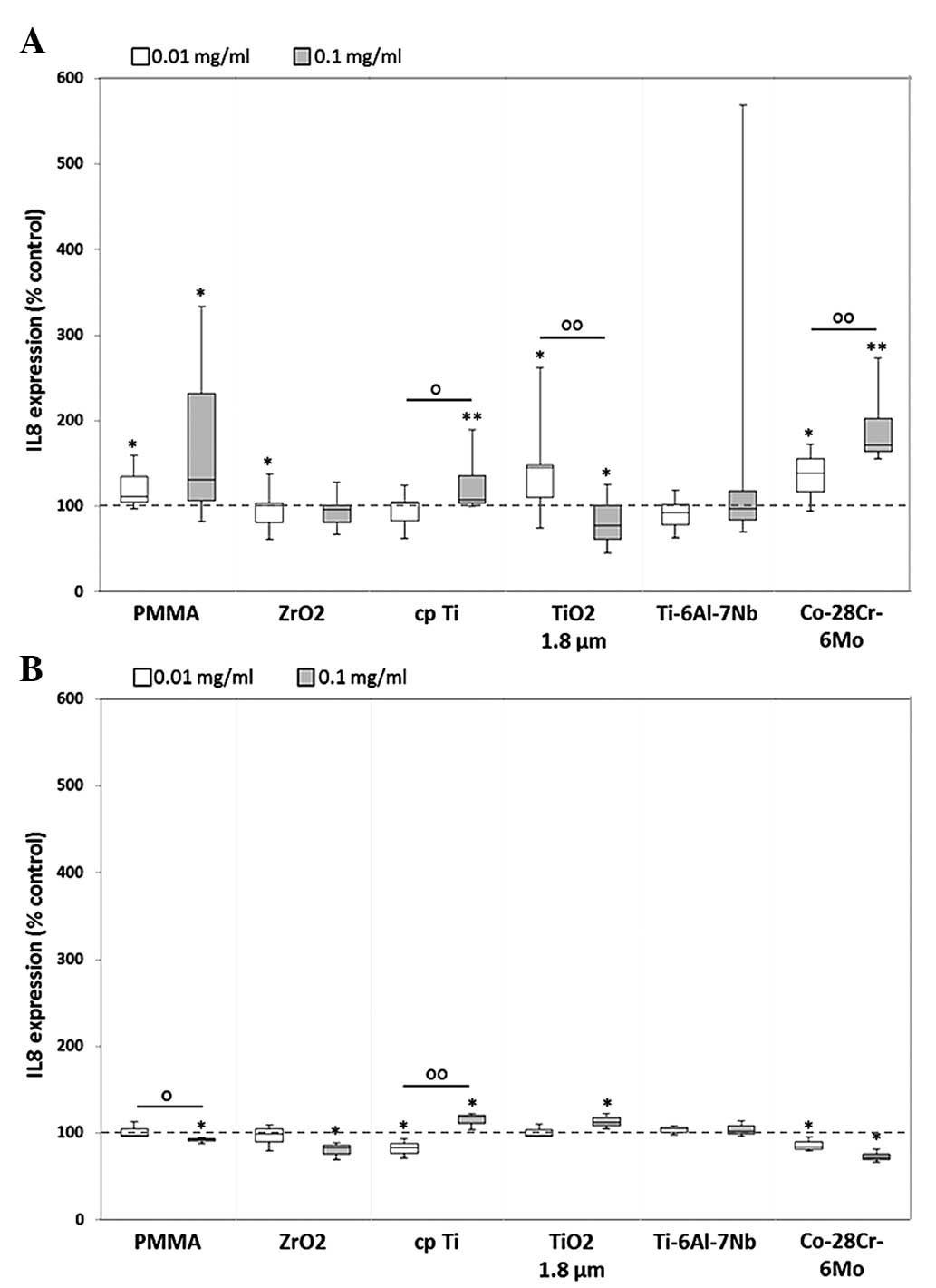
Following particle exposure, significant differences
in IL-8 protein levels between particle concentrations were shown
for cpTi [1.0-fold (0.01 mg/ml) vs. 1.1-fold increase (0.1 mg/ml),
P= 0.013], TiO2 [1.5-fold increase (0.01 mg/ml) vs.
0.8-fold decrease (0.1 mg/ml), P=0.009] and Co-28Cr-6Mo [1.4-fold
increase (0.01 mg/ml) vs. 1.7-fold increase (0.1 mg/ml), P=0.006]
(Fig. 6A). In human macrophages, a
tendency towards IL-8 downregulation was visible, with the
exception of significant increases in the titanium references
(cpTi: 1.2-fold, P=0.037; and TiO2: 1.1-fold, P=0.037)
following 0.1 mg/ml particle treatment. Furthermore, significant
differences between the levels induced by different doses of PMMA
and cpTi particles were observed (P=0.05; Fig. 6B).
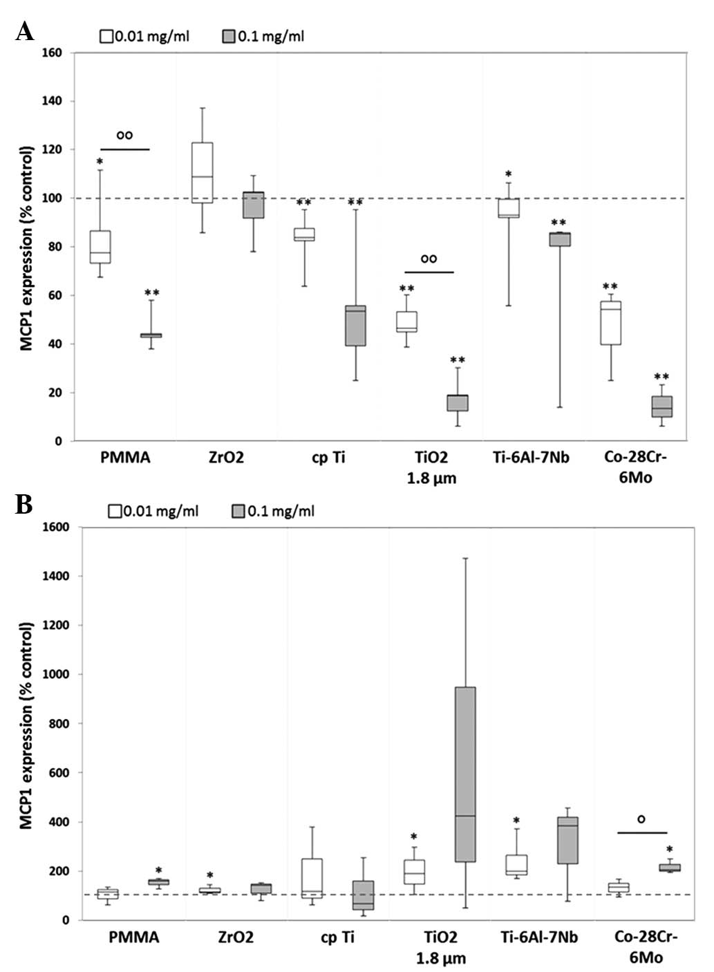
MCP1 protein synthesis was downregulated in human
osteoblasts, whereas upregulation was visible in human macrophages.
For all particle types, dose-dependent differences were observed in
osteoblastic cells, with significant differences between the doses
of PMMA [0.8-fold decrease (0.01 mg/ml) vs. 0.4-fold decrease (0.1
mg/ml), P=0.002] and TiO2 [0.5-fold decrease (0.01
mg/ml) vs. 0.2-fold decrease (0.1 mg/ml), P=0.002] (Fig. 7A). Notably, treatment with the
higher particle concentration resulted in markedly lower protein
contents after 48 h. This was in contrast to that in human
macrophages, wherein exposure to 0.1 mg/ml particle suspension
generally led to a higher induction of MCP1 protein (with the
exception of cpTi) (Fig. 7B).
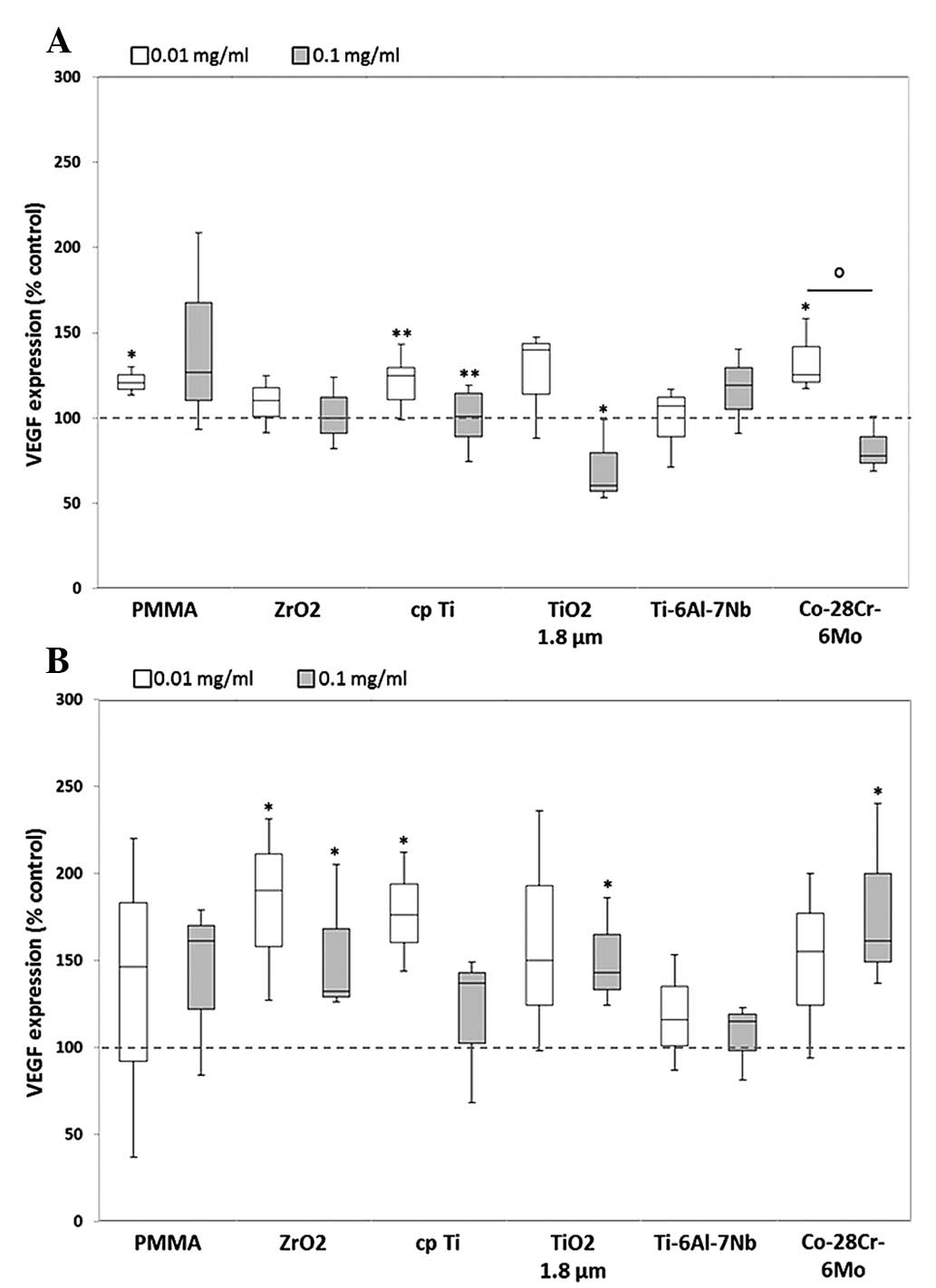
Apart from its function in the developmental
regulation of blood vessels, VEGF is also involved in the migration
of monocytes and macrophages (4).
In human osteoblasts, VEGF expression levels were mainly increased
following exposure to lower particle concentration. However, the
higher particle dose led to a downregulation of VEGF, with the
exception of PMMA and Ti-6Al-7Nb. Significant differences between
the protein levels induced by the two concentrations were only
shown for Co-28Cr-6Mo, whereby the lower particle dose induced
greater expression of VEGF protein [1.3-fold (0.01 mg/ml) vs.
0.8-fold (0.1 mg/ml), P=0.05] (Fig.
8A). In human macrophages, marked differences between the doses
were observed for each particle, however, these were not identified
to be statistically significant. Furthermore, compared with the
unstimulated cells, VEGF release was more strongly induced
following exposure to the lower particle concentration of the
references ZrO2 (1.9-fold, P=0.037), cpTi (1.8-fold,
P=0.037) and TiO2 (1.5-fold).
Discussion
In recent decades, the development of modern
endoprosthetic implants and novel bearing surfaces has been
promoted in orthopedic surgery (24,25).
Nevertheless, the generation of prosthetic wear particles resulting
in osteolytic processes has not yet been overcome. The specific
biological reactions are dependent on particle type, shape, size
and quantity, as well as the patient's individual genetic
variations (17,24). Aside from the monocyte/macrophage
cell lineage, a number of other cell types, such as fibroblasts,
lymphocytes, osteoprogenitor cells, and osteoblasts, have been
found in the periprosthetic tissue of patients, constituting a
chronic inflammatory signal (25).
The aim of the present study was to identify the
initial effects within the first 48 h of particle exposure on
cytokine release and matrix degradation by human osteoblasts and
macrophages. Therefore, cells were exposed to cemented hip
stem-derived particles of Co-28Cr-6Mo and Ti-6Al-7Nb, which were
generated in a special wear simulator by micromotions between hip
stem and cement mantle (1).
Furthermore, reference particles of cpTi, TiO2, PMMA
containing ZrO2, and ZrO2 alone were used.
Comparability of all particles was achieved due to their similar
mean size range of 1.75 µm (ZrO2) to 4.4
µm (Ti-6Al-7Nb). Additionally, the generated test particle
conglomerates contained similar contents of metallic and bone
cement particles (20).
In contrast to our previous studies that
investigated the particle effects on osteoblastic matrix production
and cytokine release (4,17), in the current study, it was
determined which cell type (osteoblasts and macrophages)
contributes the most to matrix degradation and inflammation
following exposure to wear particles.
As mentioned above, the effect of different types of
metallic and bone cement particles have previously been determined
on collagen type 1 production in human osteoblasts (4,17).
In these instances, exposure to wear particles resulted in
decreased collagen type 1 synthesis and increased MMP1 production.
Additionally, cell viability was clearly restricted and apoptosis
induced compared with the unstimulated osteoblasts (4,17).
These results indicated that bony cells are already involved in
matrix degradation, resulting in limited implant integration into
the bone stock. Thus, the present study examined the release of
different MMPs by osteoblasts and macrophages, in order to
determine their contribution to matrix degradation. In addition,
the expression of TIMPs was also analyzed. The four TIMPs have been
identified to bind in a 1:1 stoichiometry to all MMPs to inhibit
their action. The balance between activated MMPs and free TIMPs
defines overall MMP activity (5).
It was previously shown that specific MMPs and TIMPs were
upregulated in the periprosthetic tissue participating in bone
resorption and therefore implant failure (5). In accordance with our previous
studies (4,17), high levels of MMP1 were detected to
be released by human osteoblasts following exposure to Ti-6Al-7Nb
hip stem particles. Additionally, in the present study, MMP1
expression was also induced in human macrophages following
treatment with Ti-6Al-7Nb particles as well as cpTi and
TiO2. Furthermore, particles from Ti-6Al-7Nb and cpTi
induced the highest MMP8 expression levels in the two cell types.
MMP1 and MMP8 are collagenases that are known to exhibit a direct
effect on collagen type 1 disruption (5). Additionally, in macrophages, high
MMP3 and MMP10 expression rates were determined following exposure
to hip stem-derived particles and cpTi. These matrix degrading
stromelysins are known to bind glycoproteins (e.g. proteoglycans,
fibronectin and elastin) of the extracellular matrix (5). Both lysosomal enzymes (collagenases
and stromelysins) were identified as bone resorbing mediators by
their involvement in bone catabolism and reorganization of the
organic extracellular matrix (2,24).
The results of the present study indicate that matrix degradation
could be promoted by the two cell types; however the effect of the
released MMPs was not verified. Using multiplex technology for the
detection of several MMPs, differences between zymogens and active
forms of MMPs were not detected. Therefore, cleavage experiments
are required to confirm the degradation effects of the MMPs on the
ECM. Nevertheless, activated forms of MMP1 and MMP13 have
previously been identified in the periprosthetic tissue (5) suggesting a direct effect of MMP1
released by osteoblastic cells on collagen type 1 disruption,
wherein particles from titanium materials have the strongest
effects on degradation processes, which is consistent with results
from other studies (15,26). Sasaki et al (27). determined a significant
upregulation of TIMP1, 2 and 3 mRNA, and decreased TIMP4 mRNA in
the periprosthetic tissue. In the present study, decreased levels
of TIMP1 and 2 were verified in human osteoblasts and macrophages
following particle exposure. Notably, in human osteoblasts, the two
TIMPs were significantly downregulated after treatment with all hip
stem-derived particles as well as cpTi particles whereas in
macrophages, only a significant decreased TIMP2 expression rate was
determined as a response of metallic particle exposure.
Furthermore, no TIMP3 or TIMP4 protein could be detected in
particle-treated and control cell cultures (osteoblasts and
macrophages). These results indicate that the activity of MMPs is
not inhibited by TIMPs, thus resulting in unrestricted matrix
degradation.
Particle-induced communication between macrophages,
osteoblasts, fibroblasts and other cells is mediated by the release
of several inflammatory mediators including IL-6, IL-8 and MCP1
(24,25). When using self-generated particles
there is nevertheless the possibility of a contamination with
lipopolysaccharides (LPS) ensuring an undefined differentiation
process in macrophages. As a result, LPS-mediated cytokine release
by macrophages may overlap with the effects of particle exposure.
Avoiding a contamination with LPS was thus important in the present
study. Therefore, sterile commercially available hip stems as well
as bone cement that had not been in contact with human tissue were
used in order to minimize the possibility of contamination with
LPS. Additionally, sterilization of particles with gamma radiation
should subsequently ensure less toxicity (28).
Of the cytokines that have a chemotactic effect on
peripheral monocytes and macrophages, MCP1 is one of the most
important (24). Additionally, the
release of VEGF by cells into the periprosthetic tissue can
activate the monocyte migration and fibroblast proliferation
(29). In a previous study, high
VEGF levels were determined following particle exposure in
osteoblastic cells, but only a weak expression rate of MCP1
(4). In the current study, the
increase in VEGF levels were confirmed; however a difference in
MCP1 levels was identified with MCP1 upregulation as it was
determined in human macrophages. As a result, the release of the
proinflammatory cytokines may be upregulated exponentially by high
levels of MCP1 (30). These
results indicate that macrophages contribute principally to
monocyte/macrophage recruitment whereas osteoblasts may contribute
indirectly by the release of VEGF.
The two cells types demonstrated increased levels of
IL-6 expression following particle exposure. As a result of high
IL-6 levels, the gene expression of procollagens in osteoblasts is
suppressed (30). As osteoblasts
exhibited decreased procollagen type I expression following
particle exposure (4,17), this suggested a correlation between
high IL-6 and low procollagen levels. Macrophages may therefore
have an additive effect on the inhibition of procollagen production
as they were also shown to upregulate the levels of IL-6 protein
synthesis. Concurrent with the results of Fritz et al
(31,32), increased levels of IL-8 were
identified in human osteoblasts. As a result, in the periprosthetic
tissue, granulocytes may be activated, directly promoting bone
degradation by the release of elastase and cathepsin G (30). Aside from the release of diverse
MMPs and downregulation of TIMPs, osteoblasts and macrophages
directly influence matrix degradation and therefore osteolysis by
the sustained release of proinflammatory cytokines following
particle exposure. Nevertheless, it was demonstrated that hip
stem-derived particles of Ti-6Al-7Nb and Co-28Cr-6Mo as well as
cpTi as a reference have a significant, positive regulatory effect
on cytokine production. Notably, the results suggest that lower
particle concentrations often have a greater effect on mediator
release, indicating that cells react sensitively to the presence of
low particle densities. Due to particle agglomeration at higher
concentrations, cells were not able to phagocytose these particles
to induce cellular inflammatory cascades. In this context, the
particles should not exhibit >10 µm in size (25).
In conclusion, the results suggest that human
osteoblasts are directly involved in the proinflammatory cascade of
bone matrix degradation. Additionally, the simultaneous activation
and recruitment of monocytes/macrophages may boost osteolytic
processes in the periprosthetic tissue. By downregulation of TIMPs
production and the concomitant upregulation of MMPs, bone matrix
will be destroyed. Nevertheless, in future studies, it should be
distinguished whether released MMPs will be already activated to
function as matrix-degrading enzymes. Once they have been
activated, bone formation around implants may be suppressed,
resulting in implant failure.
Acknowledgments
This study was supported by the FORUN program of the
University Medical Center Rostock. The authors would like to
acknowledge Dr Petra Müller (Department of Cell Biology, University
Medical Center Rostock) who provided technical support for the
Bio-Plex assays.
References
|
1
|
Bader R, Steinhauser E, Holzwarth U,
Schmitt M and Mittelmeier W: A novel test method for evaluation of
the abrasive wear behaviour of total hip stems at the interface
between implant surface and bone cement. Proc Inst Mech Eng H.
218:223–230. 2004. View Article : Google Scholar : PubMed/NCBI
|
|
2
|
Jacobs JJ, Roebuck KA, Archibeck M, Hallab
NJ and Glant TT: Osteolysis: Basic science. Clin Orthop Relat Res.
393:71–77. 2001. View Article : Google Scholar
|
|
3
|
Willert HG and Semlitsch M: Tissue
reactions to plastic and metallic wear products of joint
endoprostheses. Clin Orthop Relat Res. 333:4–14. 1996. View Article : Google Scholar : PubMed/NCBI
|
|
4
|
Lochner K, Fritsche A, Jonitz A, Hansmann
D, Mueller P, Mueller-Hilke B and Bader R: The potential role of
human osteoblasts for periprosthetic osteolysis following exposure
to wear particles. Int J Mol Med. 28:1055–1063. 2011.PubMed/NCBI
|
|
5
|
Syggelos SA, Aletras AJ, Smirlaki I and
Skandalis SS: Extracellular matrix degradation and tissue
remodeling in periprosthetic loosening and osteolysis: Focus on
matrix metalloproteinases, their endogenous tissue inhibitors and
the proteasome. Biomed Res Int. 2013:2308052013. View Article : Google Scholar
|
|
6
|
Tuan RS, Lee FY, T Konttinen Y, Wilkinson
JM and Smith RL; Implant Wear Symposium 2007 Biologic Work Group:
What are the local and systemic biologic reactions and mediators to
wear debris and what host factors determine or modulate the
biologic response to wear particles? J Am Acad Orthop Surg.
16(Suppl 1): 42–48. 2008. View Article : Google Scholar
|
|
7
|
Nakashima Y, Sun DH, Maloney WJ, Goodman
SB, Schurman DJ and Smith RL: Induction of matrix metalloproteinase
expression in human macrophages by orthopaedic particulate debris
in vitro. J Bone Joint Surg Br. 80:694–700. 1998. View Article : Google Scholar : PubMed/NCBI
|
|
8
|
Diehl P, Hantke B, Hennig M, Tschesche H,
Mittelmeier W, Schmitt M and Muehlenweg B: Protein expression of
MMP-13, uPA and PAI-1 in pseudocapsular and interface tissue around
implants of loose artificial hip joints and in osteoarthritis. Int
J Mol Med. 13:711–715. 2004.PubMed/NCBI
|
|
9
|
Nich C, Takakubo Y, Pajarinen J, Ainola M,
Salem A, Sillat T, Rao AJ, Raska M, Tamaki Y, Takagi M, et al:
Macrophages-Key cells in the response to wear debris from joint
replacements. J Biomed Mater Res A. 101:3033–3045. 2013. View Article : Google Scholar : PubMed/NCBI
|
|
10
|
Rooprai HK, Rucklidge GJ, Panou C and
Pilkington GJ: The effects of exogenous growth factors on matrix
metalloproteinase secretion by human brain tumour cells. Br J
Cancer. 82:52–55. 2008. View Article : Google Scholar
|
|
11
|
Tasaki K, Shintani Y, Saotome T, Andoh A,
Fujiyama Y, Hozawa S and Bamba T: Pro-inflammatory cytokine-induced
matrix metalloproteinase-1 (MMP-1) secretion in human pancreatic
periacinar myofibroblasts. Pancreatology. 3:414–421. 2003.
View Article : Google Scholar : PubMed/NCBI
|
|
12
|
Andersson MK, Lundberg P, Ohlin A, Perry
MJ, Lie A, Stark A and Lerner UH: Effects on osteoclast and
osteoblast activities in cultured mouse calvarial bones by synovial
fluids from patients with a loose joint prosthesis and from
osteoarthritis patients. Arthritis Res Ther. 9:R182007. View Article : Google Scholar : PubMed/NCBI
|
|
13
|
Kular J, Tickner J, Chim SM and Xu J: An
overview of the regulation of bone remodelling at the cellular
level. Clin Biochem. 45:863–873. 2012. View Article : Google Scholar : PubMed/NCBI
|
|
14
|
Koreny T, Tunyogi-Csapó M, Gál I, Vermes
C, Jacobs JJ and Glant TT: The role of fibroblasts and
fibroblast-derived factors in periprosthetic osteolysis. Arthritis
Rheum. 54:3221–3232. 2006. View Article : Google Scholar : PubMed/NCBI
|
|
15
|
Yao J, Cs-Szabó G, Jacobs JJ, Kuettner KE
and Glant TT: Suppression of osteoblast function by titanium
particles. J Bone Joint Surg Am. 79:107–112. 1997.PubMed/NCBI
|
|
16
|
Goodman S, Aspenberg P, Song Y, Knoblich
G, Huie P, Regula D and Lidgren L: Tissue ingrowth and
differentiation in the bone-harvest chamber in the presence of
cobalt-chromium-alloy and high-density-polyethylene particles. J
Bone Joint Surg Am. 77:1025–1035. 1995.PubMed/NCBI
|
|
17
|
Schulze C, Lochner K, Jonitz A, Lenz R,
Duettmann O, Hansmann D and Bader R: Cell viability, collagen
synthesis and cytokine expression in human osteoblasts following
incubation with generated wear particles using different bone
cements. Int J Mol Med. 32:227–234. 2013.PubMed/NCBI
|
|
18
|
Trindade MC, Lind M, Sun D, Schurman DJ,
Goodman SB and Smith RL: In vitro reaction to orthopaedic
biomaterials by macrophages and lymphocytes isolated from patients
undergoing revision surgery. Biomaterials. 22:253–259. 2001.
View Article : Google Scholar : PubMed/NCBI
|
|
19
|
Keeney M, Waters H, Barcay K, Jiang X, Yao
Z, Pajarinen J, Egashira K, Goodman SB and Yang F: Mutant MCP-1
protein delivery from layer-by-layer coatings on orthopedic
implants to modulate inflammatory response. Biomaterials.
34:10287–9525. 2013. View Article : Google Scholar : PubMed/NCBI
|
|
20
|
Bader R, Mittelmeier W, Steinhauser E,
Brehm P, Brem R, Tübel J, Choungthong P, Winklmair D, Schmitt M and
Holzwarth U: Abriebverhalten von zementierten
Hüftendoprothesen-Stielen-Einfluss der
Knochenzement-Zusammensetzung. Materialprüfung. 47:175–180. 2005.In
German.
|
|
21
|
Lenz R, Mittelmeier W, Hansmann D, Brem R,
Diehl P, Fritsche A and Bader R: Response of human osteoblasts
exposed to wear particles generated at the interface of total hip
stems and bone cement. J Biomed Mater Res A. 89:370–378. 2009.
View Article : Google Scholar
|
|
22
|
Park EK, Jung HS, Yang HI, Yoo MC, Kim C
and Kim KS: Optimized THP-1 differentiation is required for the
detection of responses to weak stimuli. Inflamm Res. 56:45–50.
2007. View Article : Google Scholar : PubMed/NCBI
|
|
23
|
Brew K, Dinakarpandian D and Nagase H:
Tissue inhibitors of metalloproteinases: Evolution, structure and
function. Biochem Biophys Acta. 1477:267–283. 2000.PubMed/NCBI
|
|
24
|
Bitar D and Parvizi J: Biological response
to prosthetic debris. World J Orthop. 6:172–189. 2015. View Article : Google Scholar : PubMed/NCBI
|
|
25
|
Goodman SB, Gibon E and Yao Z: The basic
science of periprosthetic osteolysis. Instr Course Lect.
62:201–206. 2013.PubMed/NCBI
|
|
26
|
Vermes C, Chandrasekaran R, Jacobs JJ,
Galante JO, Roebuck KA and Glant TT: The effects of particulate
wear debris, cytokines and growth factors on the functions of MG-63
osteoblasts. J Bone Joint Surg. 83-A:201–211. 2001.
|
|
27
|
Sasaki K, Takagi M, Mandelin J, Takei I,
Santavirta S, Ida H, Ogino T and Konttinen YT: Quantitative
analysis of mRNA expression of TIMPs in the periprosthetic
interface tissue of loose hips by real-time PCR system. J Biomed
Mater Res. 58:605–612. 2001. View Article : Google Scholar : PubMed/NCBI
|
|
28
|
Naidu MD, Chander R and Nair PM: Effect of
gamma irradiation on chemical and biological properties of
lipopolysaccharides from Salmonella typhimurium. Indian J Exp Biol.
36:588–92. 1998.PubMed/NCBI
|
|
29
|
Zittermann SI and Issekutz AC: Endothelial
growth factors VEGF and bFGF differentially enhance monocyte and
neutrophil recruitment to inflammation. J Leukoc Biol. 80:247–257.
2006. View Article : Google Scholar : PubMed/NCBI
|
|
30
|
Otto M, Kriegsmann J, Gehrke T and Bertz
S: Wear particles: Key to aseptic prosthetic loosening? Pathologe.
27:447–460. 2006.In German. View Article : Google Scholar : PubMed/NCBI
|
|
31
|
Fritz EA, Glant TT, Vermes C, Jacobs JJ
and Roebuck KA: Titanium particles induce the immediate early
stress responsive chemokines IL-8 and MCP-1 in osteoblasts. J
Orthop Res. 20:490–498. 2002. View Article : Google Scholar : PubMed/NCBI
|
|
32
|
Fritz EA, Jacobs JJ, Glant TT and Roebuck
KA: Chemokine IL-8 induction by particulate wear debris in
osteoblasts is mediated by NF-kappaB. J Orthop Res. 23:1249–1257.
2005. View Article : Google Scholar : PubMed/NCBI
|