Introduction
Dimethylnitrosamine (DMN) is a representative
chemical of a family of N-nitroso compounds, and has been
found in industrial products. It is a potent hepatotoxin,
carcinogen and mutagen (1). DMN
exerts carcinogenic effects, central necrosis, inflammation and
hemorrhage, and induces hepatic necrosis through metabolic
activation by cytochrome P450 2E1 (CYP2E1) in experimental animals
(2,3). The activation of nitrosamine by
CYP2E1 in the mouse liver stimulates Kuffer cells, leading to the
generation of superoxide anion radicals and other reactive oxygen
species (ROS) capable of damaging liver cells (4). In addition, single and repeated
exposure to DMN causes acute and chronic liver injury leading to
necrosis, fibrosis, hypertrophy and nodular regeneration (5–7).
Oxidative stress and ROS, which contribute to the
physiological disturbances in the redox status of biological
molecules, have been suggested to be closely associated with
various pathological conditions (8). ROS induce liver fibrosis,
cholestasis, hepatic inflammation and necrosis of liver cells
(9,10). These pathologic changes are also
responsible for stimulating the production of cytokines, including
interleukin (IL)-1β, IL-2, IL-4, IL-6, IL-10, IL-12, tumor necrosis
factor-α (TNF-α), interferon-γ (IFN-γ) and granulocyte/macrophage
colony-stimulating factor (GM-CSF) (11–13).
Furthermore, antioxidant enzymes, including superoxide dismutase
(SOD), catalase (CAT) and glutathione peroxidase (GPx), may provide
protection against the deleterious effects of ROS (14).
Centella asiatica (L.) Urban, known in the
United States as Gotu kola, is widely used as a traditional herbal
medicine in Chinese or Indian Pennywort. It is a perennial
herbaceous creeper of the family Apiaceae and is commonly found in
abundance on moist, sandy or clay soils. The efficacy of
Centella asiatica is comprehensive and has anti-inflammatory
effects, improves memory, and has antitumor activity and
anti-gastric ulcer effects (15–18).
In several studies, Centella asiatica has been reported to
have anti-lipid peroxidative and free radical scavenging activities
(19,20). Consequently, the present study
investigated whether Centella asiatica was capable of
preventing DMN-induced liver injury. The investigation focused on
functional and morphological improvements through the increasing of
anti-oxidant enzymes and attenuation of inflammatory mediators, and
evaluating DMN-induced liver injury in a rat model using ethanol
(EtOH) extract obtained from Centella asiatica leaves.
Materials and methods
Preparation of extracts from Centella
asiatica
A 20 g sample of Centella asiatica leaf
(Martin Bauer GmbH & Co. KG, Vestenbergsgreuth, Germany) was
extracted using the dipping method in 320 ml of 75% EtOH at 30°C
for 22 h and filtered using fabric filter. The filtrate was
vaporized by an evaporator (Eyela, Tokyo, Japan) at 60°C (yield
45%; Brix 54).
Experimental animals
A total of 40 male Sprague-Dawley rats (6-week-old,
weighing 180–200 g) were obtained from ORIENT-BIO Laboratory Animal
Research Center Co., Ltd. (Gyeonggi-do, Korea). Animal care and all
experimental procedures were performed in accordance with the Guide
for Animal Experiments by the Korean Academy of Medical Sciences
and Inha Research Institute for Medical Sciences (Incheon, Korea;
approval ID: INHA 130107-184). All animals were fed standard rat
chow with access to tap water ad libitum under 12 h
light-dark cycles at 21°C.
Animal treatment
The rats were randomly distributed into five
experimental groups, each containing eight rats. The treatment
groups were treated with Centella asiatica at concentrations
of 100 or 200 mg/kg in distilled water (D.W) or with silymarin (200
mg/kg in D.W.; Sigma-Aldrich; Merck Millipore, Darmstadt, Germany)
by oral administration each day for 5 days following
intraperitoneal injections of 30 mg/kg DMN (Tokyo Chemical Industry
Co., Ltd., Tokyo, Japan). The DMN (vehicle control) group was
treated with DMN and equivalent volumes of D.W. The negative
control group was treated with saline and D.W. The day following
the final administration, all rats were sacrificed under
ketamine/xylazine anesthesia, and blood was collected and
centrifuged at 1,500 × g for 10 min at 4°C. Liver samples were
rapidly obtained and weighed, and biochemical parameters were
measured immediately. For the remaining experiments, the serum and
liver tissue samples were stored at −80°C.
Biochemical analysis
The enzymatic activities and levels of serum
aspartate transaminase (AST), alanine transaminase (ALT), albumin,
total protein, alkaline phosphatase (ALP), total bilirubin
(T-bilirubin), total protein and albumin were analyzed using an
auto-analyzer (Beckman Counter AU 480; Beckman Coulter, Fullerton,
CA, USA).
Histopathological examinations
For histopathological analyses, the liver tissues
were fixed in 10% buffered formaldehyde and embedded in paraffin.
Subsequently, 4–5 µm thick sections were stained with hematoxylin
and eosin for histological observation using a light microscope
(Olympus Corporation, Tokyo, Japan). The histological observations
were scored using a previously described criteria (21).
Liver tissue preparation
The liver tissue from each rat was homogenized in 50
mM of cold potassium phosphate buffer (pH 7.4) containing 1 mM
EDTA. The tissue homogenates were sonicated twice at 30-sec
intervals. Homogenization and sonication were performed at 4°C.
Following sonication, the homogenates for lipid peroxidation and
biochemical analysis were centrifuged at 13,000 g for 15 min.
Aliquots of the supernatants were used for subsequent
experiments.
Levels of malondialdehyde (MDA) in
liver tissues
A thiobarbituric acid reactive substance (TBARS)
assay kit (ZeptoMetrix Corporation, Buffalo, NY, USA) was used to
measure the lipid peroxidation products, MDA equivalents. The
formation of lipid peroxides was measured in the homogenates of the
hepatic tissues. The formation of MDA, an end product of fatty acid
peroxidation, was measured spectrophotometrically at 532 nm using a
TBARS assay, and levels of MDA were expressed as nmol/mg
tissue.
Levels of SOD in liver tissues
The levels of SOD in the liver tissue homogenates
were measured using a commercial kit (Dojindo Laboratories,
Kumamoto, Japan) according to the manufacturer's protocol. The
assay kit utilizes mitochondrial activity, producing a
water-soluble formazan dye upon reduction with the superoxide
anion, and the rate of reduction with a superoxide anion is
linearly correlated with the activity of xanthine oxidase (XO) and
is inhibited by SOD. Thus, the inhibition rate of XO activity,
determined by a colorimetric method, was used to reflect the levels
of SOD in the present study. The reaction was measured at an
absorbance at 450 nm on a spectrophotometer, with the levels of SOD
expressed as U/mg tissue.
Levels of GPx in liver tissues
The levels of GPx in the liver tissue homogenates
were measured using the GPx kit (Enzo Life Sciences, Inc.,
Farmingdale, NY, USA) according to the manufacturer's protocol. GPx
catalyzes the reduction of hydroperoxides, including H2O2, by
reducing glutathione; it functions to protect the cell from
oxidative damage. The reaction was measured at an absorbance at 340
nm on a spectrophotometer, with the levels of GPx expressed as U/mg
tissue.
Levels of CAT in liver tissues
The levels of CAT in the liver tissue homogenates
were measured using a commercial kit (Cayman Chemical Co., Ann
Arbor, MI USA) according to the manufacturer's protocol. Total
nitrate/nitrite (NOx), an index of nitric oxide (NO) production,
was measured based on the reduction of NOx by vanadium trichloride
combined with detection using the acidic Griess reaction according
to the method of Miranda et al (22). The reaction was measured at an
absorbance at 540 nm on a spectrophotometer, with the levels of CAT
expressed as U/mg tissue.
Levels of serum cytokines
The serum cytokines were measured using a
Multi-Analyte ELISArray kit (cat. no. MER-004A; SABiosciences;
Qiagen, Inc, Valencia, CA, USA) according to the manufacturer's
protocol. Samples were added to the array, which included the
following specific cytokine capture antibodies from the
above-mentioned kit: IL-1β, IL-2, IL-4, IL-6, IL-10, IL-12, TNF-α,
IFN-γ and GM-CSF. The reaction was measured by changes at 450 and
540 nm on a spectrophotometer.
Statistical analysis
All values are expressed as the mean ± standard
error of the mean. The statistical significance of differences
among groups were examined using a Mann-Whitney U test. P<0.05
was considered to indicate a statistically significant difference.
Statistical calculations were performed using SPSS software for the
MS Windows operating system (Version 19.0; IBM SPSS, Armonk, NY,
USA).
Results
Effects of Centella asiatica on gross
morphology, body weights, liver weights and relative liver weights
in rats with DMN-induced liver injury
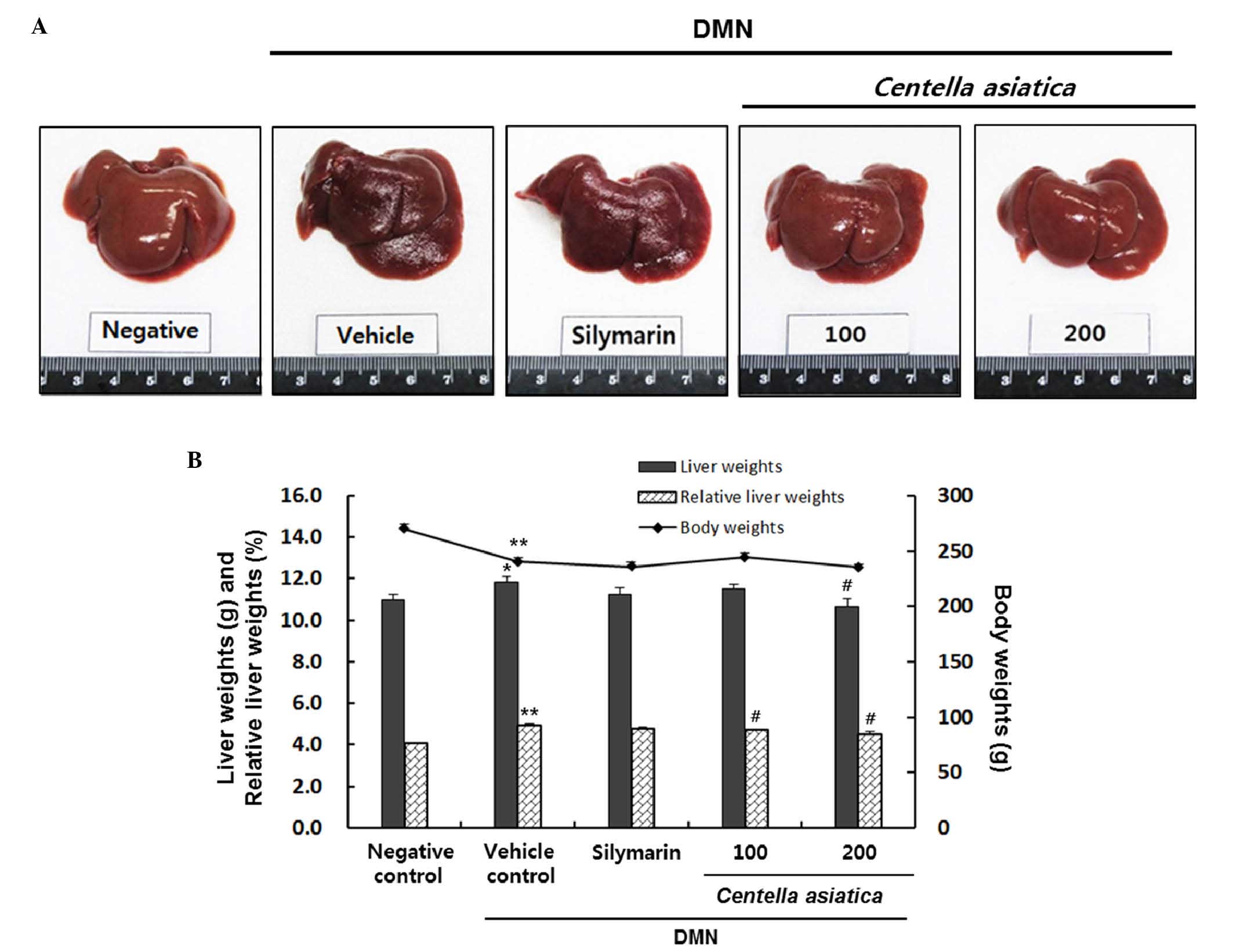
As shown in Fig.
1A, the surface of the negative control liver tissue was smooth
and brown with an evident gloss and a soft texture. By contrast,
the surface of the vehicle control liver tissue was rough and
reddish-brown, and the texture was hard. In addition, treatment
with DMN significantly decreased the body weights of the rats, by
~89%, compared with the rats in the negative control group
(Fig. 1B). This result is in
contrast with the vehicle control group, in which the liver weights
and relative liver weights were increased by 7.6 and 20.9%,
respectively (liver weight: 11.0±0.26, vs. 11.8±0.28 g; relative
liver weight: 4.1±0.05, vs. 4.9±0.08%). The results showed that
Centella asiatica decreased the liver weights by 11.5±0.22 g
and 10.6±0.37 g at doses of 100 and 200 mg/kg, respectively. In
particular, 200 mg/kg of Centella asiatica significantly
decreased liver weights, and significantly decreased relative liver
weights by 4.7±0.04 and 4.5±0.11% at doses of 100 and 200 mg/kg,
respectively.
Effects of Centella asiatica on serum
cytokine levels in rats with DMN-induced liver injury
The effects of Centella asiatica on
DMN-induced liver injury were evaluated by determining the levels
of AST, ALT, ALP, T-bilirubin, total protein and albumin. As shown
in Table I, the levels of AST
(145.2±4.5 IU/l), ALT (72.9±4.0 IU/l), ALP (694.1±27.5 IU/l) and
T-biliubin (0.281±0.018 mg/dl) were significantly elevated
following DMN treatment, compared with the negative control group
(AST, 123.8±3.0 IU/l; ALT, 49.9±2.0 IU/l; ALP, 466.0±18.7 IU/l;
T-biliubin, 0.110±3.0 mg/dl). By contrast, Centella asiatica
ameliorated this increase significantly for AST, ALT, ALP and
T-biliubin, compared with the vehicle control group. Silymarin
significantly decreased the levels of AST (118.4±6.4 IU/l) and ALP
(530.6±38.7 IU/l), however, no significant changes in ALT (68.1±3.9
IU/l) or T-biliubin (0.269±0.019 mg/dl) were observed.
 | Table I.Effects of Centella asiatica
on serum parameters in rata with DMN-induced liver injury. |
Table I.
Effects of Centella asiatica
on serum parameters in rata with DMN-induced liver injury.
|
|
| DMN |
|---|
|
|
|
|
|---|
|
|
|
|
| Centella
asiatica (mg/kg) |
|---|
|
|
|
|
|
|
|---|
| Parameter | Negative
control | Vehicle
control | Silymarin | 100 | 200 |
|---|
| AST (IU/l) | 123.8±3.0 |
145.2±4.5a |
118.4±6.4b |
108.3±5.9b |
99.2±3.7b |
| ALT (IU/l) | 49.9±2.0 |
72.9±4.0a | 68.1±3.9 | 66.8±3.4 |
58.0±2.4b |
| ALP (IU/l) | 466.0±18.7 |
694.1±27.5a |
530.6±38.7b |
556.4±39.6c |
495.3±27.0b |
| T-biliubin
(mg/dl) | 0.110±0.008 |
0.281±0.018a | 0.269±0.019 | 0.241±0.016 |
0.224±0.012c |
| Total protein
(g/dl) | 5.9±0.2 | 6.07±0.09 | 5.56±0.12 | 5.85±0.12 | 5.80±0.08 |
| Albumin (g/dl) | 3.3±0.1 | 3.41±0.05 | 3.16±0.06 | 3.29±0.06 | 3.25±0.04 |
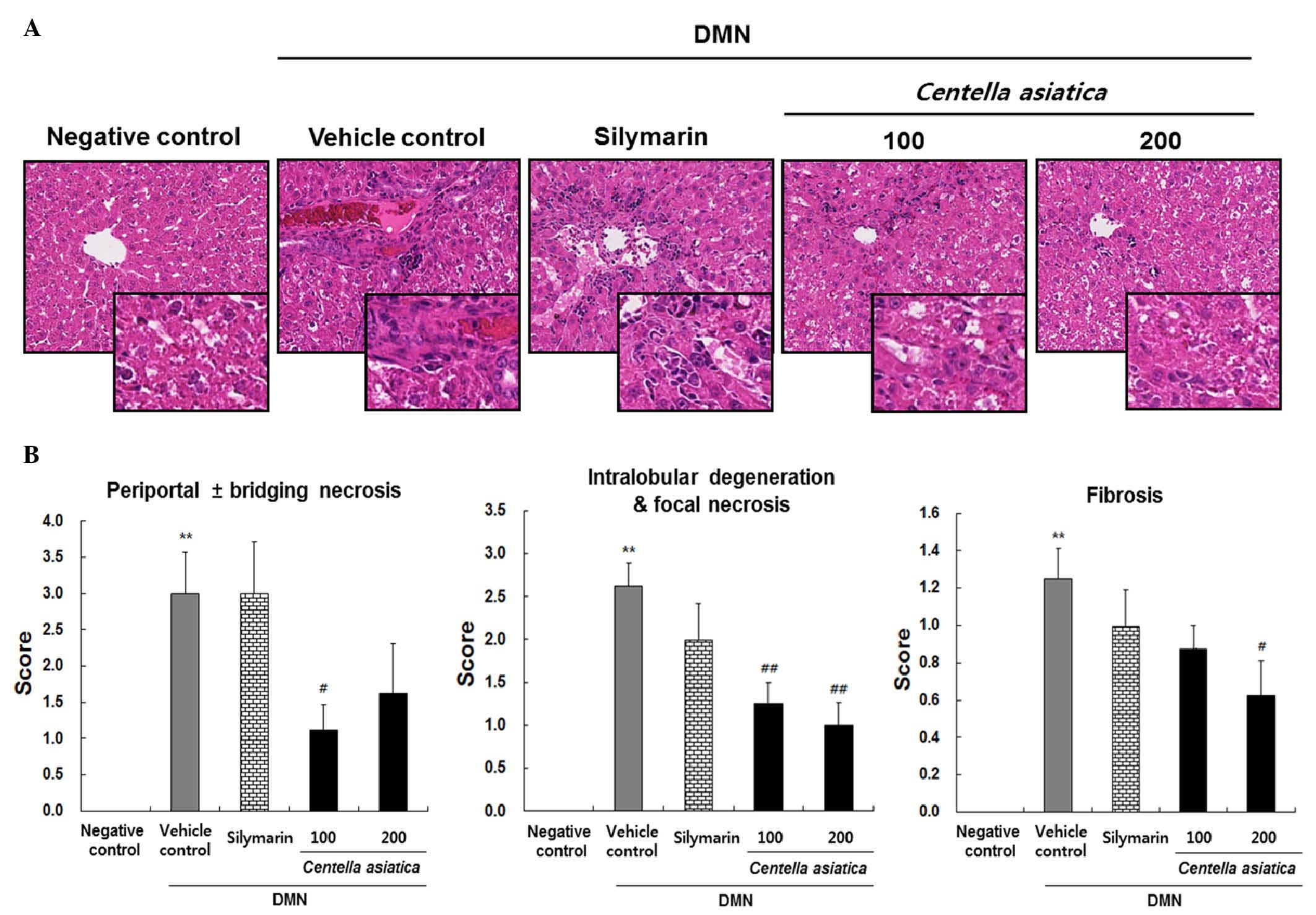
Histologic analysis of the effects of
Centella asiatica in rats with DMN-induced liver injury
Representative histological images of the pancreas
are shown in Fig. 2A and the
quantitative histological scoring of periportal ± bridging
necrosis, intralobular degeneration and focal necrosis, and
fibrosis are shown in Fig. 2B. The
liver tissue from the vehicle control group induced by DMN showed
significant increases in mass periportal ± bridging necrosis,
intralobular degeneration and focal necrosis, and fibrosis. By
contrast, Centella asiatica reduced the periportal ±
bridging necrosis at the dose of 200 mg/kg, and a significant
effect was shown in the rats treated with 100 mg/kg. Centella
asiatica also significantly decreased intralobular degeneration
and focal necrosis at the doses of 100 and 200 mg/kg. Centella
asiatica reduced liver fibrosis at the dose of 100 mg/kg, and a
significant effect was shown in the rats treated with 200 mg/kg.
However, treatment with silymarin caused no significant
improvements in periportal ± bridging necrosis, intralobular
degeneration and focal necrosis or fibrosis. These results
suggested that Centella asiatica led to reductions in liver
tissue damage, which was characterized by significant amelioration
of liver injury, measured by histological scores, compared with
those of the vehicle control group.
Effects of Centella asiatica on
hepatic levels of MDA, SOD, GPx and CAT in rats with DMN-induced
liver injury
Oxidative stress was quantified through the levels
of MDA, SOD, GPx and CAT in liver tissue homogenates, as indicators
of lipid peroxidation and antioxidant enzyme activity. As shown in
Fig. 3A, the level of MDA was
markedly increased to 0.5±0.1 nmol/mg tissue in the vehicle control
group. However, Centella asiatica significantly decreased
the level of MDA to 0.3±0.03 nmol/mg and 0.2±0.01 nmol/mg tissue at
the doses of 100 and 200 mg/kg, respectively. The effect of 200
mg/kg of Centella asiatica was more marked, compared with
that of silymarin. The level of SOD in the liver in the vehicle
control group was significantly lower, compared with that in the
negative control group, whereas the level of SOD level in the 200
mg/kg Centella asiatica treated group was 1.6-fold higher,
compared with that in the vehicle control group (Fig. 3B). In addition, the level of GPx in
the liver was markedly decreased to 243.7±18.9 U/mg tissue in the
vehicle control group; however, Centella asiatica increased
the level of GPx at a dose of 100 mg/kg, with a significant effect
in the rats treated with 200 mg/kg (Fig. 3C). Furthermore, the levels of CAT
in the liver decreased considerably to 485.6±92.6 U/mg tissue in
the vehicle control group. However, Centella asiatica
increased the level of CAT at a dose of 100 mg/kg and had a
significant effect in the rats treated with 200 mg/kg (Fig. 3D).
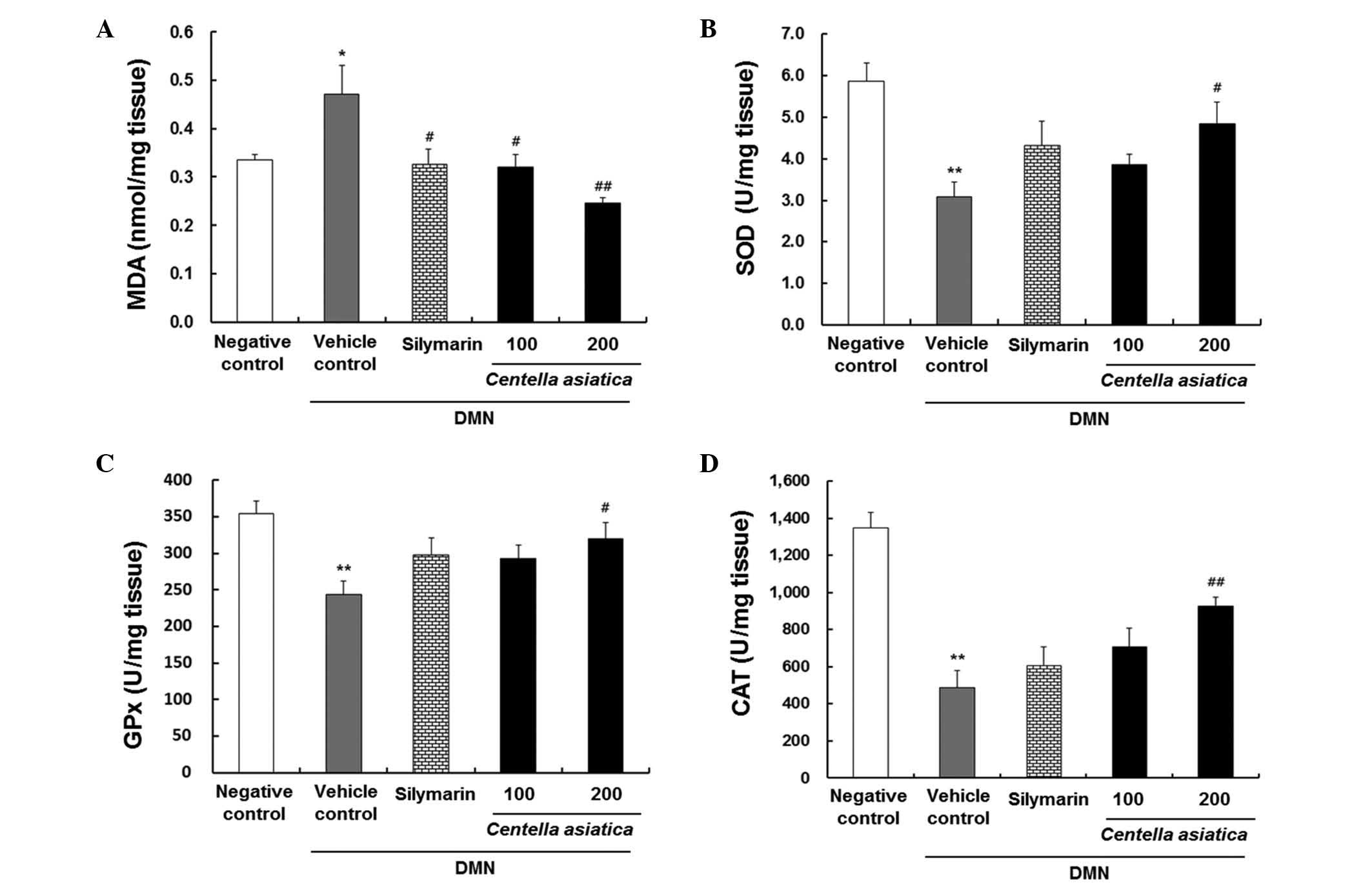 | Figure 3.Effects of Centella asiatica
on DMN-induced hepatic levels in liver tissue. (A) MDA (nmol/mg
tissue), (B) SOD (U/mg tissue), (C) GPx (U/mg tissue) and (D) CAT
(U/mg tissue). The animals (n=8/group) were orally administrated
with Centella asiatica at 100 or 200 mg/kg. DMN in D.W was
used as a vehicle control. Saline in D.W was used as a negative
control. Values are presented as the mean ± standard error of the
mean. **P<0.01 and *P<0.05, compared with the negative
control; ##P<0.01 and #P<0.05, compared
with the vehicle control. DMN, dimethylnitrosamine; MDA,
malondialdehyde; SOD, superoxide dismutase; GPx, glutathione
peroxidase; CAT, catalase; D.W, distilled water. |
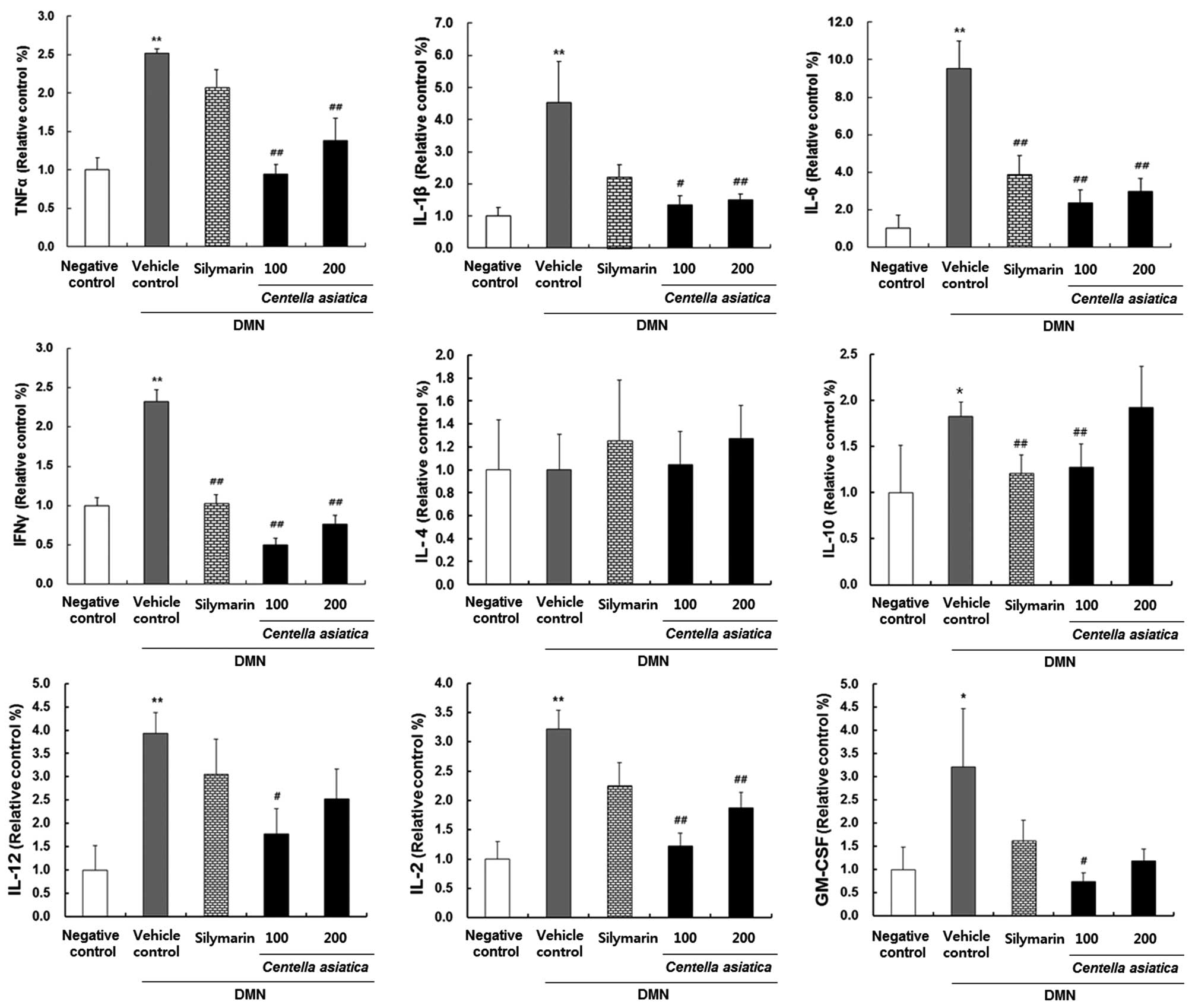
Effects of Centella asiatica on
cytokine levels in rats with DMN-induced liver injury
To identify whether Centella asiatica had an
effect on inflammation, the present study investigated the cytokine
levels of IL-1β, IL-2, IL-4, IL-6, IL-10, IL-12, TNF-α, IFN-γ and
GM-CSF in rats with DMN-induced liver injury. As shown in Fig. 4, the cytokine levels of TNF-α,
IL-1β, IL-6, IFN-γ, IL-10, IL-12, IL-2 and GM-CSF were
significantly elevated in the vehicle control group, compared to
the negative control group. When the rats were treated with 100 or
200 mg/kg Centella asiatica, the levels of TNF-α, IL-1β,
IL-6, IFN-γ, IL-10, IL-12, IL-2 and GM-CSF were significantly
decreased, compared with those in the vehicle control group.
Discussion
DMN is reported to be involved in liver necrosis and
carcinogenesis, exhibited through a number of mechanisms (23). The present study showed that
Centella asiatica had a protective effect against liver
injury induced by DMN.
In the present study, no significant changes in body
weights were observed in the different experimental groups, with
the exception of the vehicle control. The measurement of the
relative liver weights provides a more accurate approach to
demonstrate the changes in liver size, compared with the
measurement of liver weights alone, as the liver weight depends
primarily on the size of the rat (24). The enlargement of the livers in the
DMN-treated rats signified hepatic lesions and liver damage
associated with the induction of injury by DMN. These significant
changes in the liver weights may be attributed to the accumulation
of collagen and extracellular matrix protein in liver tissue
(25). In the present study, liver
enlargement was significantly reduced in the rats treated with
Centella asiatica.
Hepatocellular damage is evidenced by marked
elevation in the activities of serum AST, ALT, ALP and T-bilirubin.
Serum AST, ALT, ALP and T-bilirubin are the most sensitive markers
used in the diagnosis of liver damage as they are cytoplasmic in
location and are leaked into the circulation following cellular
injury (26).
Among the serum liver biomarkers, ALT and AST are
considered to be indicators of hepatotoxic effects, where their
elevation is considered as a more sensitive indicator. The levels
of enzymes, which leak into the blood stream indicate the severity
of hepatic injury (27,28). In the present study, the rats with
DMN-induced liver injury were found to have significantly higher
serum biomarker levels, compared with the negative control rats.
However, Centella asiatica exhibited hepatoprotective
effects by attenuating the elevated serum parameters.
ROS, the natural byproducts of oxidative energy
metabolism, are considered to be a physiologic modulator of a
number of intracellular signaling pathways (29). Therefore, the defense mechanisms
against oxidative damage are an important element of the cellular
stress response, during which a diverse array of electrophilic and
oxidative toxicants can be either eliminated or inactivated prior
to them causing damage critical macromolecules (30). In addition, the enhancement of
oxidative stress and the peroxidation process have been implicated
in DMN-induced liver injury in rats (31).
Centella asiatica has been reported to
enhance antioxidant enzymes, including SOD, GPx and CAT in
adriamycin-induced cardiomyopathy and pentylenetetrazole-induced
oxidative stress, and in experimentally induced parkinsonism in
rats (32–34). As expected, the present study
demonstrated that Centella asiatica increased the levels of
SOD, GPx and CAT with concomitant decreases in the level of MDA in
rats with DMN-induced liver injury.
ROS are important cytotoxic and signaling mediators
in the pathophysiology of inflammatory liver injury (35). Among those, pro-inflammatory and
fibrogenic responses are mediated by the action of cytokines,
including IL-1β, IL-6 and TNF-α (36). In addition, IFN-γ is reported to be
involved in various types of inflammatory diseases (37). A study by Zoheir et al
reported that treatment with DMN alone caused a significant
increase in the expression of IFN-γ (38). In addition, a study by Thompson
et al (39) reported that
IL-10 is expressed during macrophage activation in liver injury,
and that IL-10 is capable of downregulating various aspects of
pro-inflammatory macrophage function. The enhanced production of
IL-2 and IL-12 are also involved in cell-mediated cytotoxic
activity (40–42) and GM-CSF can induce the secretion
of IL-1 and TNF-α cytokines (43).
In the present study, Centella asiatica decreased the
expression of proinflammatory cytokines, including IL-1β, IL-6,
TNF-α, and other inflammatory cytokines and mediators in parallel
with decreasing the levels of inflammatory cytokines in DMN-induced
liver injury. Histologically, DMN administration in the present
study produced severe periportal ± bridging necrosis, intralobular
degeneration and focal necrosis, and fibrosis in the liver tissues,
whereas Centella asiatica ameliorated hepatocyte necrosis
and fibrosis in the rats with DMN-induced liver injury.
In conclusion, the present study demonstrated that
Centella asiatica exhibited hepatoprotective effects through
increasing the levels of antioxidant enzymes and reducing the
levels of inflammatory mediators in rats with DMN-induced liver
injury. Therefore, Centella asiatica may be useful in
preventing liver damage in the future.
Acknowledgements
This study was supported by grants from the Korean
Health Industry Development Institute and the National Center of
Efficacy Evaluation for the Development of Health Products
Targeting Digestive Disorders (grant no. HI15C0989).
References
|
1
|
George J, Rao KR, Stern R and Chandrakasan
G: Dimethylnitrosamine-induced liver injury in rats: The early
deposition of collagen. Toxicology. 156:129–138. 2001. View Article : Google Scholar : PubMed/NCBI
|
|
2
|
Guengerich FP, Kim DH and Iwasaki M: Role
of human cytochrome P-450 IIE1 in the oxidation of many low
molecular weight cancer suspects. Chem Res Toxicol. 4:168–179.
1991. View Article : Google Scholar : PubMed/NCBI
|
|
3
|
Jin YL, Enzan H, Kuroda N, Hayashi Y,
Nakayama H, Zhang YH, Toi M, Miyazaki E, Hiroi M, Guo LM and
Saibara T: Tissue remodeling following submassive hemorrhagic
necrosis in rat livers induced by an intraperitoneal injection of
dimethylnitrosamine. Virchows Arch. 442:39–47. 2003.PubMed/NCBI
|
|
4
|
Teufelhofer O, Parzefall W, Kainzbauer E,
Ferk F, Freiler C, Knasmüller S, Elbling L, Thurman R and
Schulte-Hermann R: Superoxide generation from kupffer cells
contributes to hepatocarcinogenesis: Studies on NADPHoxidase
knockout mice. Carcinogenesis. 26:319–329. 2005. View Article : Google Scholar : PubMed/NCBI
|
|
5
|
Wills PJ, Suresh V, Arun M and Asha VV:
Antiangiogenic effect of lygodium flexuosum against
N-nitrosodiethylamine-induced hepatotoxicity in rats. Chem Biol
Interact. 164:25–38. 2006. View Article : Google Scholar : PubMed/NCBI
|
|
6
|
Pradeep K, Mohan CV, Gobianand K and
Karthikeyan S: Effect of Cassia fistula linn. Leaf extract on
diethylnitrosamine induced hepatic injury in rats. Chem Biol
Interact. 167:12–18. 2007. View Article : Google Scholar : PubMed/NCBI
|
|
7
|
Jayakumar S, Madankumar A, Asokkumar S,
Raghunandhakumar S, dhas K Gokula, Kamaraj S, Divya MG and Devaki
T: Potential preventive effect of carvacrol against
diethylnitrosamine-induced hepatocellular carcinoma in rats. Mol
Cell Biochem. 360:51–60. 2012. View Article : Google Scholar : PubMed/NCBI
|
|
8
|
Caro AA and Cederbaum AI: Oxidative
stress, toxicology and pharmacology of CYP2E1. Annu Rev Pharmacol
Toxicol. 44:27–42. 2004. View Article : Google Scholar : PubMed/NCBI
|
|
9
|
Cressman DE, Greenbaum LE, DeAngelis RA,
Ciliberto G, Furth EE, Poli V and Taub R: Liver failure and
defective hepatocyte regeneration in interleukin-6-deficient mice.
Science. 274:1379–1383. 1996. View Article : Google Scholar : PubMed/NCBI
|
|
10
|
Tilg H and Diehl AM: Cytokines in
alcoholic and nonalcoholic steatohepatitis. N Engl J Med.
343:1467–1476. 2000. View Article : Google Scholar : PubMed/NCBI
|
|
11
|
Guan L, Fu PY, Li PD, Li ZN, Liu HY, Xin
MG and Li W: Mechanisms of hepatic ischemia-reperfusion injury and
protective effects of nitric oxide. World J Gastrointest Surg.
6:122–128. 2014. View Article : Google Scholar : PubMed/NCBI
|
|
12
|
Schümann J, Prockl J, Kiemer AK, Vollmar
AM, Bang R and Tiegs G: Silibinin protects mice from T
cell-dependent liver injury. J Hepatol. 39:333–340. 2003.
View Article : Google Scholar : PubMed/NCBI
|
|
13
|
Radaeva S, Sun R, Jaruga B, Nguyen VT,
Tian Z and Gao B: Natural killer cells ameliorate liver fibrosis by
killing activated stellate cells in NKG2D-dependent and tumor
necrosis factor-related apoptosis-inducing ligand-dependent
manners. Gastroenterology. 130:435–452. 2006. View Article : Google Scholar : PubMed/NCBI
|
|
14
|
Halliwell B and Gutteridge JM: Role of
free radical and catalytic metal ions in human disease: An
overview. Methods Enzymol. 186:1–85. 1990. View Article : Google Scholar : PubMed/NCBI
|
|
15
|
Suguna L, Sivakumar P and Chandrakasan G:
Effects of Centella asiatica extract on dermal wound healing in
rats. Indian J Exp Biol. 34:1208–1211. 1996.PubMed/NCBI
|
|
16
|
Kumar Veerendra MH and Gupta YK: Effect of
different extracts of Centella asiatica on cognition and markers of
oxidative stress in rats. J Ethnopharmacol. 79:253–260. 2002.
View Article : Google Scholar : PubMed/NCBI
|
|
17
|
Babu TD, Kuttan G and Padikkala J:
Cytotoxic and anti-tumour properties of certain taxa of
umbelliferae with special reference to Centella asiatica (L.)
urban. J Ethnopharmacol. 48:53–57. 1995. View Article : Google Scholar : PubMed/NCBI
|
|
18
|
Sairam K, Rao CV and Goel RK: Effect of
Centella asiatica linn on physical and chemical factors induced
gastric ulceration and secretion in rats. Indian J Exp Biol.
39:137–142. 2001.PubMed/NCBI
|
|
19
|
Katare SS and Ganachari MS: Effect of
Centella asiatica on hypoxia induced convulsions and
lithium-pilocarpine induced status epilepticus and antilipid
peroxidation activity. Ind J Pharmacol. 33:1282001.
|
|
20
|
Jayashree G, Muraleedhara G Kurup,
Sudarslal S and Jacob VB: Anti-oxidant activity of Centella
asiatica on lymphoma-bearing mice. Fitoterapia. 74:431–434. 2003.
View Article : Google Scholar : PubMed/NCBI
|
|
21
|
Brunt EM: Grading and staging the
histopathological lesions of chronic hepatitis: The knodell
histology activity index and beyond. Hepatology. 31:241–246. 2000.
View Article : Google Scholar : PubMed/NCBI
|
|
22
|
Miranda KM, Espey MG and Wink DA: A rapid,
simple spectrophotometric method for simultaneous detection of
nitrate and nitrite. Nitric Oxide. 5:62–71. 2001. View Article : Google Scholar : PubMed/NCBI
|
|
23
|
Ray SD, Sorge CL, Kamendulis LM and
Corcoran GB: Ca (++)-activated DNA fragmentation and
dimethylnitrosamine-induced hepatic necrosis: Effects of ca
(++)-endonuclease and poly (ADP-ribose) polymerase inhibitors in
mice. J Pharmacol Exp Ther. 263:387–394. 1992.PubMed/NCBI
|
|
24
|
Saad RA, EL-Bab MF and Shalaby AA:
Attenuation of acute and chronic liver injury by melatonin in rats.
J Taibah Univ Sci. 7:88–96. 2013. View Article : Google Scholar
|
|
25
|
Pinzani M and Rombouts K: Liver fibrosis:
From the bench to clinical targets. Dig Liver Dis. 36:231–242.
2004. View Article : Google Scholar : PubMed/NCBI
|
|
26
|
Sallie R, Tredger JM and Williams R: Drugs
and the liver. Part 1: Testing liver function. Biopharm Drug
Dispos. 12:251–259. 1991. View Article : Google Scholar : PubMed/NCBI
|
|
27
|
Zimmerman HJ: Hepatotoxicity. Dis Mon.
39:675–787. 1993.PubMed/NCBI
|
|
28
|
Nkosi CZ, Opoku AR and Terblanche SE:
Effect of pumpkin seed (Cucurbita pepo) protein isolate on the
activity levels of certain plasma enzymes in CCl4-induced liver
injury in low-protein fed rats. Phytother Res. 19:341–345. 2005.
View Article : Google Scholar : PubMed/NCBI
|
|
29
|
Lander HM: An essential role for free
radicals and derived species in signal transduction. FASEB J.
11:118–124. 1997.PubMed/NCBI
|
|
30
|
Rushmore TH and Kong AN: Pharmacogenomics,
regulation and signaling pathways of phase I and II drug
metabolizing enzymes. Curr Drug Metab. 3:481–490. 2002. View Article : Google Scholar : PubMed/NCBI
|
|
31
|
Vendemiale G, Grattagliano I, Caruso ML,
Serviddio G, Valentini AM, Pirrelli M and Altomare E: Increased
oxidative stress in dimethylnitrosamine-induced liver fibrosis in
the rat: Effect of N-acetylcysteine and interferon-alpha. Toxicol
Appl Pharmacol. 175:130–139. 2001. View Article : Google Scholar : PubMed/NCBI
|
|
32
|
Gnanapragasam A, Ebenezar KK, Sathish V,
Govindaraju P and Devaki T: Protective effect of Centella asiatica
on antioxidant tissue defense system against adriamycin induced
cardiomyopathy in rats. Life Sci. 76:585–597. 2004. View Article : Google Scholar : PubMed/NCBI
|
|
33
|
Gupta YK, Kumar MH Veerendra and
Srivastava AK: Effect of Centella asiatica on
pentylenetetrazole-induced kindling, cognition and oxidative stress
in rats. Pharmacol Biochem Behav. 74:579–585. 2003. View Article : Google Scholar : PubMed/NCBI
|
|
34
|
Haleagrahara N and Ponnusamy K:
Neuroprotective effect of Centella asiatica extract (CAE) on
experimentally induced parkinsonism in aged sprague-dawley rats. J
Toxicol Sci. 35:41–47. 2010. View Article : Google Scholar : PubMed/NCBI
|
|
35
|
Jaeschke H: Reactive oxygen and mechanisms
of inflammatory liver injury. J Gastroenterol Hepatol. 15:718–724.
2000. View Article : Google Scholar : PubMed/NCBI
|
|
36
|
Duffield JS, Forbes SJ, Constandinou CM,
Clay S, Partolina M, Vuthoori S, Wu S, Lang R and Iredale JP:
Selective depletion of macrophages reveals distinct, opposing roles
during liver injury and repair. J Clin Invest. 115:56–65. 2005.
View Article : Google Scholar : PubMed/NCBI
|
|
37
|
Ishida Y, Maegawa T, Kondo T, Kimura A,
Iwakura Y, Nakamura S and Mukaida N: Essential involvement of
IFN-gamma in clostridium difficile toxin A-induced enteritis. J
Immunol. 172:3018–3025. 2004. View Article : Google Scholar : PubMed/NCBI
|
|
38
|
Zoheir KM, Amara AA, Ahmad S, et al: Study
of the therapeutic effects of lactobacillus and α-lipoic acid
against dimethylnitrosamine-induced liver fibrosis in rats. J Genet
Eng Biotechnol. 12:135–142. 2014. View Article : Google Scholar
|
|
39
|
Thompson K, Maltby J, Fallowfield J,
McAulay M, Millward-Sadler H and Sheron N: Interleukin-10
expression and function in experimental murine liver inflammation
and fibrosis. Hepatology. 28:1597–1606. 1998. View Article : Google Scholar : PubMed/NCBI
|
|
40
|
Dennert G: Cloned lines of natural killer
cells. Nature. 287:47–49. 1980. View Article : Google Scholar : PubMed/NCBI
|
|
41
|
Trinchieri G: Interleukin-12: A cytokine
at the interface of inflammation and immunity. Adv Immunol.
70:83–243. 1998. View Article : Google Scholar : PubMed/NCBI
|
|
42
|
Zeh HJ III, Hurd S, Storkus WJ and Lotze
MT: Interleukin-12 promotes the proliferation and cytolytic
maturation of immune effectors: Implications for the immunotherapy
of cancer. J Immunother Emphasis Tumor Immunol. 14:155–161. 1993.
View Article : Google Scholar : PubMed/NCBI
|
|
43
|
Hamilton JA: Colony stimulating factors,
cytokines and monocyte-macrophages-some controversies. Immunol
Today. 14:18–24. 1993. View Article : Google Scholar : PubMed/NCBI
|