Introduction
Infertility is a devastating problem of human
reproduction, and male infertility contributes to 50% of all
infertility cases (1,2). In recent years, an increasing number
of reports have demonstrated a global trend indicating a
significant deterioration in male reproductive function (3–5). In
parallel, obesity rates are increasing rapidly worldwide (6,7),
which is not only associated with an increased risk of developing
chronic diseases, but has also been demonstrated to increase the
risk of developing reproductive disorders (8). Due to the reported association
between obesity and reproductive disorders, a greater clinical
awareness and understanding of the underlying mechanisms of the
effects of obesity on fertility are urgently required in order to
determine the appropriate interventions.
Studies have determined that the natural polyphenol,
resveratrol, has a similar molecular structure to
diethylstilbestrol and estradiol (E2), and is produced in several
plants in response to injury, stress, bacterial or fungal
infection, ultraviolet (UV) radiation or exposure to ozone
(9–11). Resveratrol is known for its
anti-inflammatory, antioxidant, analgesic, cardioprotective,
anti-aging and neuroprotective roles (11–13).
Studies have demonstrated that resveratrol may inhibit cell
apoptosis, thereby providing protection from numerous diseases,
including atherosclerosis, cerebral ischemia and myocardial
ischemic reperfusion injury (14,15).
Juan et al (16) reported
that resveratrol is able to decrease germ cell apoptosis in mice
and rats, and serves a protective role in the male reproductive
tract, as well as enhancing blood testosterone levels, testicular
sperm count and epididymis sperm motility in rabbits.
The aim of the present study was to investigate the
association between obesity and a number of sperm parameters, and
to assess the protective effects of resveratrol on preventing the
harmful effects of obesity on spermatozoa. This may lead to the
development of strategies that facilitate the maintenance of
fertility.
Materials and methods
Study population
The study population consisted of 324 men (mean age,
37.2±0.89 years) referred to the Reproductive Medicine Center of
Shanxi Women and Infants Hospital (Taiyuan, China) between January
2013 and January 2015. All subjects were of Han ethnicity and from
the Shanxi Province in northern China. The present study was
approved by the Reproductive Medicine Ethics Committee of Shanxi
Women and Infants Hospital (Taiyuan, China) and written informed
consent was obtained from all subjects and their partners prior to
enrollment.
The inclusion criteria for the study were as
follows: i) Subjects had to be male and part of a couple that had
been unable to conceive for >1 year; ii) the couple had regular
intercourse, and iii) they were receiving infertility treatment at
the Reproductive Medicine Center, Shanxi Women and Infants Hospital
over the study period. Primary infertility was diagnosed after the
following medical assessments were performed: The patient's medical
history was examined; a clinical examination; semen analysis; semen
culture for mycoplasma ureliticum and chlamydia detection; analysis
of follicle-stimulating hormone (FSH), luteinizing hormone (LH),
testosterone and E2 levels; a prolactin assay; and sonography of
the genitalia. Each subject underwent a careful anamnesis to
exclude systemic diseases, alcohol consumption, smoking,
occupational chemical exposure, a history of (or presence of)
endocrine disorders, testicular diseases (for example,
cryptorchidism, orchitis and varicocelle), infectious genital
diseases, leukocytospermia (seminal white blood cell count
>1×10⁶/ml), azoospermia and treatment with drugs or
the use of antioxidant supplements within 3 months prior to
enrollment.
A brief medical background for each subject was
collected by conducting an informal interview, using clinical notes
or using a self-report questionnaire. The male subjects were
divided into the following four groups according to the
International Association for the Study of Obesity criteria
(17): Underweight [body mass
index, (BMI)<18.5 kg/m2], normal weight
(18.5≤BMI<23 kg/m2), overweight (23≤BMI<25
kg/m2) and obese (BMI≥25 kg/m2) groups.
Serum collection and analysis
Blood samples were obtained between 8:00 and 9:00
a.m. following a 12-h overnight fast, and were immediately drawn
and collected in a tube containing ethylenediamine tetra-acetic
acid. Samples were separated by centrifugation at 18,00 × g
for 15 min at room temperature and the serum and buffy coat were
separated. The serum was stored at −80°C until downstream analyses
were performed.
The hormone levels, including FSH (reference value,
2.10–18.6 mIU/ml), LH (reference value, 1.7–11.2 mIU/ml), E2
(reference value, ≤77 pg/ml), progesterone (reference value, ≤0.46
ng/ml), testosterone (reference value, 262.0–870.0 ng/dl) and
prolactin (PRL; reference value: 2.10–18.60 mIU/ml) were measured
using an AIA-2000 ST Automated Immunoassay Analyzer (Tosoh
Corporation, Tokyo, Japan).
Semen collection and analysis
Semen samples were obtained by masturbation
following 2–7 days of abstinence from sexual intercourse, in order
to conduct a routine sperm count according to the World Health
Organization (WHO, 2010) criteria (18) for sperm concentration, motility,
morphology and viability. Samples were collected in sterile
containers and allowed to liquefy at 37°C for 20 min. Briefly,
ejaculated volumes were estimated by specimen weight, and a semen
density of 1.0 g/ml was assumed. Sperm concentration, motility and
viability were assessed using a Sperm Class Analyzer CASA System
(Microoptic S.L, Barcelona, Spain). Sperm motility was analyzed
according to the WHO (2010) guidelines (18) for determining progressive motility,
non-progressive motility and immotility. Sperm morphology was
assessed using David's classification system (19).
Sperm viability analysis
Sperm viability was assessed within 30 min of
ejaculation using eosin Y staining. This was achieved by dissolving
1 g eosin with 1 g fresh sperm. The percentage of viable sperm
(unstained sperm heads) and non-viable sperm (stained sperm heads)
was assessed by counting a minimum of 200 spermatozoa for each
sample. Each sample was analyzed in duplicate.
UV spectrophotometric assay for
spermatozoa acrosin activity
Spermatozoa samples from each group were analyzed
for acrosin activity using the Human Spermatozoa Acrosin Activity
Quantitative assay kit (Huakang Biotechnology Development Co.,
Ltd., Changsha, China) according to the manufacturer's protocol.
The required amount of semen (7.5×10⁶ sperm) was transferred into a
plastic tube and centrifuged at 2,000 × g for 20 min at room
temperature. The supernatant was removed, and the tube was inverted
on absorbent paper to remove residual seminal plasma. A total of
100 µl inhibitor solution (0.3 g/ml HEPES) and 1 ml reactive liquid
(0.6 g/ml N-α-benzoyl-L-arginine-p-nitroanilide) were added to the
sample and control tubes, prior to the addition of 100 µl stop
solution (8.5 g/ml benzamidine) to the control tube. Sample and
control tubes were then thoroughly mixed and incubated at 24°C for
1 h. Stop solution (100 µl) was added to the control tube and mixed
thoroughly, and subsequently both tubes were centrifuged at 2,000 ×
g for 10 min at 24°C. At this temperature, the amount of
substrate that hydrolyzes 1.0 µmol
N-benzoyl-DL-arginine-4-nitroanilide hydrochloride/min is defined
as 1 IU acrosin activity. Acrosin activity was determined using the
following formula: Acrosin activity (µIU/106 spermatozoa)=[sample
optical density (OD)-control OD] × [2/(495×7.5)] ×106.
Colorimetric assay for seminal plasma
zinc
The zinc concentrations in seminal fluid samples for
each group were analyzed using the Seminal Plasma Zinc Quantitative
assay kit (Huakang Biotechnology Development Co., Ltd.) according
to the manufacturer's protocol. Seminal fluid (1 ml) was
centrifuged at 1,800 × g for 10 min at 24°C. The supernatant
was subsequently transferred into a separate test tube for seminal
plasma analysis. Physiological saline solution (1 ml) was used to
wash the sediment, prior to the mixing of samples using a
vortex-type mixer for 30 sec, and centrifugation for at 1,800 ×
g for 10 min at 24°C. The supernatant was removed, and the
sediment was used to determine the zinc concentration instead of
using 200 µl of liquid sample. The absorbance of the sample
solutions was read at 490 nm. Sample zinc concentrations were
calculated using the following formula: Seminal plasma zinc (µmol)
= zinc concentration (mmol/l) × semen volume (ml).
Detection of sperm DNA integrity
DNA integrity analysis of sperm in fresh semen
samples was performed using a the Sperm DNA Fragments Staining kit
(Huakang Biotechnology Development Co., Ltd.), which is based on
the emission of fluorescence signals from individual sperm stained
with acridine orange (AO). The AO molecules intercalate into
double-stranded DNA, and green fluorescence is emitted from the
sperm nuclei. The DNA in sperm with immature nuclei can be
denatured into single strands, which leads to the aggregation of AO
molecules in the nuclei and emission of an orange-red fluorescence
signal. The cell suspension was pipetted onto a glass slide and
observed under a BX51 fluorescence microscope (Olympus Corporation,
Tokyo, Japan) with a 480–490 nm filter. The percentage of green
(normal DNA integrity) and orange-red (abnormal DNA integrity)
spermatozoa/200 spermatozoa in each sample was determined by a
single investigator. An abnormal sperm nuclear DNA integrity was
considered to be when >34% of sperm nuclei emitted orange-red
fluorescence signals following AO staining.
Resveratrol treatment and dose
preparation
The sperm suspensions from obese patients with
astenospermia (60 cases) were pooled and divided randomly into the
following three drug treatment groups: i) the control group, where
spermatozoa were treated with Quinn's Advantage™ Fertilization
(HTF) Medium (SAGE-In vitro Fertilization, Inc., Trumbull,
CT, USA); ii) the negative control group, where spermatozoa were
treated with Quinn's Advantage™ Fertilization (HTF) Medium plus
0.1% dimethyl sulfoxide (DMSO); and iii) the experimental group,
which was subdivided into six subgroups based on the concentration
of resveratrol (2.6, 6, 15, 30, 50, 100 µmol/l; Sigma-Aldrich;
Merck Millipore, Darmstadt, Germany) added to the medium, which was
maintained at 37°C, 5% CO2 and 95% humidity. Samples were incubated
at 37°C for 30 min with resveratrol before sperm motility, seminal
plasma zinc concentration and spermatozoa acrosin activity were
analyzed.
Statistical analysis
Statistical analyses were performed using the SPSS
software program (version 17.0; SPSS, Inc., Chicago, IL, USA).
Normally distributed data were expressed as the mean ± standard
deviation. To verify the normality of the distribution, the
Shapiro-Wilk test was performed, and one-way analysis of variance
was used to compare the mean among the different groups. Variables
with a non-normal distribution were analyzed using a Mann-Whitney U
test or Kruskal-Wallis variance analysis test. P<0.05 was
considered to indicate a statistically significant difference.
Results
Distribution of male infertility
frequency over the BMI groups
A total of 324 men (mean age, 37.2 years) were
recruited to the study. Based on the analysis results of the semen
parameters, 139 males (42.90%) were classified as fertile and 185
(57.10%) were classified as infertile. The distribution of male
fertility in all BMI groups is shown in Table I. The general characteristics of
the participants were stratified according to the four BMI groups,
where 56/73 (76.71%) of underweight males were infertile, 32/82
(39.02%) of males with a normal weight were infertile, 47/95
(49.47%) of overweight males were infertile and 50/74 (67.57%) of
obese males were infertile.
 | Table I.Distribution of male infertility
frequency across the BMI groups. |
Table I.
Distribution of male infertility
frequency across the BMI groups.
| BMI
groupa | Fertile men (%) | Infertile men
(%) | Total |
|---|
| BMI<18.5 | 17
(23.29) | 56
(76.71) | 73 |
| 18.8≤BMI<23 | 50
(60.98) | 32
(39.02) | 82 |
| 23≤BMI<25 | 48
(50.53) | 47
(49.47) | 95 |
| BMI≥25 | 24
(32.43) | 50
(67.57) | 74 |
| Total | 139 (42.90) | 185 (57.10) | 324 |
Comparison of routine semen parameters
and serum sex hormone levels among BMI groups
As shown in Table
II, routine semen parameters and serum sex hormone levels were
assessed in abnormal weight groups, and the results were compared
with those of the normal weight group. No significant difference in
FSH, LH and PRL levels were observed among the abnormal weight
groups and the normal weight group (P>0.05). In addition, the
underweight group demonstrated no significant alterations in semen
volume when compared with the normal weight group (P>0.05),
whereas overweight and obese groups exhibited significantly lower
semen volumes (P=0.0248 and P=0.0142, respectively). The percentage
of sperm with progressive motility in the overweight group was not
significantly different when compared with the normal weight group
(P>0.05), whereas the percentage of sperm with progressive
motility in the underweight and obese groups was significantly
decreased (P=0.0009 and P=0.0419 respectively). When compared with
the normal weight group, the sperm concentration (underweight vs.
normal weight, P<0.0001; overweight vs. normal weight, P=0.0185;
obese vs. normal weight, P=0.0034), the percentage of sperm with a
normal morphology (underweight vs. normal weight, P<0.0001;
overweight vs. normal weight, P=0.0396; obese vs. normal weight,
P=0.0004) and the testosterone levels (underweight vs. normal
weight, P=0.0011; overweight vs. normal weight, P<0.0001; obese
vs. normal weight, P<0.0001) in abnormal weight groups were
significantly decreased. By contrast, E2 levels were significantly
increased in underweight, overweight and obese groups when compared
with the normal weight group (P=0.0003, P<0.0001 and
P<0.0001, respectively).
| Table II.Comparison of routine semen parameters
and serum sex hormone levels among BMI groups. |
Table II.
Comparison of routine semen parameters
and serum sex hormone levels among BMI groups.
| Parameter | Normal weight
(18.8≤BMI<23) | Underweight
(BMI<18.5) | Overweight
(23≤BMI<25) | Obese (BMI≥25) |
|---|
| Semen volume
(ml) | 3.56±1.74 | 3.54±1.68 |
3.10±0.88a |
3.02±0.73a |
| Sperm concentration
(×106/ml) | 68.39±8.54 |
59.42±8.16b |
65.39±8.22a |
64.39±8.19b |
| Progressive motility
(%) | 40.28±12.98 |
33.62±11.31b | 39.56±11.74 |
36.39±10.39a |
| Morphology (%
normal) | 12.11±3.59 |
7.63±1.33b |
11.08±3.32a |
10.21±2.9b |
| Follicle stimulating
hormone (mIU/ml) | 6.98±2.55 | 4.71±1.83 | 5.11±2.24 | 5.49±1.79 |
| Luteinizing hormone
(mIU/ml) | 9.35±2.35 | 9.1±1.32 | 8.74±1.66 | 8.63±1.29 |
| Estradiol
(pg/ml) | 29.32±7.90 |
34.11±8.27b |
36.63±7.53b |
37.21±8.94b |
| Testosterone
(ng/dl) | 386.58±21.32 |
398.24±22.19b |
369.76±19.38b |
354.71±19.23b |
| Prolactin
(mIU/ml) | 12.28±4.87 | 12.26±3.48 | 12.38±4.25 | 12.45±4.71 |
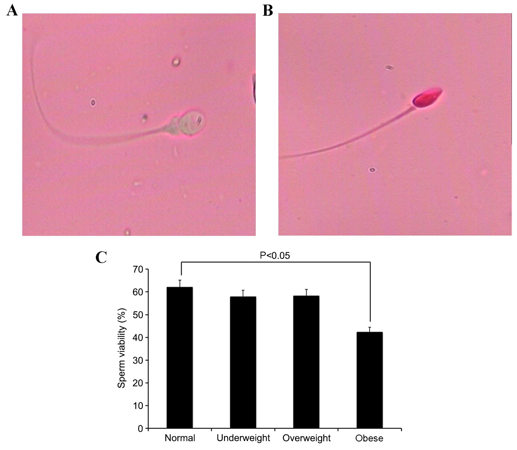
Comparison of sperm viability among
BMI groups
Sperm viability was analyzed by eosin Y staining.
The number of viable sperm (Fig.
1A) and non-viable sperm (Fig.
1B) in each group were examined using an Olympus CX31
microscope. When compared with the normal weight group, the obese
group demonstrated a significant reduction in sperm viability
(P=0.0297; Fig. 1C).
Comparison of the plasma zinc
concentration, spermatozoa acrosin activity and DNA fragmentation
rates among BMI groups
As shown in Fig.
2A, the seminal plasma zinc concentration was significantly
reduced in the obese group compared with the normal weight group
(P=0.0233). Spermatozoa acrosin activity was analyzed using a UV
spectrophotometric assay. Compared with normal weight group, the
overweight and obese groups demonstrated a significant decrease in
spermatozoa acrosin activity (P=0.0215 and P=0.0193, respectively;
Fig. 2B). DNA fragmentation rates
were analyzed by AO staining. As shown in Fig. 2C-E, spermatozoa emitting green
(normal DNA integrity) and orange-red (abnormal DNA integrity)
fluorescence signals were visualized and counted using an Olympus
BX51 fluorescence microscope with a 480–490 nm filter. When
compared with the normal weight group, the underweight, overweight
and obese groups demonstrated a significant increase in DNA
fragmentation rates (P=0.0347, P=0.0339 and P=0.0208 respectively;
Fig. 2F).
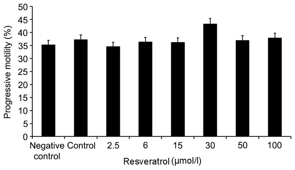
Alterations in the progressive
motility of sperm following treatment with increasing
concentrations of resveratrol
As shown in Fig. 3,
semen samples from obese patients with astenospermia (60 cases)
treated with 0–100 µmol/l resveratrol for 30 min, exhibited varying
degrees of improvement in sperm motility. When semen samples were
exposed to 30 µmol/l resveratrol, sperm motility was observed to
increase compared with the other doses of resveratrol. Therefore,
30 µmol/l resveratrol was used for subsequent experiments.
Effect of resveratrol on plasma zinc
concentration and spermatozoa acrosin activity
Semen samples were treated with quinn's 1020
nutrient solution (control), quinn's 1020 nutrient solution plus
0.1% DMSO (negative control) or 30 µmol/l resveratrol for 30 min,
and the plasma zinc concentration and spermatozoa acrosin activity
was analyzed. As shown in Fig. 4A,
the seminal plasma zinc concentration increased significantly in
the resveratrol group compared with the control group (P=0.0199).
In addition, the resveratrol group demonstrated a significant
increase in spermatozoa acrosin activity when compared with the
control group (P=0.0248; Fig.
4B).
Discussion
Excess body weight and obesity, defined by the WHO
as the abnormal or excessive accumulation of fat, is posing a
growing threat to health in countries worldwide (20,21).
Excessive weight is not only linked to an increased risk of
developing chronic disease, but has also been demonstrated to
increase the risk of developing reproductive disorders (8,22,23).
The association between excessive weight and infertility has been
established in females, but not in males. Females who are
overweight or obese are more likely to experience ovulatory and
menstrual disorders, and may experience delayed childbearing as a
consequence (8). Several studies
have investigated the association between BMI and different sperm
parameters (24). There is
evidence to suggest that sperm concentration and total motile sperm
count are detrimentally affected by a high BMI (23–25).
In addition, previous studies have demonstrated that low (<18.5
kg/m2) and high (≥25 kg/m2) BMIs are
associated with reduced testicular volume and reduced semen
quality, which suggests an impairment of spermatogenesis (24,25).
Similarly to females, a sex hormone imbalance may influence
reproduction in men, and excessive weight may affect male hormone
levels (24,25). It has been reported that obesity is
closely associated with endocrine disorders, such as sex hormone
abnormalities (26,27). It has been hypothesized that
extended periods of obesity may induce alterations in the secretion
and/or metabolism of hormones, leading to altered hormone-mediated
effects on target organs (28).
These associations are well-documented effects of excess body
weight on the production and function of sex hormones (29). Thus, it is necessary to evaluate
the effects of excess body weight and obesity on male
infertility.
In the present study, males in the excess weight and
obese groups demonstrated a significant decrease in semen volume
compared with those of the normal weight group, whereas males in
the underweight group did not exhibit any significant alterations.
The progressive motility of sperm in the underweight and obesity
groups was significantly reduced compared with the normal weight
group, whereas no significant alteration was observed in the
overweight group. When compared with the normal weight group, the
sperm concentration, normal sperm morphology and testosterone
levels in the abnormal weight groups demonstrated a significant
decrease, whereas the E2 levels were significantly increased. No
significant difference in FSH, LH and PRL levels among the abnormal
weight groups and the normal weight group were observed. The
seminal plasma zinc concentration was significantly reduced in the
obese group compared with normal weight group. Spermatozoa acrosin
activity was analyzed using a UV spectrophotometric assay. Compared
with normal weight group, the overweight and obese groups
demonstrated a significant decrease in spermatozoa acrosin
activity. When compared with the normal weight group, the
underweight, overweight and obese groups exhibited a significant
increase in DNA fragmentation rates. These results are consistent
with those reported in previous studies (6–8).
Taking into account that obesity may lead to a
reduction in sperm quality, the aim of the present study was to
determine whether resveratrol may improve the sperm quality in
obese patients. Resveratrol (trans-3,5,4′-trihydroxystilbene), a
phytoalexin that functions to protect plants from parasites or
environmental stress (30), is a
polyphenolic compound present in grapes, peanuts, berries and wine.
A number of studies have reported that resveratrol possesses a wide
variety of health-promoting properties, including anticancer,
anti-inflammatory, antimicrobial, cardioprotective and antioxidant
properties. Recent studies involving animal models have reported
that resveratrol demonstrates therapeutic and protective effects
against testicular damage, abnormal hormonal levels and semen
parameters (11–13).
In the present study, following the treatment of
semen samples from obese patients with astenospermia with 0–100
µmol/l resveratrol for 30 min, sperm motility was improved to
varying degrees. Among these concentrations of resveratrol,
treatment with 30 µmol/l was associated with the most significant
improvement in sperm motility when compared with controls. In
addition, the seminal plasma zinc concentration increased
significantly in resveratrol-treated group compared with control
group. Furthermore, the resveratrol group demonstrated a
significant increase in spermatozoa acrosin activity when compared
with control and negative control groups. These data suggest that
male obesity negatively impacts on fertility by directly altering
sperm function and the molecular composition. Therefore,
resveratrol may demonstrate therapeutic and protective effects
against obesity-induced abnormal semen parameters. The results of
the present study have not revealed the precise mechanisms
underlying these protective effects; however, it is possible that
the combined antioxidant and anti-apoptotic properties of
resveratrol may serve an important role. Further studies are
required to examine the molecular pathways responsible for the
mechanisms underlying the effects of resveratrol on improving sperm
motility, acrosin activity and plasma zinc concentrations.
Acknowledgements
The present study was supported by the Research Fund
of National Health and Family Planning Commission of China (grant
no. RFNHFPCC, 201402004).
References
|
1
|
Drobnis EZ and Johnson M: The question of
sperm DNA fragmentation testing in the male infertility work-up: A
response to Professor Lewis' commentary. Reprod Biomed Online.
31:138–139. 2015. View Article : Google Scholar : PubMed/NCBI
|
|
2
|
Whitfield M, Pollet-Villard X, Levy R,
Drevet JR and Saez F: Posttesticular sperm maturation, infertility,
and hypercholesterolemia. Asian J Androl. 17:742–748.
2015.PubMed/NCBI
|
|
3
|
Hammoud AO, Wilde N, Gibson M, Parks A,
Carell DT and Meikle AW: Male obesity and alteration in sperm
parameters. Fertil Steril. 90:2222–2225. 2008. View Article : Google Scholar : PubMed/NCBI
|
|
4
|
Ring JD, Lwin AA and Köhler TS: Current
medical management of endocrine-related male infertility. Asian J
Androl. 18:357–363. 2016. View Article : Google Scholar : PubMed/NCBI
|
|
5
|
Cissen M, Bensdorp A, Cohlen BJ, Repping
S, de Bruin JP and van Wely M: Assisted reproductive technologies
for male subfertility. Cochrane Database Syst Rev.
2:CD0003602016.PubMed/NCBI
|
|
6
|
Dağ ZÖ and Dilbaz B: Impact of obesity on
infertility in women. J Turk Ger Gynecol Assoc. 16:111–117. 2015.
View Article : Google Scholar : PubMed/NCBI
|
|
7
|
Katib A: Mechanisms linking obesity to
male infertility. Cent European J Urol. 68:79–85. 2015.PubMed/NCBI
|
|
8
|
Esmaeilzadeh S, Andarieh MG, Ghadimi R and
Delavar MA: Body mass index and gonadotropin hormones (LH &
FSH) associate with clinical symptoms among women with polycystic
ovary syndrome. Glob J Health Sci. 7:101–106. 2014.PubMed/NCBI
|
|
9
|
Zhou Y, Chen K, He L, Xia Y, Dai W, Wang
F, Li J, Li S, Liu T, Zheng Y, et al: The protective effect of
resveratrol on concanavalin-A-Induced acute hepatic injury in mice.
Gastroenterol Res Pract. 2015:5063902015. View Article : Google Scholar : PubMed/NCBI
|
|
10
|
Lang F, Qin Z, Li F, Zhang H, Fang Z and
Hao E: Apoptotic cell death induced by resveratrol is partially
mediated by the autophagy pathway in human ovarian cancer cells.
PLoS One. 10:e01291962015. View Article : Google Scholar : PubMed/NCBI
|
|
11
|
Orihuela-Campos RC, Tamaki N, Mukai R,
Fukui M, Miki K, Terao J and Ito HO: Biological impacts of
resveratrol, quercetin, and N-acetylcysteine on oxidative stress in
human gingival fibroblasts. J Clin Biochem Nutr. 56:220–227. 2015.
View Article : Google Scholar : PubMed/NCBI
|
|
12
|
Jiao Y, Li H, Liu Y, Guo A, Xu X, Qu X,
Wang S, Zhao J, Li Y and Cao Y: Resveratrol inhibits the invasion
of Glioblastoma-Initiating cells via Down-Regulation of the
PI3K/Akt/NF-kappaB signaling pathway. Nutrients. 7:4383–4402. 2015.
View Article : Google Scholar : PubMed/NCBI
|
|
13
|
Liu J, Yi L, Xiang Z, Zhong J, Zhang H and
Sun T: Resveratrol attenuates spinal cord injury-induced
inflammatory damage in rat lungs. Int J Clin Exp Pathol.
8:1237–1246. 2015.PubMed/NCBI
|
|
14
|
Sharma P, Huq AU and Singh R:
Cypermethrin-induced reproductive toxicity in the rat is prevented
by resveratrol. J Hum Reprod Sci. 7:99–106. 2014. View Article : Google Scholar : PubMed/NCBI
|
|
15
|
Eleawa SM, Alkhateeb MA, Alhashem FH,
Bin-Jaliah I, Sakr HF, Elrefaey HM, Elkarib AO, Alessa RM, Haidara
MA, Shatoor AS and Khalil MA: Resveratrol reverses cadmium
chloride-induced testicular damage and subfertility by
downregulating p53 and Bax and upregulating gonadotropins and Bcl-2
gene expression. J Reprod Dev. 60:115–127. 2014. View Article : Google Scholar : PubMed/NCBI
|
|
16
|
Juan ME, González-Pons E, Munuera T,
Ballester J, Rodríguez-Gil JE and Planas JM: trans-Resveratrol, a
natural antioxidant from grapes, increases sperm output in healthy
rats. J Nutr. 135:757–760. 2005.PubMed/NCBI
|
|
17
|
Asia-Pacific Perspective, . Redefining
obesity and its treatment. Health Communications Australia;
Melbourne: 2000
|
|
18
|
World Health Organization, . WHO
laboratory manual for the examination and processing of human
semen. 5th. Geneva: World Health Organization; 2010
|
|
19
|
Auger J, Eustache F, Andersen AG, Irvine
DS, Jørgensen N, Skakkebaek NE, Suominen J, Toppari J, Vierula M
and Jouannet P: Sperm morphological defects related to environment,
lifestyle and medical history of 1001 male partners of pregnant
women from four European cities. Hum Reprod. 16:2710–2717. 2001.
View Article : Google Scholar : PubMed/NCBI
|
|
20
|
McTigue KM, Hess R and Ziouras J:
Diagnosis and treatment of obesity in the elderly. AHRQ Technology
Assessments; Rockville (MD): 2003
|
|
21
|
Goulão B, Santos O and Carmo Id: The
impact of migration on body weight: A review. Cad Saude Publica.
31:229–245. 2015. View Article : Google Scholar : PubMed/NCBI
|
|
22
|
Jungheim ES, Schon SB, Schulte MB,
DeUgarte DA, Fowler SA and Tuuli MG: IVF outcomes in obese donor
oocyte recipients: A systematic review and meta-analysis. Hum
Reprod. 28:2720–2727. 2013. View Article : Google Scholar : PubMed/NCBI
|
|
23
|
Sermondade N, Faure C, Fezeu L, Shayeb AG,
Bonde JP, Jensen TK, Van Wely M, Cao J, Martini AC, Eskandar M, et
al: BMI in relation to sperm count: An updated systematic review
and collaborative meta-analysis. Hum Reprod Update. 19:221–231.
2013. View Article : Google Scholar : PubMed/NCBI
|
|
24
|
Samavat J, Natali I, Degl'Innocenti S,
Filimberti E, Cantini G, Di Franco A, Danza G, Seghieri G, Lucchese
M, Baldi E, et al: Acrosome reaction is impaired in spermatozoa of
obese men: A preliminary study. Fertil Steril. 102:1274–1281.e2.
2014. View Article : Google Scholar : PubMed/NCBI
|
|
25
|
Macdonald AA, Stewart AW and Farquhar CM:
Body mass index in relation to semen quality and reproductive
hormones in New Zealand men: A cross-sectional study in fertility
clinics. Hum Reprod. 28:3178–3187. 2013. View Article : Google Scholar : PubMed/NCBI
|
|
26
|
Hammoud AO, Meikle AW, Peterson CM,
Stanford J, Gibson M and Carrell DT: Association of
25-hydroxy-vitamin D levels with semen and hormonal parameters.
Asian J Androl. 14:855–859. 2012. View Article : Google Scholar : PubMed/NCBI
|
|
27
|
Eskandar M, Al-Asmari M, Chaduvula S Babu,
Al-Shahrani M, Al-Sunaidi M, Almushait M, Donia O and Al-Fifi S:
Impact of male obesity on semen quality and serum sex hormones. Adv
Urol. 2012:4076012012. View Article : Google Scholar : PubMed/NCBI
|
|
28
|
Yan WJ, Mu Y, Yu N, Yi TL, Zhang Y, Pang
XL, Cheng D and Yang J: Protective effects of metformin on
reproductive function in obese male rats induced by high-fat diet.
J Assist Reprod Genet. 32:1097–1104. 2015. View Article : Google Scholar : PubMed/NCBI
|
|
29
|
Hofny ER, Ali ME, Abdel-Hafez HZ, Kamal
Eel-D, Mohamed EE, Abd El-Azeem HG and Mostafa T: Semen parameters
and hormonal profile in obese fertile and infertile males. Fertil
Steril. 94:581–584. 2010. View Article : Google Scholar : PubMed/NCBI
|
|
30
|
Trotta V, Lee WH, Loo CY, Haghi M, Young
PM, Scalia S and Traini D: In vitro biological activity of
resveratrol using a novel inhalable resveratrol spray-dried
formulation. Int J Pharm. 491:190–197. 2015. View Article : Google Scholar : PubMed/NCBI
|