Introduction
Cataracts are the leading cause of blindness.
Although cataract surgery has a high success rate, it cannot meet
the great need for treatment, particularly in developing countries.
At present, no effective drugs to cure cataracts are available,
thus the identification and development of novel drugs to cure or
relieve cataract-related vision loss is required. MicroRNAs
(miRNAs) are endogenous, small, non-coding, regulatory RNAs
approximately 22 nucleotides in size, which partially or completely
bind to complementary recognition sequences of mRNA, resulting in
translational repression or cleavage and degradation of mRNA, thus
regulating the expression of mRNA targets (1). A previous study using miRNA chips
demonstrated increased expression of miRNA 34a (miR-34a) in
cataracts compared with transparent lenses (2). Previous studies have identified that
miR-34a is involved in apoptosis and senescence (3–5), and
E2F3 is known to be a regulatory factor of the cell cycle, which
can induce quiescent lens cells to re-enter the cell cycle.
Inappropriate cell cycle re-entry can be accompanied by programmed
cell death (6–8) and can also occur in human lens
epithelial cells, which is associated with cataractogenesis
(9). Various cataractogenesis
models, including those based on UV (10,11),
selenite (12), hypergalactosemia
(13–15), N-methyl-N-nitrosourea (15), naphthalene (16), CPT-11 (17) and muscarinic receptor antagonists
(18), indicate that the apoptosis
of lens epithelial cells largely accounts for cataract formation.
Therefore, the current study investigated the effect of miR-34a on
human lens epithelial cells and hypothesized that the effect takes
place through the E2F3 pathway.
Materials and methods
Capsule sample
Three transparent lens capsule samples were
collected from eyes that were obtained 8 to 24 h postmortem from
the Eye Bank of Zhongshan Ophthalmic Center (Guangzhou, China) and
3 cataractous capsule samples were collected from age-associated
cataract patients during surgery subsequent to obtaining written
informed consent from each patient. All lens specimens were
centered anterior capsules with a ~5–6mm diameter obtained using
anterior continuous curvilinear capsulorhexis. These samples were
randomly divided into 3 groups for testing miR-34a expression, with
each group consisting of one pair of individual transparent and
cataractous lens capsules. The research was performed in accordance
with the Declaration of Helsinki for Research Involving Human
Tissue and with the approval of the Sun Yat-Sen
University-Zhongshan Ophthalmic Center-Institutional Review Board
(Guangzhou, China) (SYSU-ZOC-IRB).
RNA extraction and quantification of
miR-34a by stem-loop reverse transcription-quantiative polymerase
chain reaction (RT-qPCR)
All tissue samples were homogenized in TRIzol
reagent (Invitrogen; Thermo Fisher Scientific, Inc., Waltham, MA,
USA), and the total RNA was isolated according to the
manufacturer's instructions. A total of 1 µg RNA was reverse
transcribed into first strand cDNA using the PrimeScript RT Reagent
kit (RR037A; Takara Biotechnology Co., Ltd., Dalian, China) in a 20
µl reaction mixture containing the following: 5X PrimeScript buffer
(4 µl), PrimeScript RT Enzyme Mix I (1 µl), Oligo Dt Primer (1 µl),
stem-loop R (1 µl) and total RNA (1 µg), made up to 20 µl with
RNase-free dH2O. The RT reaction was performed as
follows: 37°C for 15 min, 85°C for 5 sec, then terminated at 4°C.
RNA concentration was determined by absorption at 260 nm.
Quantification of miR-34a by stem-loop RT-qPCR was performed as
described previously (2). Primers
were as follows: miR-34a, FACACTCCAGCTGGGTGGCAGTGTCTTAGCT,
stem-loop RCTCAACTGGTGTCGTGGAGTCGGCAATTCAGTTGAGACAACCAG and general
reverse TGGTGTCGTGGAGTCG; U6,
FCTCGCTTCGGCAGCACAandRAACGCTTCACGAATTTGCGT.
miRNA target prediction
The candidate targets for miR-34a were identified
using the miRWalk 1.0 database algorithm for miRNA target
prediction (http://zmf.umm.uni-heidelberg.de/apps/zmf/mirwalk2/index.html),
selecting miRanda, miRWalk, PicTar5 and TargetScan as the
prediction programs.
Cell culture
The 293T cells were obtained from the Human Virus
Institute of the Medical College of Sun Yat-Sen University. The
293T cells were cultured in high glucose Dulbecco's modified
Eagle's medium (DMEM; Gibco® Invitrogen; Thermo Fisher
Scientific, Inc.) supplemented with 10% fetal bovine serum (FBS;
Gibco® Invitrogen; Thermo Fisher Scientific, Inc.), 100
U/ml penicillin and 100 U/ml streptomycin (Gibco; Thermo Fisher
Scientific, Inc.). The cells were incubated in a humidified 37°C
incubator containing 5% CO2. The SRA01/04 cells were
obtained from the from the Cancer Institute of Chinese Academy of
Medical Sciences (Beijing, China). The SRA01/04 cells were cultured
in low glucose DMEM (Gibco® Invitrogen; Thermo Fisher
Scientific, Inc.) supplemented with 10% FBS, 100 U/ml penicillin
and 100 U/ml streptomycin. The cells were incubated in a humidified
37°C incubator containing 5% CO2.
Clone and miRNA
The potential binding sites of miR-34a on the
3′-untranslated region (UTR) of human E2F3 were cloned into a dual
luciferase vector psiCHECK2 (Promega Corporation, Madison, WI, USA)
named psiCHECK-E2F3. A mutant 3′-UTR fragment of E2F3 with
mutations in miR-34a seed binding sites was generated, called
psiCHECK-E2F3 mut. The miRNA mimics/small interfering (si)E2F3 were
purchased from Guangzhou RiboBio Co., Ltd. (Guangzhou, China), and
were transfected at a concentration of 50 nM using RNAimax
(Invitrogen; Thermo Fisher Scientific, Inc.).
Luciferase assay
The 293T cells were plated onto the 24-well plate.
The cells were transfected with psiCHECK-E2F3 or psiCHECK-E2F3 mut,
in the presence miR-34a or the mimic control (Guangzhou RiboBio
Co., Ltd.). After 48 h, the firefly and Renilla luciferase
activities were assayed using the Dual-Glo Luciferase Assay System
(Promega Corporation) in a Tecan Safire Microplate Reader II (Tecan
Group, Ltd., Männedorf, Switzerland). The ratio of the luminescent
signals from Renilla vs. firefly represents the target
specificity of miR-34a. All experiments were performed in
triplicate.
Flow cytometry
The SRA01/04 cells were plated into a 12-well plate;
subsequent to transfection for 120 h, the SRA01/04 cells were
collected and then subjected to an apoptosis assay. Apoptosis was
determined by Annexin V-fluorescein isothiocyanate/propidium iodide
staining with the apoptosis detection kit (Dojindo Molecular
Technologies, Inc., Kumamoto, Japan). Subsequent to treatment
according to the manufacturer's instructions, the specimens were
assessed by flow cytometry (BD Biosciences, San Jose, CA, USA).
Cell proliferation assay
Cell proliferation was determined by the Cell
Counting Kit-8 (CCK-8; Dojindo Molecular Technologies, Inc.)
according to the manufacturer's instructions. Briefly, SRA1/04
cells were seeded into a 96-well flat-bottomed plate, grown at 37°C
for 8 h, and transfected with (50 nM) miRNA mimics. Subsequently 10
µl CCK-8 dye was added to each well, cells were incubated at 37°C
for 0.5 h and the absorbance was measured at 450 nm in the
microplate reader.
Nuclear staining
The nuclei were stained with DAPI (Beyotime
Institute of Biotechnology, Inc., Jiangsu, China) according the
manufacturer's instructions. Fluorescence images were captured
using a confocal microscope (Leica Microsystems GmbH, Wetzlar,
Germany).
TUNEL
The TUNEL (Roche Diagnostics, Basel, Switzerland)
method was used to label the 3′ end of apoptotic cell fragmented
DNA. SRA01/04 cells were plated onto chamber slides in 24-well
plates and were treated as described above. Subsequent to
transfection for 120 h, cells were washed with phosphate-buffered
saline (PBS) and were fixed with 4% paraformaldehyde for 30 min at
room temperature, and were treated with 0.1% Triton X-100 for 2 min
on ice. Chamber slides were rinsed in PBS and incubated for 60 min
at 37°C with 50 µl TUNEL reaction mixture. Subsequent to washing
with PBS, the glass coverslips were analyzed by confocal
microscopy.
Immunofluorescence
SRA01/04 cells were plated onto chamber slides in
24-well plates and were treated as described above. Subsequent to
transfection for 120 h, the cells were washed twice with ice cold
PBS. Cells were fixed with 4% paraformaldehyde for 15 min at room
temperature, then 1% bovine serum albumin solution (Sigma-Aldrich;
Merck Millipore, Darmstadt, Germany) was added as the blocking
buffer for 1 h at 37°C. Subsequent to washing with PBS twice, cells
were incubated overnight at 4°C with the anti-active caspase 3
(Asp175) antibody (#9661; 1:400; Cell Signaling Technology, Inc.,
Danvers, MA, USA). Subsequent to washing in PBS, the Cy3-conjugated
secondary antibody (111-165-046; 1:500; Jackson ImmunoResearch,
Inc., PA, USA) was added. Following incubation with the antibodies,
the nuclei were stained with DAPI (Beyotime Institute of
Biotechnology and the images were captured using the confocal
microscope.
Western blotting
SDS-PAGE and western blot analysis were performed
according to standard procedures. The cell pellets were collected
at 72 h post-transfection for protein extraction. The cells were
extracted with lysis buffer containing 150 mM NaCl, 1% NP-40, 0.1%
SDS, 2 mg/ml aprotinin and 1 mM PMSF for 30 min at 4°C. The protein
extracts were separated on 10% SDS-PAGE gels and transferred onto
PVDF membranes. Subsequent to blocking in Tris-buffered saline with
Tween-20 (TBST) containing 25 mmol/l Tris-HCl, pH 7.5, 137 mmol/l
NaCl, 2.7 mmol/l KCl and 0.05% Tween-20 with 5% nonfat milk for 1 h
at 37°C, the membranes were incubated with the primary antibody
against E2F3 (sc-878; 1:200; Santa Cruz Biotechnology, Inc.,
Dallas, TX, USA) or GAPDH (ab9485; 1:2,500; Abcam, Cambridge, MA,
USA) in TBST with 5% nonfat milk at 4°C overnight. The membranes
were extensively washed three times with TBST and incubated with
the goat anti-rabbit IgG secondary antibody conjugated with
horseradish peroxidase (ab205718; 1:2,000; Abcam) at room
temperature for 1 h. Subsequent to additional washes with TBST, the
proteins were visualized with an ECL kit (Beyotime Institute of
Biotechnology).
Statistical analysis
Statistical comparisons were made using Student's
t-test for unpaired data or using one-way analysis of variance with
the Bonferroni post-hoc test for multiple comparisons (GraphPad
Prism, version 5.01; GraphPad Software, Inc., La Jolla, CA, USA).
P<0.05 was considered to indicate a statistically significant
difference.
Results
miR-34a is upregulated in the
cataractous lens
Previous miRNA chip data indicated higher expression
of miR-34a in cataractous lenses when compared with transparent
lenses (2), and these microarray
results were confirmed by stem-loop RT-qPCR. The results indicated
that miR-34a was expressed greater than 90-fold in cataractous
lenses compared with transparent lenses (Fig. 1).
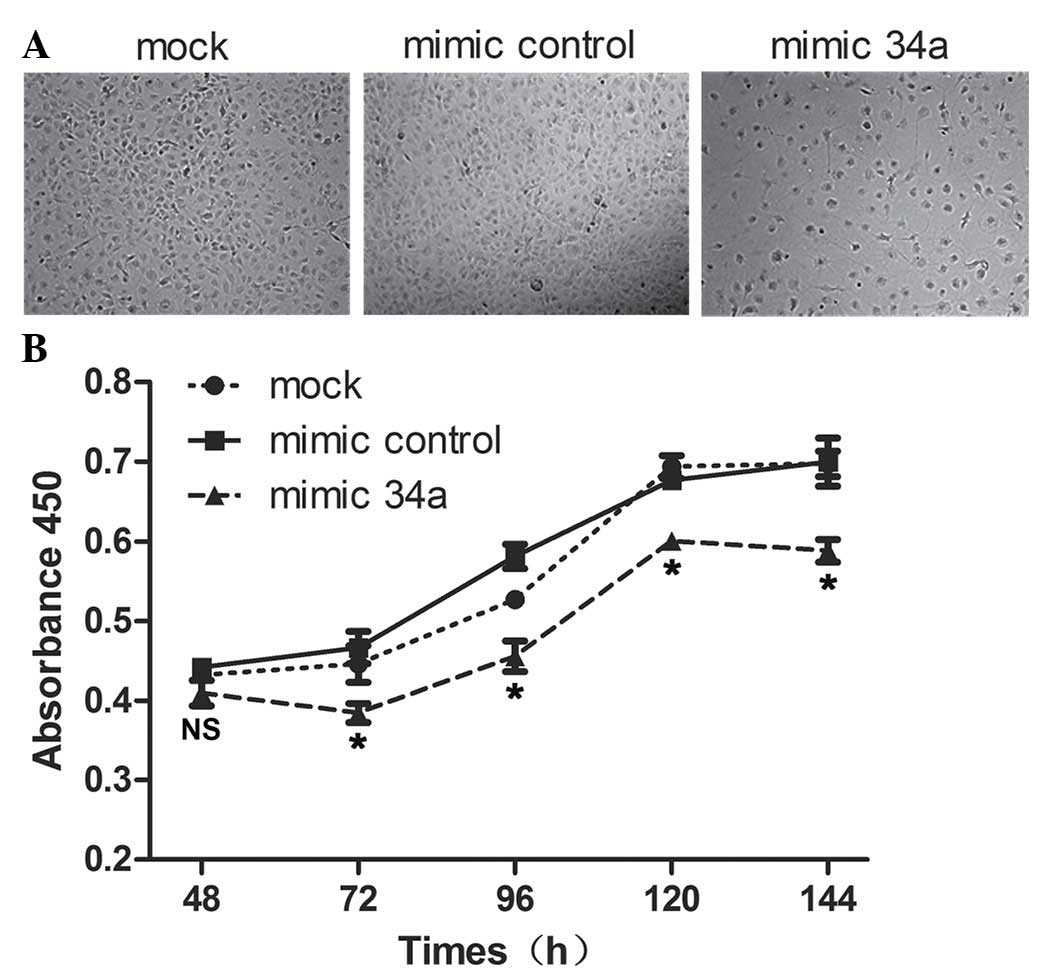
miR-34a suppresses SRA01/04
proliferation and induces apoptosis
To investigate the effect of miR-34a on SRA01/04
human lens epithelium cell proliferation and apoptosis, miR-34a
mimics were transfected into SRA01/04 cells and it was identified
that cell apoptosis and slow growth occurred subsequent to
transfection (Fig. 2A). The CCK-8
assay detected the growth conditions of SRA01/04, the absorbance at
450 nm was observed to be reduced in the SRA01/04 cells transfected
with miR-34a mimics compared with the mimic control at 72, 96, 120
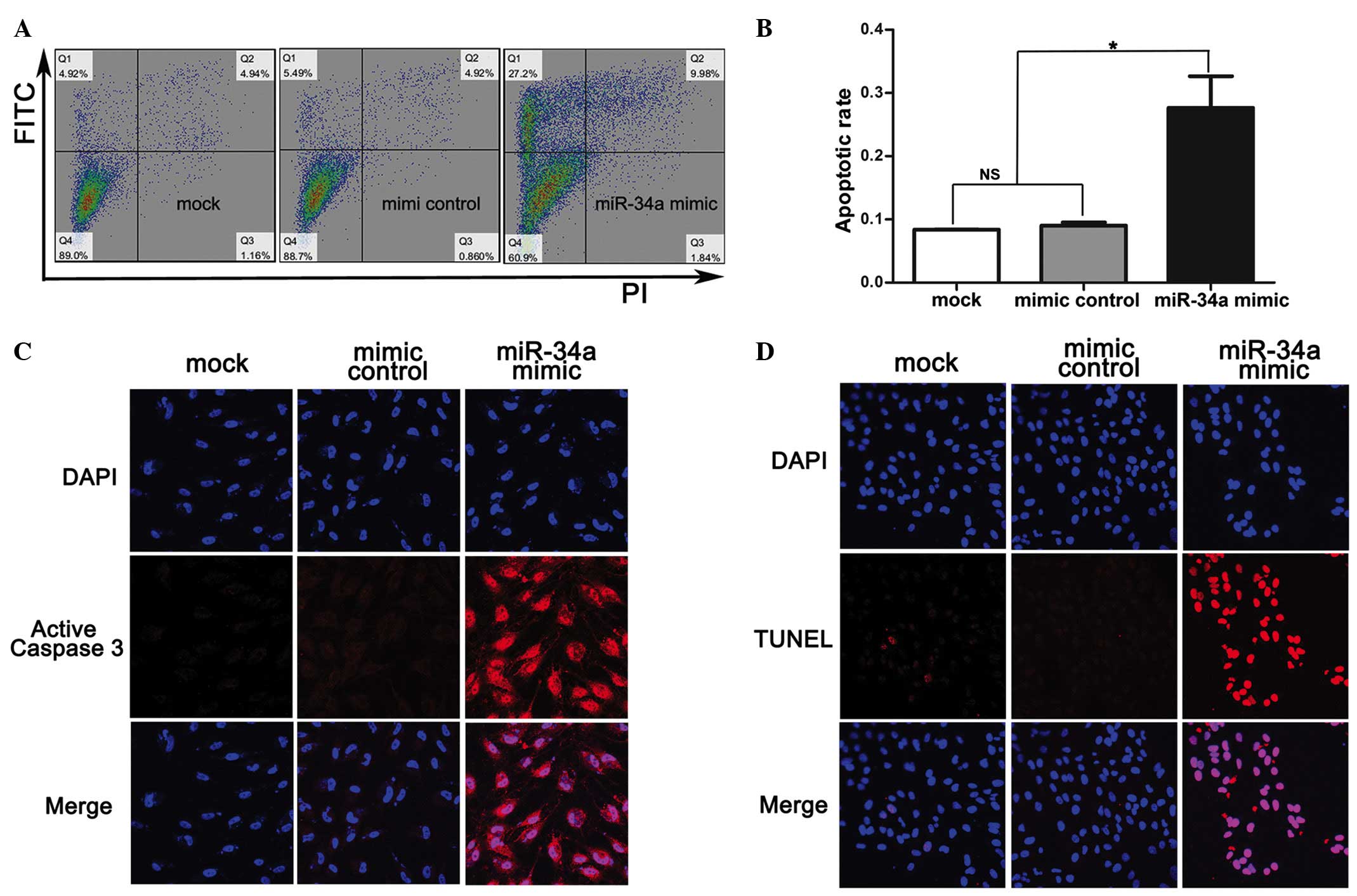
and 144 h following transfection with miRNA mimics (Fig. 2B). The apoptotic rate measured by
flow cytometry was significantly increased in SRA01/04 cells
transfected with miR-34a mimics (27.63±5%) compared with the mimic
control (9±0.5%) and mock (8.37±0.06%) (Fig. 3A and B). In addition, apoptotic
indicators TUNEL and active caspase 3 were observed to be highly
expressed in SRA01/04 cells transfected with miR-34a mimics
compared with the mimic control and mock (Fig. 3C and D).
Identification of candidate target
genes for miR-34a
To determine the potential targets of miR 34a in
SRA01/04 cells, the miRNA target prediction tool miRWalk (selecting
miRanda, miRWalk, PicTar5 and TargetScan) was used, and this
identified 20 putative target messenger RNAs of miR-34a (TOPORS,
PGRMC2, UHRF2, CPEB2, CTNND2, CAMSAP1, EML5, DBC1, DCX, JAG1, E2F3,
E2F5, ACSL1, RAB21, POGZ, RTF1, CAMTA1, SATB2, ZDHHC17 and ZNF281).
Of these candidates, E2F3 was selected due to its involvement in
cell apoptosis and the cell cycle (6–8), and
possible association with cataractogenesis (9).
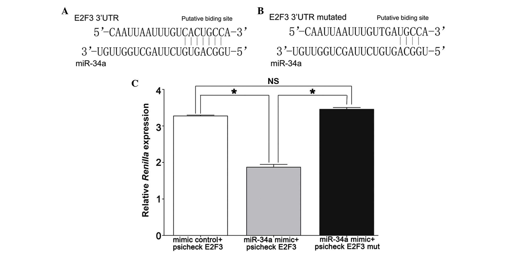
miR-34a targets the 3′-UTR of E2F3
mRNA
Using TargetScan to analyze the 3′-UTR of E2F3 to
identify potential binding sites for miR-34a, a single recognition
sequence containing a conserved 8-mer exact seed match at positions
2730–2737 bp (Fig. 4A and B) was
identified in the E2F3 3′UTR. This indicated that miR-34a may
directly bind to E2F3 3′UTR to regulate E2F3 expression at the
transcriptional level.
To validate E2F3 as a direct target of miR-34a,
luciferase reporter constructs (psiCHECK E2F3) were generated, in
which the E2F3 3′-UTR containing the miR-34a seed target was placed
behind the Renilla luciferase gene of the plasmid psiCHECK2.
miR-34a mimics inhibited Renilla luciferase activity
(1.87±0.13), whereas no effect was observed when the miR-34a target
site was mutated (3.46±0.09), which was similar to that of the
mimic control (3.27±0.04) (Fig.
4C).
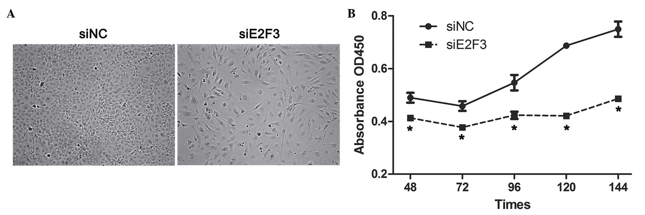
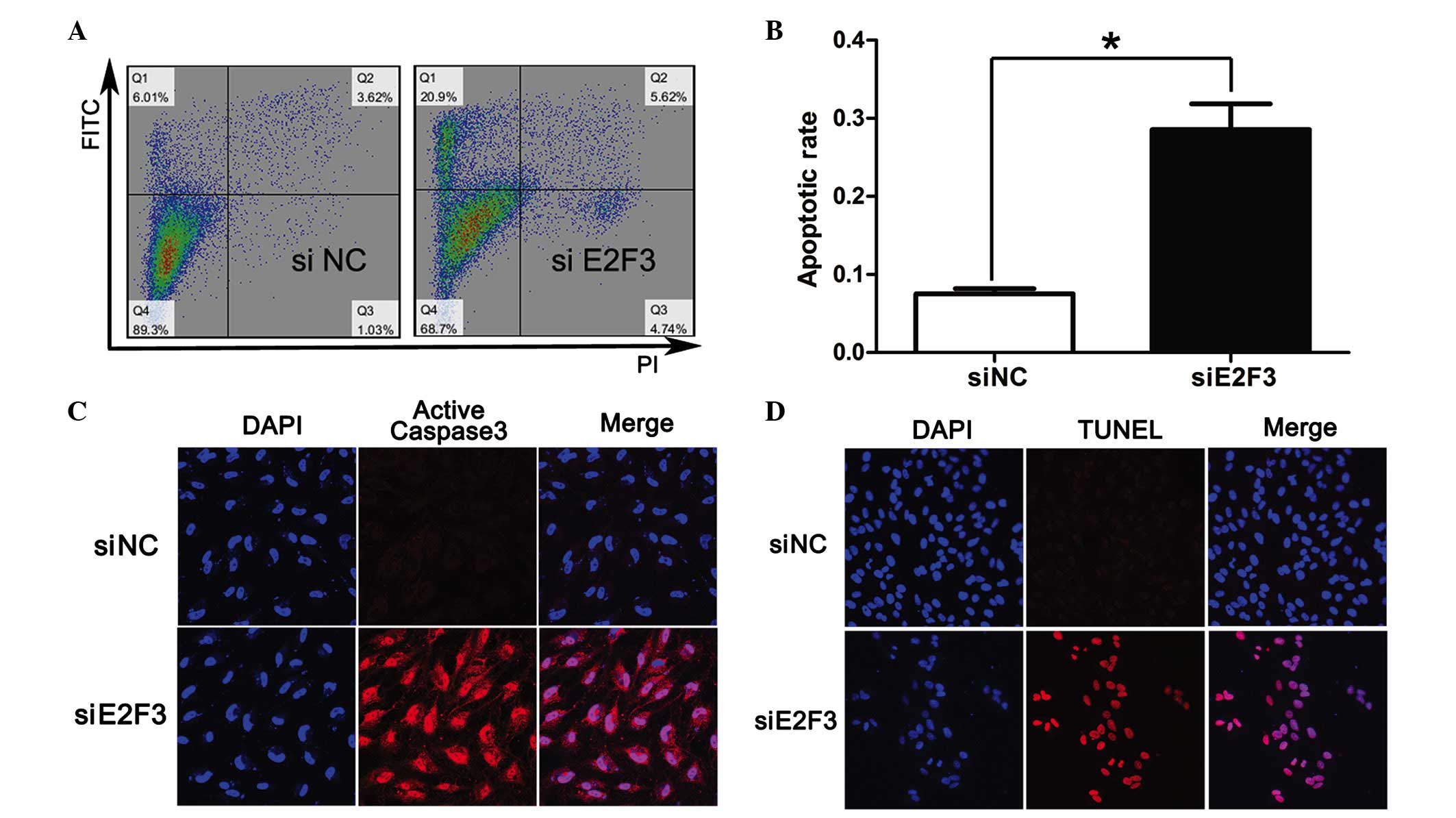
E2F3 siRNA (siE2F3) suppresses
SRA01/04 proliferation and induces apoptosis
In order to determine the role of E2F3 in
proliferation and apoptosis of SRA01/04 cells, siE2F3 were
transfected into SRA01/04 cells and this was observed to
significantly suppress proliferation of SRA01/04 cells at 48, 72,
96, 120 and 144 h subsequent to siE2F3 transfection compared with
the negative control (siNC) (Fig.
5). In addition, the results indicated a higher apoptotic rate
in SRA01/04 cells transfected with siE2F3 (28.5±1.9%) compared with
siNC (7.5±0.4%) (Fig. 6A and B).
In addition, apoptotic indicators TUNEL and active caspase 3 were
highly expressed in SRA01/04 cells transfected with siE2F3 when
compared with siNC (Fig. 6C and
D).
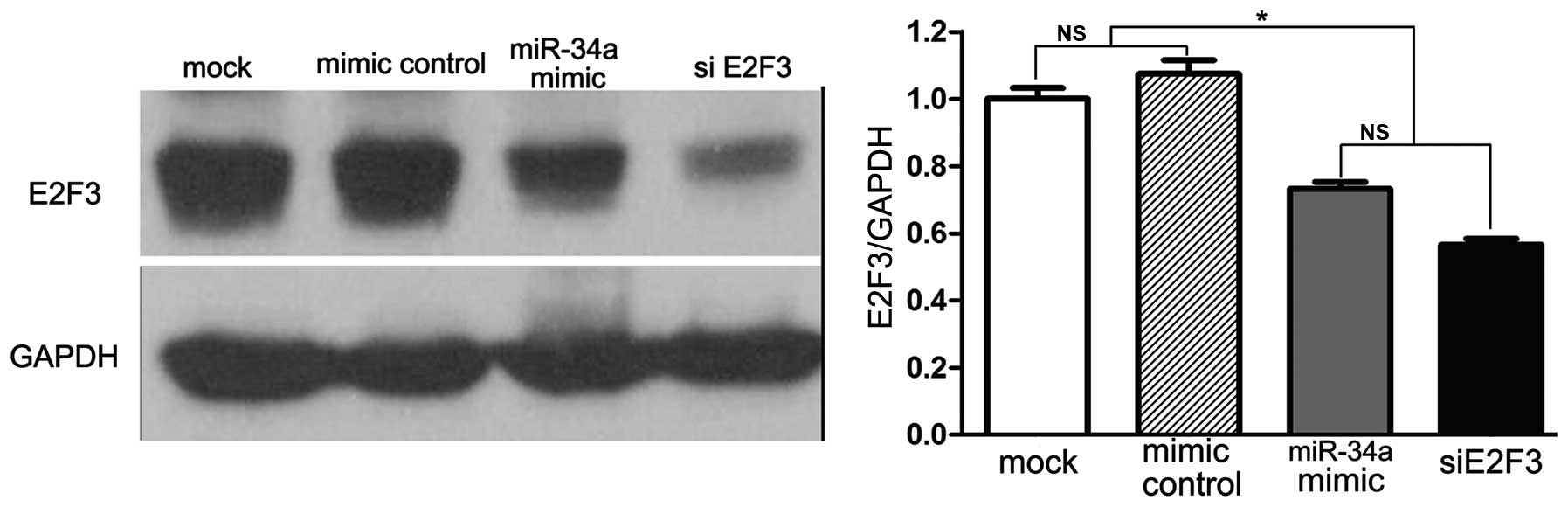
miR-34a and siE2F3 reduce E2F3
expression
Subsequent to verifying that miR-34a and siE2F3
inhibit proliferation and induce apoptosis of SRA01/04 cells, it
was investgated whether miR-34a and siE2F3 reduce the expression of
E2F3. Results indicate that miR-34a mimics and siE2F3 significantly
inhibited E2F3 protein expression (Fig. 7).
Discussion
miRNAs are endogenous and non-coding RNAs,
approximately 22 nucleotides in length, which perform important
regulatory roles by targeting mRNAs for cleavage or translational
repression (1). Greater than one
third of human genes appear to be conserved miRNA targets (19). Numerous diseases have been
indicated to involve changes in the miRNA expression profile. As
miRNAs are involved in the regulation of cellular proliferation,
differentiation and apoptosis, it is suggested that they take part
in complex, multifactorial and environmentally influenced cellular
processes, leading to the aging of cells and organisms, and human
disease (20).
According to a previous study (2), miR-34a is markedly increased in
cataracts compared with transparent lenses. This led to the
hypothesis of the current study, that miR-34a may participate in
cataractogenesis and its mechanisms. Tazawa et al (21) and Welch et al (22) previously demonstrated that
introducing miR-34a inhibited cancer cell proliferation and induced
apoptosis, accompanying downregulated E2F3 expression. In addition,
E2F3 is associated with cell proliferation, differentiation and
apoptosis (23). Therefore, using
the miRNA prediction software, it was identified that miR-34a may
take part in cataractogenesis by inducing lens epithelial cell
apoptosis by downregulating E2F3.
miR-34a is located in the 1p36.23 chromosome region,
and it is abnormally expressed in various tumor types including
human neuroblastoma (22,24), prostate cancer, colon cancer and
pancreatic cancer (25), and it is
highly expressed in age-associated diseases including cardiac aging
(26), atherosclerotic
cardiovascular disease (27) and
cataracts (28). Ectopic
overexpression of miR-34a in numerous tumor cell lines leads to the
reactivation of the apoptotic pathway, which suggests that miR-34a
may be a potent tumor suppressor gene (29). In addition, miR-34a has been
reported to be involved in senescence. In a premature senescence
model, induction by hydrogen peroxide resulted in significant
upregulation of miR-34a (30),
however, antisense inhibition of miR-34a blocked the onset of
replicative senescence (31).
Chien et al (28)
demonstrated positive correlations between miR-34a levels and lens
opacity severity, which suggested that miR-34a may serve a role in
lens senescence. Ito et al (27) identified that miR-34a expression
increased in senescent human umbilical cord vein endothelial cells
and in the heart and spleen of older mice. In addition, Zhao et
al (32) identified that the
overexpression of miR-34a in bone-marrow-derived endothelial
progenitor cells from rats led to the induction of senescence by
suppressing the sirtuin 1 gene.
The results indicated that miR-34a was markedly
increased in cataracts compared with transparent lenses, which was
in agreement with the results of Chien et al (28). Cataractogenesis has been reported
to be closely associated with lens epithelial cell apoptosis, and
it is widely accepted that the normal lens epithelial cells are
essential for maintenance of metabolic homeostasis and transparency
of the lens (33). If the lens
epithelial cells lose function, then the microenvironment of the
lens gets disturbed and the lens becomes opaque (34). In order to explain the association
between lens epithelial cell apoptosis and cataractogenesis, there
is direct evidence that depletion or stress-induced apoptotic death
of lens epithelial cells can initiate cataract formation in adult
lenses (9). In addition, in
vitro and in vivo studies have demonstrated that
cataract formation as a result of treatment of adult lenses with
stress factors, such as calcimycin and H2O2,
induced the apoptosis of lens epithelial cells (35,36).
A previous study (37) implicated
that normal lens epithelial cells exhibited reduced apoptosis
compared with that of cataracts, which suggests that lens
epithelial cell apoptosis may be a common cellular basis for the
initiation of non-congenital cataract formation.
Guggenmoos-Holtzmann et al (38) and Balaram et al (39) identified that the number of lens
epithelial cells gradually reduced in line with aging. Therefore,
it is suggested that through the different stages of growth and
differentiation in the adult lens, various stress conditions can
induce apoptosis of the lens epithelial cells, resulting in
eventual non-congenital cataractogenesis (40), including age-associated
cataracts.
The E2F transcription factor family that contains 8
members (E2F1-8) serves a crucial role in the regulation of
cellular proliferation, differentiation and apoptosis (23). The ablation of E2F3 in mouse
embryonic fibroblasts (MEFs) markedly reduce E2F target gene
expression (41) and significantly
compromise the proliferation of MEFs (42), which suggests that E2F3 serves a
critical role in the control of cell proliferation. In the current
study, the suppression of E2F3 by miR-34a and siE2F3 suppressed
SRA01/04 cell proliferation and induced cell apoptosis, which
suggested that miR-34a acted via through E2F3.
In summary, the results of the present study
indicated that miR-34a was highly expressed in cataractous lens
epithelial cells, and miR-34a suppresses proliferation and induces
apoptosis of SRA01/04 cells in a similar way to siE2F3. In
addition, miR-34a and siE2F3 significantly reduced E2F3 protein
expression. All of these results demonstrated that miR-34a induced
SRA01/04 human lens epithelial cell apoptosis through E2F3, and may
be closely involved in the pathogenesis of cataracts. These
observations may aid in the development of novel therapeutic
strategies.
Acknowledgements
The current study was supported by the National
Natural Science Foundation of China (grant nos. 81270980 and
81000389), the Pearl River Science and Technology New Star Project
of Guangzhou City (grant no. 2014J2200060), the Guangdong
Provincial Natural Science Foundation for Distinguished Young
Scholars of China (grant no. 2014A030306030) and the Science and
Technology Program of Guangdong Province (grant no.
2013B021800054).
References
|
1
|
Bartel DP: MicroRNAs: Genomics,
biogenesis, mechanism, and function. Cell. 116:281–297. 2004.
View Article : Google Scholar : PubMed/NCBI
|
|
2
|
Wu C, Lin H, Wang Q, Chen W, Luo H, Chen W
and Zhang H: Discrepant expression of microRNAs in transparent and
cataractous human lenses. Invest Ophthalmol Vis Sci. 53:3906–3912.
2012. View Article : Google Scholar : PubMed/NCBI
|
|
3
|
Christoffersen NR, Shalgi R, Frankel LB,
Leucci E, Lees M, Klausen M, Pilpel Y, Nielsen FC, Oren M and Lund
AH: p53-independent upregulation of miR-34a during oncogene-induced
senescence represses MYC. Cell Death Differ. 17:236–245. 2010.
View Article : Google Scholar : PubMed/NCBI
|
|
4
|
He L, He X, Lim LP, de Stanchina E, Xuan
Z, Liang Y, Xue W, Zender L, Magnus J, Ridzon D, et al: A microRNA
component of the p53 tumour suppressor network. Nature.
447:1130–1134. 2007. View Article : Google Scholar : PubMed/NCBI
|
|
5
|
Hermeking H: MicroRNAs in the p53 network:
Micromanagement of tumour suppression. Nat Rev Cancer. 12:613–626.
2012. View
Article : Google Scholar : PubMed/NCBI
|
|
6
|
Wenzel PL, Chong JL, Sáenz-Robles MT,
Ferrey A, Hagan JP, Gomez YM, Rajmohan R, Sharma N, Chen HZ, Pipas
JM, et al: Cell proliferation in the absence of E2F1-3. Dev Biol.
351:35–45. 2011. View Article : Google Scholar : PubMed/NCBI
|
|
7
|
Chen Q, Liang D, Yang T, Leone G and
Overbeek PA: Distinct capacities of individual E2Fs to induce cell
cycle re-entry in postmitotic lens fiber cells of transgenic mice.
Dev Neurosci. 26:435–445. 2004. View Article : Google Scholar : PubMed/NCBI
|
|
8
|
Chong JL, Tsai SY, Sharma N, Opavsky R,
Price R, Wu L, Fernandez SA and Leone G: E2f3a and E2f3b contribute
to the control of cell proliferation and mouse development. Mol
Cell Biol. 29:414–424. 2009. View Article : Google Scholar : PubMed/NCBI
|
|
9
|
Yan Q, Liu JP and Li DW: Apoptosis in lens
development and pathology. Differentiation. 74:195–211. 2006.
View Article : Google Scholar : PubMed/NCBI
|
|
10
|
Ayala M, Strid H, Jacobsson U and
Söderberg PG: p53 expression and apoptosis in the lens after
ultraviolet radiation exposure. Invest Ophthalmol Vis Sci.
48:4187–4191. 2007. View Article : Google Scholar : PubMed/NCBI
|
|
11
|
Galichanin K, Löfgren S, Bergmanson J and
Söderberg P: Evolution of damage in the lens after in vivo close to
threshold exposure to UV-B radiation: Cytomorphological study of
apoptosis. Exp Eye Res. 91:369–377. 2010. View Article : Google Scholar : PubMed/NCBI
|
|
12
|
Tamada Y, Fukiage C, Nakamura Y, Azuma M,
Kim YH and Shearer TR: Evidence for apoptosis in the selenite rat
model of cataract. Biochem Biophys Res Commun. 275:300–306. 2000.
View Article : Google Scholar : PubMed/NCBI
|
|
13
|
Murata M, Ohta N, Sakurai S, Alam S, Tsai
J, Kador PF and Sato S: The role of aldose reductase in sugar
cataract formation: Aldose reductase plays a key role in lens
epithelial cell death (apoptosis). Chem Biol Interact 130–132.
617–625. 2001. View Article : Google Scholar
|
|
14
|
Takamura Y, Kubo E, Tsuzuki S and Akagi Y:
Apoptotic cell death in the lens epithelium of rat sugar cataract.
Exp Eye Res. 77:51–57. 2003. View Article : Google Scholar : PubMed/NCBI
|
|
15
|
Yoshizawa K, Oishi Y, Nambu H, Yamamoto D,
Yang J, Senzaki H, Miki H and Tsubura A: Cataractogenesis in
neonatal Sprague-Dawley rats by N-methyl-N-nitrosourea. Toxicol
Pathol. 28:555–564. 2000. View Article : Google Scholar : PubMed/NCBI
|
|
16
|
Pandya U, Saini MK, Jin GF, Awasthi S,
Godley BF and Awasthi YC: Dietary curcumin prevents ocular toxicity
of naphthalene in rats. Toxicol Lett. 115:195–204. 2000. View Article : Google Scholar : PubMed/NCBI
|
|
17
|
Nakajima Y, Nakamura T, Enomoto T and
Murata Y: Loss of one allele of the p53 gene in the lens epithelial
tumor in transgenic mice suppresses apoptosis induced by a
topoisomerase I inhibitor (CPT-11). Cancer Lett. 179:165–173. 2002.
View Article : Google Scholar : PubMed/NCBI
|
|
18
|
Durand G, Hubert MF, Kuno H, Cook WO,
Boussiquet-Leroux C, Owen R, Fujimaki Y, Kemi M, Virat M and van
Zwieten MJ: Muscarinic receptor antagonist-induced lenticular
opacity in rats. Toxicol Sci. 66:166–172. 2002. View Article : Google Scholar : PubMed/NCBI
|
|
19
|
Lewis BP, Burge CB and Bartel DP:
Conserved seed pairing, often flanked by adenosines, indicates that
thousands of human genes are microRNA targets. Cell. 120:15–20.
2005. View Article : Google Scholar : PubMed/NCBI
|
|
20
|
Harries LW: MicroRNAs as mediators of the
ageing process. Genes (Basel). 5:656–670. 2014.PubMed/NCBI
|
|
21
|
Tazawa H, Tsuchiya N, Izumiya M and
Nakagama H: Tumor-suppressive miR-34a induces senescence-like
growth arrest through modulation of the E2F pathway in human colon
cancer cells. Proc Natl Acad Sci USA. 104:15472–15477. 2007.
View Article : Google Scholar : PubMed/NCBI
|
|
22
|
Welch C, Chen Y and Stallings RL:
MicroRNA-34a functions as a potential tumor suppressor by inducing
apoptosis in neuroblastoma cells. Oncogene. 26:5017–5022. 2007.
View Article : Google Scholar : PubMed/NCBI
|
|
23
|
Maiti B, Li J, de Bruin A, Gordon F,
Timmers C, Opavsky R, Patil K, Tuttle J, Cleghorn W and Leone G:
Cloning and characterization of mouse E2F8, a novel mammalian E2F
family member capable of blocking cellular proliferation. J Biol
Chem. 280:18211–18220. 2005. View Article : Google Scholar : PubMed/NCBI
|
|
24
|
Brodeur GM: Neuroblastoma: Biological
insights into a clinical enigma. Nat Rev Cancer. 3:203–216. 2003.
View Article : Google Scholar : PubMed/NCBI
|
|
25
|
Bagchi A and Mills AA: The quest for the
1p36 tumor suppressor. Cancer Res. 68:2551–2556. 2008. View Article : Google Scholar : PubMed/NCBI
|
|
26
|
Boon RA, Iekushi K, Lechner S, Seeger T,
Fischer A, Heydt S, Kaluza D, Tréguer K, Carmona G, Bonauer A, et
al: MicroRNA-34a regulates cardiac ageing and function. Nature.
495:107–110. 2013. View Article : Google Scholar : PubMed/NCBI
|
|
27
|
Ito T, Yagi S and Yamakuchi M:
MicroRNA-34a regulation of endothelial senescence. Biochem Biophys
Res Commun. 398:735–740. 2010. View Article : Google Scholar : PubMed/NCBI
|
|
28
|
Chien KH, Chen SJ, Liu JH, Chang HM, Woung
LC, Liang CM, Chen JT, Lin TJ, Chiou SH and Peng CH: Correlation
between microRNA-34a levels and lens opacity severity in
age-related cataracts. Eye (Lond). 27:883–888. 2013. View Article : Google Scholar : PubMed/NCBI
|
|
29
|
Tarasov V, Jung P, Verdoodt B, Lodygin D,
Epanchintsev A, Menssen A, Meister G and Hermeking H: Differential
regulation of microRNAs by p53 revealed by massively parallel
sequencing: miR-34a is a p53 target that induces apoptosis and
G1-arrest. Cell Cycle. 6:1586–1593. 2007. View Article : Google Scholar : PubMed/NCBI
|
|
30
|
Maes OC, Sarojini H and Wang E: Stepwise
up-regulation of microRNA expression levels from replicating to
reversible and irreversible growth arrest states in WI-38 human
fibroblasts. J Cell Physiol. 221:109–119. 2009. View Article : Google Scholar : PubMed/NCBI
|
|
31
|
Fujita K, Mondal AM, Horikawa I, Nguyen
GH, Kumamoto K, Sohn JJ, Bowman ED, Mathe EA, Schetter AJ, Pine SR,
et al: p53 isoforms Delta133p53 and p53beta are endogenous
regulators of replicative cellular senescence. Nat Cell Biol.
11:1135–1142. 2009. View
Article : Google Scholar : PubMed/NCBI
|
|
32
|
Zhao T, Li J and Chen AF: MicroRNA-34a
induces endothelial progenitor cell senescence and impedes its
angiogenesis via suppressing silent information regulator 1. Am J
Physiol Endocrinol Metab. 299:E110–E116. 2010. View Article : Google Scholar : PubMed/NCBI
|
|
33
|
Kinoshita JH: Mechanisms initiating
cataract formation. Proctor Lecture. Invest Ophthalmol. 13:713–724.
1974.PubMed/NCBI
|
|
34
|
Piatigorsky J: Lens differentiation in
vertebrates. A review of cellular and molecular features.
Differentiation. 19:134–153. 1981. View Article : Google Scholar : PubMed/NCBI
|
|
35
|
Li WC, Kuszak JR, Wang GM, Wu ZQ and
Spector A: Calcimycin-induced lens epithelial cell apoptosis
contributes to cataract formation. Exp Eye Res. 61:91–98. 1995.
View Article : Google Scholar : PubMed/NCBI
|
|
36
|
Li DW and Spector A: Hydrogen
peroxide-induced expression of the proto-oncogenes, c-jun, c-fos
and c-myc in rabbit lens epithelial cells. Mol Cell Biochem.
173:59–69. 1997. View Article : Google Scholar : PubMed/NCBI
|
|
37
|
Li WC, Kuszak JR, Dunn K, Wang RR, Ma W,
Wang GM, Spector A, Leib M, Cotliar AM, Weiss M, et al: Lens
epithelial cell apoptosis appears to be a common cellular basis for
non-congenital cataract development in humans and animals. J Cell
Biol. 130:169–181. 1995. View Article : Google Scholar : PubMed/NCBI
|
|
38
|
Guggenmoos-Holzmann I, Engel B, Henke V
and Naumann GO: Cell density of human lens epithelium in women
higher than in men. Invest Ophthalmol Vis Sci. 30:330–332.
1989.PubMed/NCBI
|
|
39
|
Balaram M, Tung WH, Kuszak JR, Ayaki M,
Shinohara T and Chylack LJ Jr: Noncontact specular microscopy of
human lens epithelium. Invest Ophthalmol Vis Sci. 41:474–481.
2000.PubMed/NCBI
|
|
40
|
Zhang L, Yan Q, Liu JP, Zou LJ, Liu J, Sun
S, Deng M, Gong L, Ji WK and Li DW: Apoptosis: Its functions and
control in the ocular lens. Curr Mol Med. 10:864–875. 2010.
View Article : Google Scholar : PubMed/NCBI
|
|
41
|
Humbert PO, Verona R, Trimarchi JM, Rogers
C, Dandapani S and Lees JA: E2f3 is critical for normal cellular
proliferation. Genes Dev. 14:690–703. 2000.PubMed/NCBI
|
|
42
|
Wu L, Timmers C, Maiti B, Saavedra HI,
Sang L, Chong GT, Nuckolls F, Giangrande P, Wright FA, Field SJ, et
al: The E2F1-3 transcription factors are essential for cellular
proliferation. Nature. 414:457–462. 2001. View Article : Google Scholar : PubMed/NCBI
|