Introduction
Cerebral ischemia, a condition in which blood flow
to the brain is insufficient, is one of the most serious
neurological disorders and can lead to stroke when cell death
occurs (1–3). Currently available treatments for
ischemic stroke, including anticoagulants and thrombolytics, have
not displayed substantial improvements in patients with ischemic
stroke (4). Therefore,
investigations into the pathological mechanisms of ischemic stroke,
and identification of effective neuroprotective agents that can be
utilized after ischemic stroke occurs are critical to improving
outcomes.
Cerebral ischemia is the result of transient or
permanent reduction of cerebral blood flow initiated by thrombotic
or thromboembolic arterial occlusions. Thrombolytic therapy
designed to restore cerebral perfusion in a timely fashion is
considered the primary therapeutic strategy for ischemic brain
injury (4). However, reperfusion
following thrombolytic therapy often leads to a series of cellular,
biochemical and metabolic consequences of cerebral ischemia,
including generation of intracellular reactive oxygen species
(ROS), calcium overload, mitochondrial injury and cell death, which
may ultimately lead to irreversible ischemia-reperfusion (I/R)
injury (5,6). The mechanisms of cerebral I/R injury
are complex, and include cellular responses such as oxidative
stress, inflammation and apoptosis (4–6).
Previous studies have demonstrated the involvement of oxidative
stress and intrinsic apoptotic signaling in the pathophysiology of
cerebral I/R injury (7,8). Elevated levels of ROS directly induce
damage to proteins, lipids and DNA. ROS also act as a molecular
trigger for apoptotic signaling, increasing apoptotic cell death
following ischemia (9). Intrinsic
apoptosis is regulated by mitochondrial activation and the release
of cytochrome c and second mitochondrial derived activator
of caspases (Smac), resulting in caspase-3 activation (10,11).
The intrinsic or mitochondrial-dependent mechanism of caspase
activation depends on the in B-cell lymphoma 2 apoptosis regulator
(Bcl-2) family of proteins that regulate mitochondrial outer
membrane permeabilization (9). The
Bcl family consits anti-apoptotic and pro-apoptotic factors
(11,12). The pro-apoptotic members of this
family, including Bcl-2-associated protein X (Bax) and Bcl-2
antagonist/killer 1, trigger the release of mitochondrial apoptotic
factors into the cytoplasm, leading to caspase activation, whereas
the anti-apoptotic family members, such as Bcl-2, act to prevent
apoptosis (12).
Polydatin (PD) is a monocrystalline drug isolated
from the herb Fallopia japonica, previously known as
Polygonum cuspidatum and commonly known as Japanese
Knotweed. It is used in traditional Chinese remedies to treat
sepsis, burns and I/R injury (12–14).
Previous studies have demonstrated the protective effects of PD
against acute shock-induced mitochondrial injury in neurons,
arteriolar smooth muscle cells and enterocytes, as evidenced by the
presence of ROS scavenging and prevention of mitochondrial damage
(15–17).
The aim of the present study was to evaluate the
potential therapeutic effect of PD following ischemic stroke, and
elucidate the mechanisms of PD neuroprotection with regard to
oxidative stress and intrinsic apoptotic signaling.
Materials and methods
Middle cerebral artery occlusion
(MCAO) model and drug treatment
All animal experiments were approved by the animal
experimental committee of Fujian Medical University (Fuzhou,
China). A total of 36 adult male Sprague-Dawley rats (250–270 g,
8–10 weeks) were purchased from the Experimental Animal Center at
Fujian Medical University, and were allowed to acclimate for 1 week
prior to the beginning of experimentation. The animals had ad
libitum access to rat chow and water. Animals were divided
randomly into three groups: sham-operated (sham); MCAO group
treated with 0.5 ml of normal saline (vehicle); and MCAO group
treated with 30 mg/kg of polydatin (PD; Neptunus Interlong
Bio-technique Co., Ltd., Shenzhen, Guangdong, China). Vehicle and
PD treatments were administered via caudal vein injection 10 min
before the start of the MCAO.
MCAO was produced using the intraluminal filament
technique as previously described (5). Rats were anesthetized with an
intraperitoneal injection of ketamine/xylazine (0.1 ml/100 g). The
right common carotid artery was exposed via a midline neck
incision, and was dissected from surrounding nerves and fascia up
to its bifurcation at the base of the skull. The external carotid
artery and internal carotid artery were then isolated in turn. A
nylon monofilament was inserted through the proximal external
carotid artery into the internal carotid artery and advanced until
a slight resistance was felt, indicating occlusion of the origin of
the MCA. The nylon monofilament remained in place for 2 h. The
nylon monofilament was then retracted so as to allow reperfusion of
the ischemic region. In the sham group, the procedure was followed
as for MCAO rats up to the occlusion of the MCA; however, the
filament was removed within 60 sec. All the rats were sacrificed by
decollation 24 h subsequent to MCAO or sham treatment for neuron
isolation and assessment.
Measurement of infarct volume
Three rats in each group were randomly selected for
2,3,5-triphenyltetrazolium chloride (TTC) staining. Freshly
isolated brains were stored at 20°C for 15 min. Brains were then
cut into 2 mm thick coronal sections. Sections were placed in 2%
TTC and warmed in a heated water bath at 37°C for 30 min. Sections
were then fixed with 4% paraformaldehyde overnight at
4°C. Stained sections were photographed, and the area of
ischemic brain injury was calculated by a blinded observer using
ImageJ software version 1.48 (National Institutes of Health,
Bethesda, MD, USA). Infarct volume is expressed as infarct area
percentage (%).
Evaluation of neurological
deficit
A modified neurological severity score (mNSS) test
was performed by a blinded investigator 24 h subsequent to the
induction of MCAO as described previously (4). The mNSS is a composite of motor,
sensory, balance and reflex tests, and is graded on a scale of
0–18, with a normal score being 0 and a maximal deficit score being
18. For injury severity scores, 1 point is awarded for the
inability to perform the test, or for lack of a tested reflex;
higher scores reflect a more severe injury.
Terminal deoxynucleotidyl transferase
dUTP (deoxyuridine 5-triphosphate-digoxigenin) nick end labeling
(TUNEL) staining
Cortex tissues on histopathological slides were
stained using a TUNEL kit (BioVision, Inc., Milpitas, CA, USA)
according to the manufacturer's protocols, counterstained with
Hoechst 33258, and then examined under an ECLIPSEFN1 fluorescent
microscope (Nikon Corporation, Japan). Apoptotic cells appeared
fluorescent green, and the number of apoptotic cells per 10 visual
fields was determined at ×200 magnification.
Morphological observation
Morphological changes to neuronal mitochondria were
observed by transmission electron microscopy (TEM). Cortical
tissues were fixed with 2.5% glutaraldehyde for 24 h at room
temperature and stained with cacodylate-buffered osmium tetroxide
for 4 h in 4°C. Sections (60 nm thickness) were prepared
and examined under an electron microscope at ×10,000 magnification
(Philips CM10; Philips, Eindhoven, The Netherlands).
Measurement of cytosolic cytochrome
c
The levels of cytochrome c in the cytoplasm
were evaluated with a cytochrome c ELISA kit (cat. no.
K257-100, BioVision, Inc.). Cortical tissues were dissected 24 h
after MCAO, then weighed, and lysed at 4°C for 10 min in
a cold lysis buffer (10 mM Tris-HCl, 0.3 M sucrose, 10 µM
aprotinin, 10 µM pepstatin, 10 µM leupeptin, and 1 mM PMSF, pH
7.5). Tissue homogenates were centrifuged at 10,000 × g for
60 min at 4°C, the supernatant containing the cytosolic
fraction collected, and protein concentration determined by
bicinchoninic acid (BCA) assay. Samples were then treated with a
conjugate reagent, were transferred to a cytochrome c
antibody-coated microwell plate, and incubated at room temperature
for 60 min. Wells were washed, treated with substrate and incubated
for 30 min at 4°C. Following termination of the
reaction, the optical density was read at 450 nm using a microplate
reader (SpectraMax M5; Molecular Devices LLC, Sunnyvale, CA, USA).
A serial dilution of cytochrome c calibrator was assayed in
parallel with the samples, and the concentration of cytochrome
c in the samples was determined by calibration of the
standard curve.
Measurement of caspase-3 and −9 activation. Activity
of caspase-3 and caspase-9 was evaluated using a commercial caspase
activity assay kit (cat. nos. K105-100 and K118-100, BioVision
Inc.). Cortical tissues were dissected 24 h subsequent to MCAO, and
were homogenized in lysis buffer supplemented with dithiothreitol
as described in the manufacturer's instructions. Following 15 min
incubation on ice, the supernatant was transferred into another
centrifuge tube, and centrifuged at 1,000 × g at
4°C for 5 min. Protein levels in the supernatant were
determined by BCA assay. Aliquots of each sample (20–50 µg protein)
were incubated with 90 µl of reaction buffer and 10 µl of the
substrate N-Acetyl-Asp-Glu-Val-Asp phospho-nitroanilide at 37°C for
2 h. The absorbance of the released phospho-nitroanilide was
measured at at 405 nm for caspase-3 and 505 nm for caspase-9 with a
spectrophotometer (SpectraMax M5; Molecular Devices, LLC).
Neuron isolation procedures
Adult rat cortical neurons were isolated in the
shortest time possible, with minimal neuronal hypoxia or ischemia,
according to the previously described method (16). Cortical tissues from the ischemic
hemisphere were dissected 24 h subsequent to MCAO. The cortex was
cut into fragments and the cells were dissociated by incubation
with 2 mg/ml papain in DMEM for 30 min at 37°C. To achieve optimal
purification, the immune adherence method was used to isolate
neurons. The digested cortical suspension was poured into
anti-neural cell adhesion molecule-coated petri dishes (cat. no.
AB5032, Millipore, Billerica, MA, USA) and placed in a shaker (4°C,
50 × g). Following 1 h incubation, the adhered cells were
collected. Trypan Blue staining was used to exclude non-intact
cells.
Immunofluorescent test
Isolated neurons were washed with PBS, permeabilized
with 4% formaldehyde and 0.5% Triton X-100 for 30 min in 4°C. Cells
were washed in PBS twice, blocked in 5% bovine serum albumin (Merck
Millipore, Darmstadt, Germany) for 1 h in 4°C, and incubated with
neurofilament antibody (1:500, cat. no. ab8135, Abcam, Cambridge,
UK) at 4°C overnight. Following a wash with PBS, the
cells were stained with a FITC-conjugated secondary antibody
(1:2,000, cat. no. ab6717, Abcam) for 1 h at room temperature.
Then, cells were captured using intravital upright microscope
(ECLIPSEFN1; Nikon Corporation, Tokyo, Japan). Results indicated
that the purity of neuron preparations was >90%, as required for
onward experiments.
Measurement of cell apoptosis
Cell apoptosis was detected using a FITC-Annexin V
apoptosis detection kit (BD Biosciences, San Jose, CA, USA).
Neurons were isolated, washed twice with PBS, and suspended in 1X
binding buffer at a concentration of approximately 1×10⁵
cells/ml. FITC-Annexin V (5 µl) and 10 µl of 50 µg/ml propidium
iodide (PI); (Sigma-Aldrich; Merck Millipore) were added to the
cell suspension. Following incubation at room temperature for 20
min in the dark, cell fluorescence was determined using a flow
cytometer (Becton Dickinson FACScan; BD Biosciences).
Measurement of mitochondrial membrane
potential
Mitochondrial membrane potential (∆Ψm)
was assessed by flow cytometry, using the potential-sensitive
fluorescent dye, JC-1. This dual-emission probe changes color from
red-orange to green as the mitochondrial membrane depolarizes.
Neurons were incubated with 5 µmol/l JC-1 for 15 min at 37°C. The
cells were then washed with PBS and analyzed by flow cytometry
(Becton Dickinson FACScan, BD Biosciences). Following gating using
forward scatter and side scatter with 488 nm excitation wavelength,
the fluorescence intensity were detected via FL1 (green, 530 nm)
and FL2 (red, 590 nm). A minimum of 10,000 cells were analyzed for
each sample. The cells with green fluorescence were evaluated to
reflect the percentage of cells with low ΔΨm.
Measurement of cellular ATP
Intracellular ATP was determined using a
CellTiter-Glo luciferase-based assay (Promega Corporation, Madison,
WI), according to the manufacturer's instructions. CellTiter-Glo
reagent (100 µl) was added to 10,000 isolated neurons in each well
of an opaque 96-well plate. The plates were incubated at room
temperature for 10 min, and luminescence (excitation wavelength at
488 nm, emission wavelength at 530 nm) was determined with a
microplate reader (SpectraMax M5; Molecular Devices, LLC).
Measurement of ROS levels
Intracellular ROS production was determined using a
2′-7′-dichlorofluorescein diacetate (DCFH-DA) ROS detection kit
(Sigma-Aldrich; Merck Millipore). Neurons were isolated and treated
with DCFH-DA (10 µM) for 20 min at 37°C. Following incubation, the
cells were washed and analyzed using a microplate reader (Spectra
Max, M5; Molecular Devices, LLC). The relative intensity of DCF
fluorescence compared with sham group cells was determined at a
wavelength of 535 nm.
Measurement of Bax and Bcl-2
Cortical tissues were isolated 24 h after MCAO and
homogenized in ice-cold tissue lysis buffer [3 M NaCl, 1 M Tris-HCl
(pH 7.4), 0.5 M EDTA, 100 mM PMSF, 10% Triton X-100] for 30 min.
The samples were then centrifuged at 10,000 × g for 20 min
at 4°C. Supernatant total protein concentration was
determined by bicinchoninic acid assay. Samples were then denatured
at 100°C for 5 min, and separated on 10% SDS-PAGE gels and
transferred to polyvinylidene difluoride (PVDF) membranes for
immunoblotting. Membranes were blocked with blocking solution (5%
non-fat milk diluted with PBS) at room temperature for 2 h,
followed by incubation with Bax (cat. no. ab32503), Bcl-2 (cat. no.
ab59348), or GAPDH (cat. no. ab8245) primary antibodies (1:1,000
dilution, Abcam) overnight at 4°C. Membranes were then
washed in PBS-Tween (PBST including 0.05% Tween-20), incubated with
secondary antibody (cat. no. ab6721, Abcam; 1:5,000 dilution with
PBS) for 2 h at room temperature, then washed 3 times in PBST. The
PVDF membranes were incubated with ultrasensitive enhanced
chemiluminescence (cat. no. abs920B-500, Absin Bioscience, Inc.,
Shanghai, China) for 1 min at 4°C. Next, the images of
PVDF membranes were obtained by a gel imaging system (Image Station
4000R, Kodak, Rochester, NY, USA).
Statistical analysis
Data are presented as the mean + standard deviation.
Differences between groups were determined using one-way analysis
of variance with the least significant difference
multiple-comparison test, or Student's t-test when appropriate.
P<0.05 was considered to indicate a statistically significant
difference.
Results
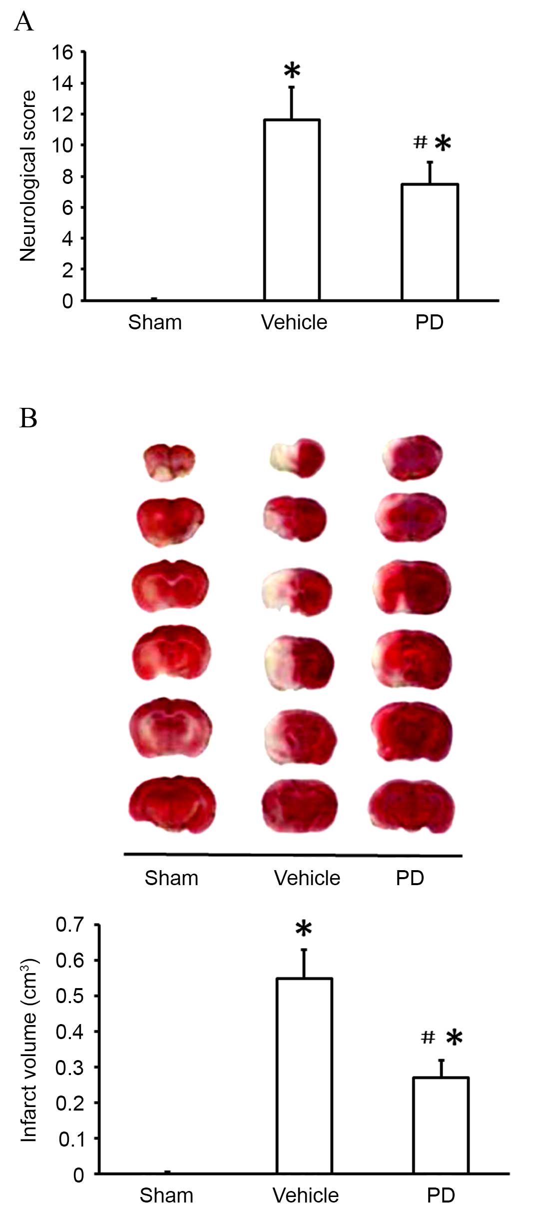
Neuroprotection by PD following
experimental stroke
To assess the protective effects of PD following
stroke, neurological scores and brain infarct volumes were
evaluated 24 h subsequent to MCAO using mNSS (Fig. 1A) and TTC staining (Fig. 1B) respectively. As demonstrated in
Fig. 1, compared with the sham
group, the vehicle group displayed significant neurological
deficits (P<0.001; Fig. 1A) and
brain infarcts (P<0.001; Fig.
1B) as evidenced by the white patches in brain tissues.
Administration of PD significantly decreased the infarct volume
(P=0.002 Fig. 1B) and ameliorated
the behavioral deficit (P Fig. 1A)
compared with the vehicle group.
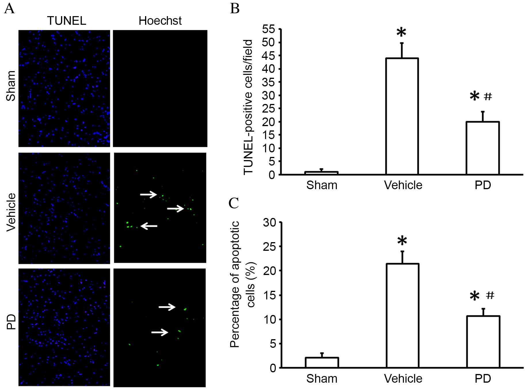
Neuroprotection of PD on I/R
injury-induced neuron apoptosis
Cell viability and apoptosis was detected by TUNEL
staining (Fig. 2A and B) and the
FITC-Annexin V/PI double staining assay (Fig. 2C) respectively. The number of
TUNEL-positive cells was significantly increased in the ischemic
region of the cortex in vehicle treated animals compared with sham
treated animals (P<0.001; Fig.
2B); this was reduced in the cerebral cortex of the PD-treated
compared with vehicle group (P=0.002 Fig. 2B). In the Annexin V-FITC assay, the
rate of cell apoptosis was significantly increased following MCAO
compared with the sham group (P<0.001; Fig. 2C); treatment with PD decreased cell
apoptosis compared with the vehicle group (P=0.011; Fig. 2C).
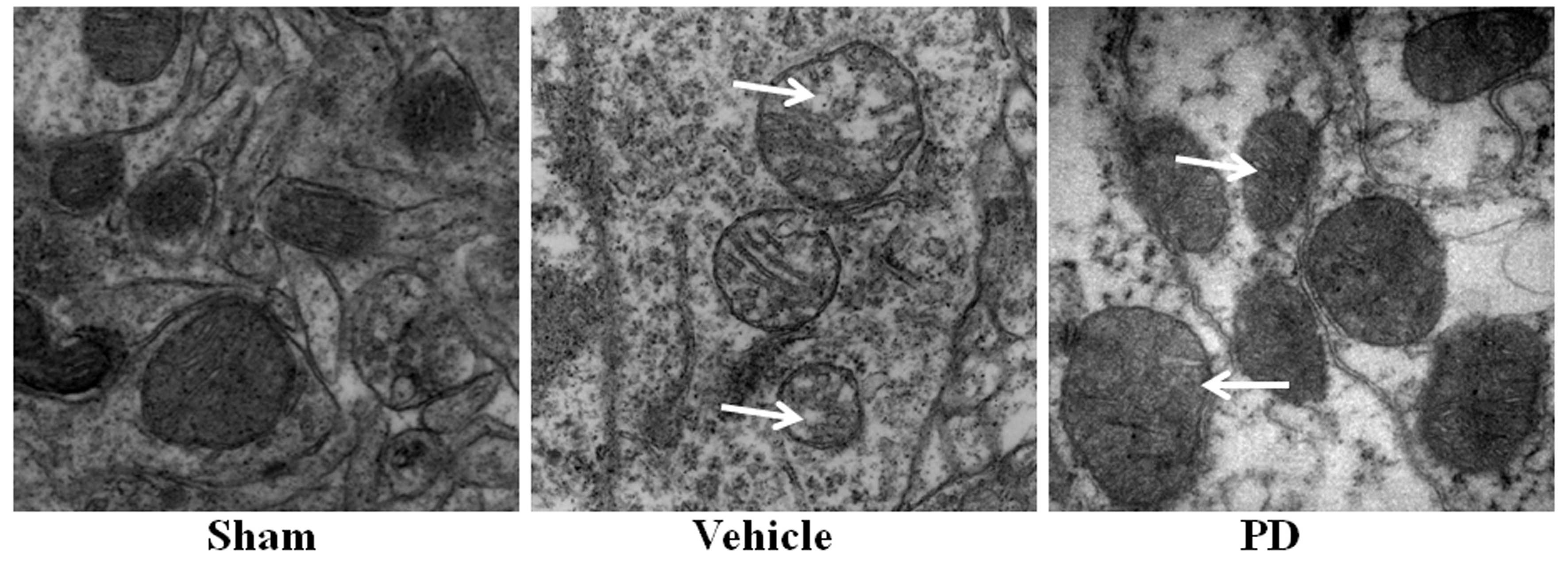
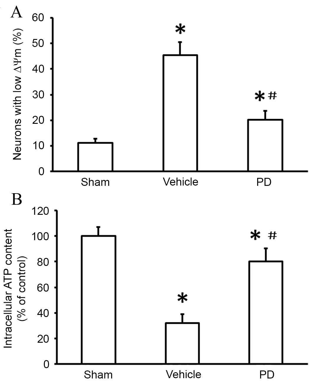
Neuroprotection of PD on I/R
injury-induced mitochondrial dysfunction
In the present study, TEM was used to examine
mitochondrial morphology. Cells in the sham group displayed normal
mitochondria with intact membranes and cristae (Fig. 3). Mitochondria from the vehicle
group appeared swollen and irregularly shaped, with disrupted and
poorly defined cristae. These mitochondrial alterations following
MCAO were partially prevented by PD treatment (Fig. 3). Mitochondrial membrane potential
(∆Ψm) was also measured using JC-1, a
potential-sensitive fluorescent dye that forms aggregates in
normally polarized mitochondria and monomers in damaged and
depolarized mitochondria. The percentage of neurons with low
∆Ψm was significantly increased in the vehicle group
compared with the sham group (P<0.001; Fig. 4A). The PD group contained
significantly fewer cells with low ∆Ψm than the vehicle
group (P=0.005; Fig. 4A).
Furthermore, intracellular ATP levels were significantly lower in
the vehicle group compared with the sham group (P<0.001;
Fig. 4B), indicating mitochondrial
dysfunction. Following PD treatment, intracellular ATP levels were
significantly increased compared with vehicle (P=0.014; Fig. 4B). These data indicate that PD
attenuates MCAO-induced mitochondrial dysfunction.
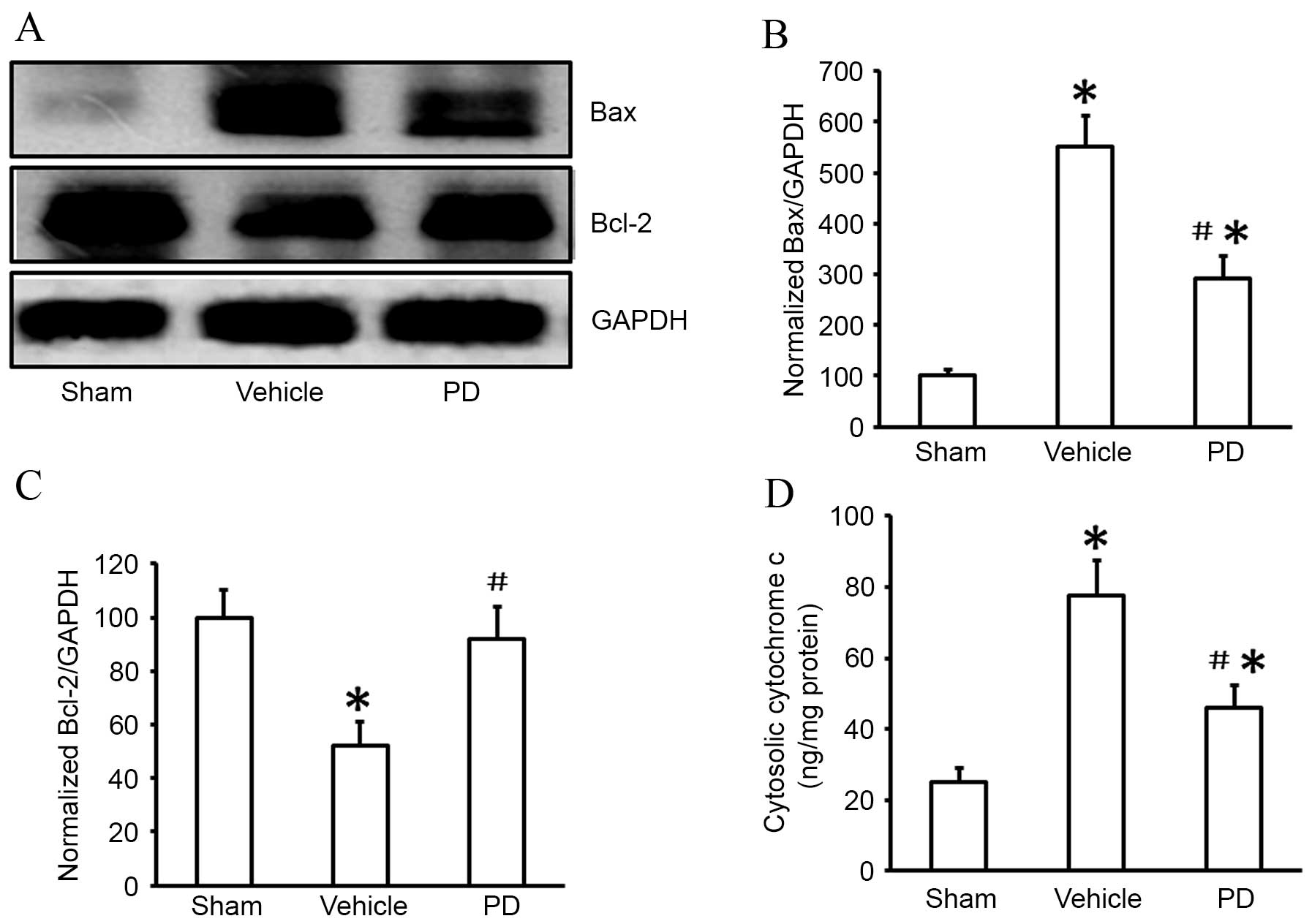
PD prevents I/R injury-induced
mitochondria-dependent apoptotic signaling
Mitochondrial regulation of apoptosis is partially
mediated by the release of cytochrome c and
apoptosis-inducing factors, which are regulated by the Bcl-2 family
of proteins (9). In the present
study, expression of the Bcl-2 family regulators, Bax and Bcl-2,
and levels of cytochrome c release, were investigated.
Compared with the sham group, vehicle treatment following MCAO
resulted in significant upregulation of Bax protein expression
levels (P<0.001; Fig. 5A and
B), and significant downregulation of Bcl-2 protein expression
levels (P<0.001; Fig. 5A and
C). PD treatment resulted in significantly lower levels of Bax
protein expression compared with vehicle (P=0.012; Fig. 5A and B) and significantly higher
levels of Bcl-2 protein expression than vehicle (P=0.019; Fig. 5A and C).
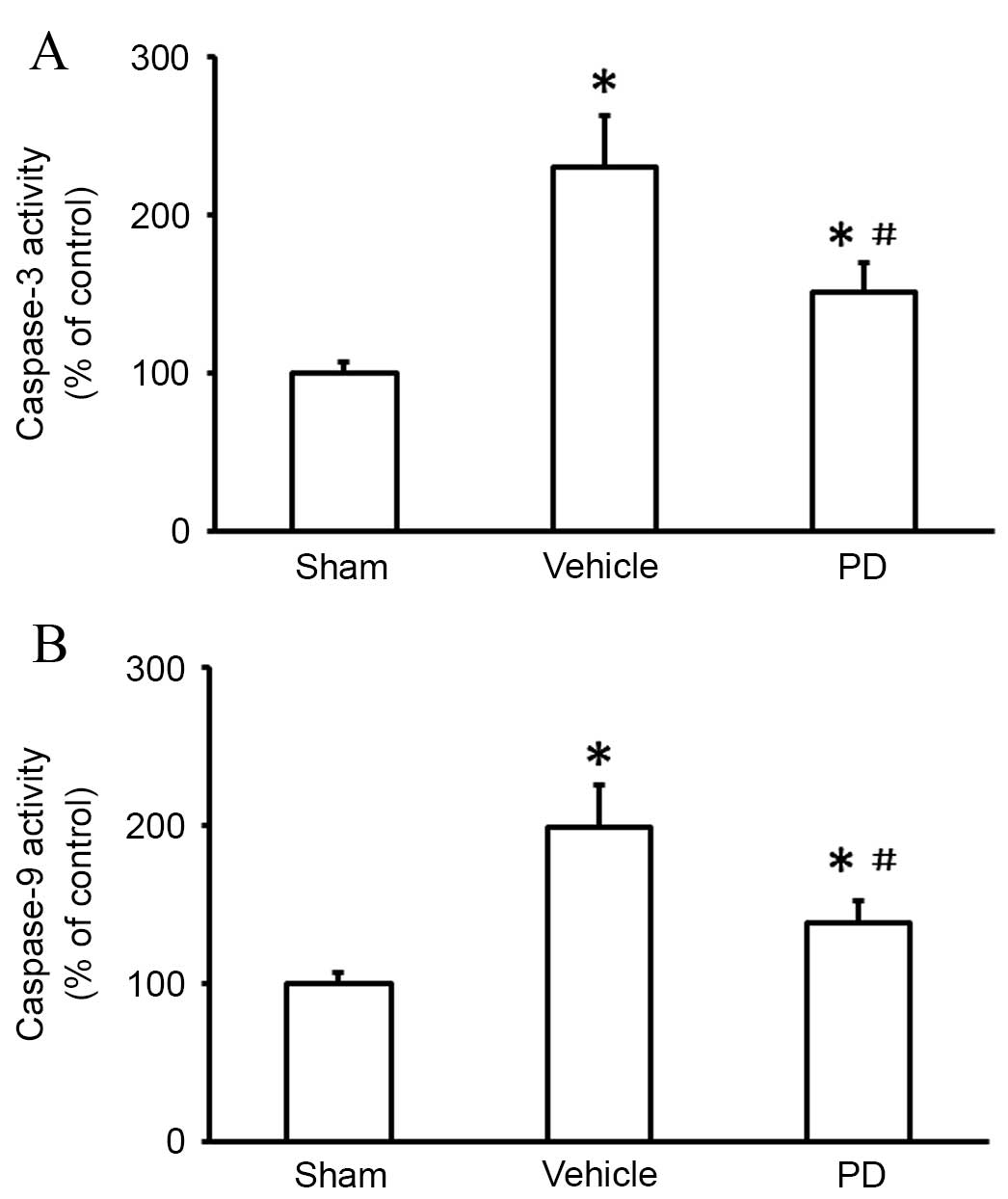
Cytochrome c release activates downstream
signaling cascades that trigger the intrinsic apoptotic pathway
(9). In the present study,
caspase-3 and caspase-9 activity were examined, as they are
critical to the apoptosis cascade (18). MCAO increased activation of
caspase-3 compared with sham (P<0.001; Fig. 6A); this upregulation was inhibited
by treatment with PD (P=0.021 vs. vehicle; Fig. 6A). A significant increase in
caspase-9 activity was observed following MCAO in the vehicle group
compared with the sham group (P<0.001; Fig. 6B), which was abrogated by PD
treatment (P=0.011 vs. vehicle; Fig.
6B).
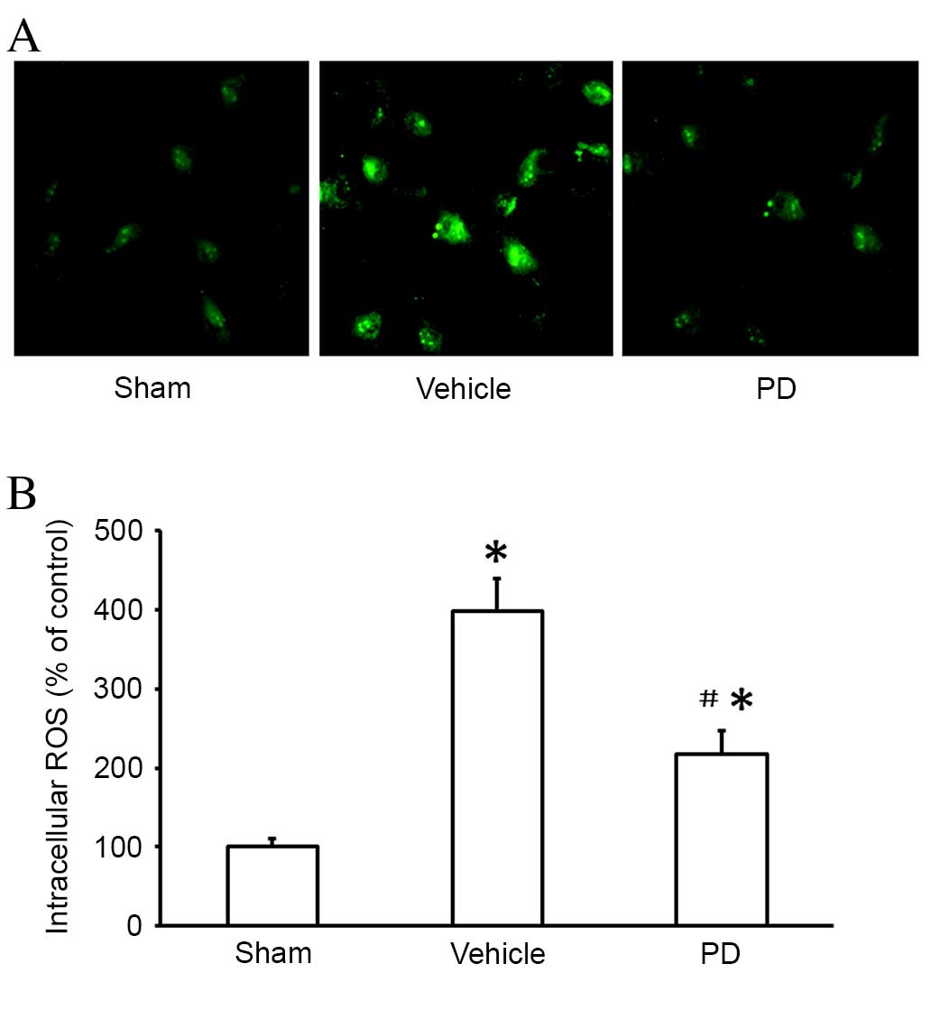
Neuroprotection of PD on I/R
injury-induced ROS production
To determine whether the neuroprotective effect of
PD was associated with its antioxidant properties, ROS production
was measured using a DCFH-DA assay 24 h after MCAO. ROS production
was significantly higher in the vehicle group than the sham group
(P<0.001, Fig. 7); this
increase was significantly lowered by PD treatment compared with
vehicle (P=0.008; Fig. 7).
Discussion
Previous studies by this group have demonstrated the
protective effects of PD on sepsis, burns and hemorrhagic shock
(12,13,16).
However, the effects of PD on the ischemic brain have, to the best
of our knowledge, not been examined previously. In the current
study, PD treatment was demonstrated to confer a neuroprotective
effect on cortical neurons in a MCAO rat model. This finding
highlighted modulation of mitochondria-dependent apoptosis as a
mechanism by which PD reduces neuronal damage and oxidative stress
following I/R injury.
Oxidative stress is a key factor in cerebral I/R
damage, with ROS contributing to apoptotic cell death in ischemia
via several pathways (19,20). Previous studies suggested that PD
acts as a free radical scavenger (14,16).
In the current study, increased ROS generation in the ischemic
brain was demonstrated to be partially reversed by PD, which could
be attributed to an antioxidant action. However, the molecular
mechanism of PD against oxidants remains to be elucidated and
should be investigated in future studies.
Mitochondria are essential to the production of
cellular energy, the generation of ROS, a by-product of normal
mitochondrial respiration is increased when the specific
respiratory chain is impaired under stress conditions, and the
regulation of apoptosis (14,15).
Therefore, mitochondria are important to neuronal survival
(18). Not only are mitochondria a
source of ROS, they are also targets of oxidative stress (16,21).
The mitochondrial transmembrane potential (∆Ψm), the
difference in potential caused by the ion concentration gradient
across the mitochondrial membrane, reflects mitochondrial function
(17). A decline in ∆Ψm
is correlated with the opening of the permeability transition pore,
leading to the release of caspase-activating proteins from the
mitochondria (21). Furthermore,
∆Ψm is reduced in response to the arrest of the activity
of respiratory complexes I–V, and contributes to inhibition of ATP
synthesis (15). Therefore,
reduced ∆Ψm, mitochondrial swelling with poorly defined
cristae, and reduced intracellular ATP content are all considered
signs of mitochondrial injury (16). In the present study, cells in the
vehicle group displayed a significant decrease in ∆Ψm
and ATP levels compared with the sham-operated group, and the
mitochondria appeared swollen and were irregularly shaped, with
disrupted and poorly defined cristae. PD treatment prevented these
morphological and functional changes, and resulted in
∆Ψm and ATP levels similar to the sham-operated group,
suggesting that PD prevents mitochondrial dysfunction following I/R
injury.
Mitochondrial damage and transmembrane potential
depolarization lead to the release of cytochrome c (22). Once released to the cytosol,
cytochrome c activates downstream caspases (9). Caspase-9 is an important initiator of
the cytochrome c-dependent caspase cascade, while caspase-3
is involved in apoptotic signaling transduction (4). In the present study, PD treatment was
demonstrated to have an effect on proteins involved in
mitochondria-dependent apoptosis. I/R injury (caused by MCAO) was
demonstrated to induce the release of cytochrome c from
mitochondria, leading to activation of caspases 9 and 3. PD
treatment inhibited these changes. These findings support the
hypothesis that PD treatment prevents the release of apoptotic
factors from mitochondria, following reducing the caspases
activation, leading to reduced apoptosis following ischemia and
reperfusion.
Mitochondria-regulated apoptosis is controlled by
members of the Bcl-2 family of proteins (23). The Bcl family consists of both
antiapoptotic (Bcl-2, Bcl-xL) and proapoptotic (BAK, BAX) factors
(24). The antiapoptotic members
of this family prevent apoptosis by sequestering proforms of
death-driving cysteine proteases or by preventing the release of
mitochondrial apoptogenic factors. Conversely, the proapoptotic
members trigger the release of mitochondrial apoptogenic factors
into the cytoplasm through the mitochondrial permeability
transition pore, thereby leading to caspases activation (12,13).
The present study demonstrated that Bcl-2 expression was
significantly increased following PD treatment and MCAO, while Bax
expression was decreased in the PD group compared with the vehicle
group following MCAO. These results indicate that the
anti-apoptotic effects of PD treatment may be mediated by changes
in Bcl-2 and Bax expression.
In conclusion, the present study has produced
evidence of significant neuroprotection in a MCAO rat model
following PD treatment. A potential mechanism of this action of PD
is through amelioration of oxidative stress and
mitochondria-dependent apoptosis. The underlying molecular
mechanisms require further clarification, more specifically
investigation of the effect of PD on brain injury and its possible
clinical application.
Acknowledgements
This work was supported by the Medical Innovation
Fund of Fujian Province, China (grant no. 2015-CX-22), the Finance
Department of Health Special Fund of Fujian Province, China (grant
no. BPB-LXZ2014), the National Natural Science Foundation of China
(grant no. 81500066) and the Natural Science Foundation of Fujian
Province (grant no. 2016J01451).
Glossary
Abbreviations
Abbreviations:
|
Bcl-2
|
B-cell lymphoma 2 apoptosis
regulator
|
|
Bax
|
Bcl-2 associated protein X apoptosis
regulator
|
|
I/R
|
ischemia/reperfusion
|
|
MCAO
|
middle cerebral artery occlusion
|
|
NF
|
neurofibromatosis
|
|
PD
|
polydatin
|
|
ROS
|
reactive oxygen species
|
|
Smac
|
second mitochondrial derived activator
of caspases
|
|
TEM
|
transmission electron microscopy
|
|
TTC
|
2,3,5-triphenyltetrazolium
chloride
|
|
TUNEL
|
terminal deoxynucleotidyl transferase
dUTP nick end labeling
|
References
|
1
|
Cai Q, Chen Z, Song P, Wu L, Wang L, Deng
G, Liu B and Chen Q: Co-transplantation of hippocampal neural stem
cells and astrocytes and microvascular endothelial cells improve
the memory in ischemic stroke rat. Int J Clin Exp Med.
8:13109–13117. 2015.PubMed/NCBI
|
|
2
|
Lin R, Lin Y, Tao J, Chen B, Yu K, Chen J,
Li X and Chen LD: Electroacupuncture ameliorates learning and
memory in rats with cerebral ischemia-reperfusion injury by
inhibiting oxidative stress and promoting p-CREB expression in the
hippocampus. Mol Med Rep. 12:6807–6814. 2015.PubMed/NCBI
|
|
3
|
Ljubisavljevic MR, Javid A, Oommen J,
Parekh K, Nagelkerke N, Shehab S and Adrian TE: The Effects of
different repetitive transcranial magnetic stimulation (rTMS)
protocols on cortical gene expression in a rat model of cerebral
ischemic-reperfusion injury. PLoS One. 10:e1398922015. View Article : Google Scholar
|
|
4
|
Yang W, Chen X, Pan J, Ge H, Yin K, Wu Z,
Li X, Sha D and Xu Y: Malibatol A protects against brain injury
through reversing mitochondrial dysfunction in experimental stroke.
Neurochem Int. 80:33–40. 2015. View Article : Google Scholar : PubMed/NCBI
|
|
5
|
Palencia G, Medrano JÁ, Ortiz-Plata A,
Farfán DJ, Sotelo J, Sánchez A and Trejo-Solís C: Anti-apoptotic,
anti-oxidant and anti-inflammatory effects of thalidomide on
cerebral ischemia/reperfusion injury in rats. J Neurol Sci.
351:78–87. 2015. View Article : Google Scholar : PubMed/NCBI
|
|
6
|
Fang L, Gao H, Zhang W, Zhang W and Wang
Y: Resveratrol alleviates nerve injury after cerebral ischemia and
reperfusion in mice by inhibiting inflammation and apoptosis. Int J
Clin Exp Med. 8:3219–3226. 2015.PubMed/NCBI
|
|
7
|
Tao T, Liu Y, Zhang J, Xu Y, Li W and Zhao
M: Therapeutic hypercapnia improves functional recovery and
attenuates injury via antiapoptotic mechanisms in a rat focal
cerebral ischemia/reperfusion model. Brain Res. 1533:52–62. 2013.
View Article : Google Scholar : PubMed/NCBI
|
|
8
|
Lu YP, Liu SY, Sun H, Wu XM, Li JJ and Zhu
L: Neuroprotective effect of astaxanthin on H(2)O(2)-induced
neurotoxicity in vitro and on focal cerebral ischemia in vivo.
Brain Res. 1360:40–48. 2010. View Article : Google Scholar : PubMed/NCBI
|
|
9
|
Li G, Li T, Li Y, Cai S, Zhang Z, Zeng Z,
Wang X, Gao Y, Li Y and Chen Z: Ulinastatin inhibits
oxidant-induced endothelial hyperpermeability and apoptotic
signaling. Int J Clin Exp Pathol. 7:7342–7350. 2014.PubMed/NCBI
|
|
10
|
Liu J, Bai J, Jiang G, Li X, Wang J, Wu D,
Owusu L, Zhang E and Li W: Anti-tumor effect of Pinus massoniana
Bark Proanthocyanidins on ovarian cancer through induction of cell
apoptosis and inhibition of cell migration. PLoS One.
10:e1421572015.
|
|
11
|
Li T, Yuan G, Zhang L, Ye L, Li S, Fan Y
and Sun J: ApoG2 inhibits the antiapoptotic protein, Mcl1 and
induces mitochondria-dependent apoptosis in human colorectal cancer
cells. Mol Med Rep. 12:6976–6984. 2015.PubMed/NCBI
|
|
12
|
Li T, Liu Y, Li G, Wang X, Zeng Z, Cai S,
Li F and Chen Z: Polydatin attenuates ipopolysaccharide-induced
acute lung injury in rats. Int J Clin Exp Pathol. 7:8401–8410.
2014.PubMed/NCBI
|
|
13
|
Li T, Cai S, Zeng Z, Zhang J, Gao Y, Wang
X and Chen Z: Protective effect of polydatin against burn-induced
lung injury in rats. Respir Care. 59:1412–1421. 2014. View Article : Google Scholar : PubMed/NCBI
|
|
14
|
Jiang X, Liu W, Deng J, Lan L, Xue X,
Zhang C, Cai G, Luo X and Liu J: Polydatin protects cardiac
function against burn injury by inhibiting sarcoplasmic reticulum
Ca2+ leak by reducing oxidative modification of ryanodine
receptors. Free Radic Biol Med. 60:292–299. 2013. View Article : Google Scholar : PubMed/NCBI
|
|
15
|
Wang X, Song R, Bian HN, Brunk UT, Zhao M
and Zhao KS: Polydatin, a natural polyphenol, protects arterial
smooth muscle cells against mitochondrial dysfunction and lysosomal
destabilization following hemorrhagic shock. Am J Physiol Regul
Integr Comp Physiol. 302:R805–R814. 2012. View Article : Google Scholar : PubMed/NCBI
|
|
16
|
Wang X, Song R, Chen Y, Zhao M and Zhao
KS: Polydatin-a new mitochondria protector for acute severe
hemorrhagic shock treatment. Expert Opin Investig Drugs.
22:169–179. 2013. View Article : Google Scholar : PubMed/NCBI
|
|
17
|
Zeng Z, Chen Z, Xu S, Song R, Yang H and
Zhao KS: Polydatin alleviates small intestine injury during
hemorrhagic shock as a SIRT1 activator. Oxid Med Cell Longev.
2015:9659612015. View Article : Google Scholar : PubMed/NCBI
|
|
18
|
Zeng Z, Chen Z, Li T, Zhang J, Gao Y, Xu
S, Cai S and Zhao KS: Polydatin: A new therapeutic agent against
multiorgan dysfunction. J Surg Res. 198:192–199. 2015. View Article : Google Scholar : PubMed/NCBI
|
|
19
|
Sun J, Li YZ, Ding YH, Wang J, Geng J,
Yang H, Ren J, Tang JY and Gao J: Neuroprotective effects of gallic
acid against hypoxia/reoxygenation-induced mitochondrial
dysfunctions in vitro and cerebral ischemia/reperfusion injury in
vivo. Brain Res. 1589:126–139. 2014. View Article : Google Scholar : PubMed/NCBI
|
|
20
|
Li J, Yu W, Li XT, Qi SH and Li B: The
effects of propofol on mitochondrial dysfunction following focal
cerebral ischemia-reperfusion in rats. Neuropharmacology.
77:358–368. 2014. View Article : Google Scholar : PubMed/NCBI
|
|
21
|
Srivastava SK, Bhardwaj A, Arora S, Tyagi
N, Singh S, Andrews J, McClellan S, Wang B and Singh AP:
MicroRNA-345 induces apoptosis in pancreatic cancer cells through
potentiation of caspase-dependent and -independent pathways. Br J
Cancer. 113:660–668. 2015. View Article : Google Scholar : PubMed/NCBI
|
|
22
|
Todd K, Ghiso J and Rostagno A: Oxidative
stress and mitochondria-mediated cell death mechanisms triggered by
the familial Danish dementia ADan amyloid. Neurobiol Dis.
85:130–143. 2015. View Article : Google Scholar : PubMed/NCBI
|
|
23
|
Cao ZH, Zheng QY, Li GQ, Hu XB, Feng SL,
Xu GL and Zhang KQ: STAT1-mediated down-regulation of Bcl-2
expression is involved in IFN-γ/TNF-α-induced apoptosis in NIT-1
cells. PLoS One. 10:e1209212015.
|
|
24
|
Kvansakul M and Hinds MG: The Bcl-2
family: Structures, interactions and targets for drug discovery.
Apoptosis. 20:136–150. 2015. View Article : Google Scholar : PubMed/NCBI
|