Introduction
Leukemia, a group of cancers, are originated from
blood-forming organs or tissues. A large number of immature and
abnormal white blood cells (WBCs) are produced by the bone marrow,
and those cells result in anemia and cause patients to be more
susceptible to infection (1).
Leukemia is the most common form of cancer in children worldwide
(2). Leukemia can be divided into
i) acute lymphocytic leukemia (ALL) derived from immature T or B
lymphocytes; ii) acute myeloid leukemia (AML) from immature myeloid
cells; iii) chronic lymphocytic leukemia (CLL) from mature
B-lymphocytes; and iv) chronic myelogenous leukemia (CML) from
granulocyte precursors (3,4). In the USA, two most prevalent types
of leukemia in children and adolescents are ALL and AML (5). In Western countries, CLL is the most
common form of leukemia (6,7).
Currently, studies have fostered the development of novel drugs
with different targets for leukemia patients. So far, the cure rate
for leukemia is still unsatisfying and thus, treatments of leukemia
remain a therapeutic challenge. The identification and development
of novel agents from natural products to induce immune function is
required.
Chitin (β-(1–4)-poly-N-acetyl-D-glucosamine), a
natural agent, is the major component of shell exoskeleton from
shrimp and crabs (8,9) and it can be converted to a
deacetylate derivative termed chitosan, which is a linear
heteropolysaccharide composed of α-(1,4)-linked-D-glucosamine (GlcN) and GlcNAc
(10). The hydrolyzed products of
chitosan, such as D-glucosamine oligomers [chitosan oligosaccharide
(COS)] are soluble in water (11)
and they have been demonstrated to have anti-bacterial, anti-tumor
and anti-oxidant effects (12–14)
and drug delivery functions (15).
It was reported that COS possesses inhibitory effects on
degranulation and cytokine generation in rat basophilic leukemia
cells in vivo (16).
Pretreatment with water-soluble chitosan in human astrocytoma cells
can lead to inhibition of secretion and expression of the
pro-inflammatory cytokines, tumor necrosis factor (TNF)-α and
interleukin (IL)-6 (17).
Previous studies have demonstrated that chitosan can
affect inflammation in vitro; however, limited information
is available regarding how chitosan affects immune responses in
vivo (18,19). Therefore, in the present study,
leukemia BLAB/c mice were generated with WEHI-3 mouse leukemia
cells, and the immune responses were subsequently evaluated in
vivo. Glutamic oxaloacetic transaminase (GOT), glutamic pyruvic
transaminase (GPT) and lactate dehydrogenase (LDH) levels in mice
following oral administration of chitosan were measured. The
results indicated that chitosan increased levels of the cell
markers cluster of differentiation (CD) 3, CD11b, CD19 and Mac-3,
increased macrophage phagocytosis and natural killer (NK) cell
activity, and affected GOT, GPT and LDH levels in leukemia BALB/c
mice in vivo.
Materials and methods
Materials and reagents
Acetic acid, concanavalin A (Con A), dimethyl
sulfoxide (DMSO), lipopolysaccharide (LPS) and YAC-1 cells were
purchased from Sigma-Aldrich; Merck KGaA (Darmstadt, Germany).
RPMI-1640 medium, fetal bovine serum (FBS), L-glutamine and
antibiotics (penicillin-streptomycin) were purchased from Gibco;
Thermo Fisher Scientific, Inc. (Waltham, MA, USA). Antibodies
against CD3, CD19, CD11b and Mac-3 were obtained from BD
Biosciences (San Jose, CA, USA). Chitosan powder with molecular
weight of ~86,000 kDa (Koyo Chemical Co., Ltd, Osaka, Japan) was
obtained from the National Taiwan University College of Medicine
Animal Medicine Center (Taipei, Taiwan, Republic of China). It was
dissolved in acetic acid, and 5 and 20 mg/kg solutions were
separately suspended in 0.2 ml acetic acid at room temperature for
1 h before use (20).
WEHI-3 cells
The WEHI-3 murine acute myelomonocytic leukemia cell
line was purchased from the Food Industry Research and Development
Institute (Hsinchu, Taiwan, Republic of China). Cells were cultured
in 75 cm2 tissue culture flasks containing RPMI 1640
medium supplemented with 10% FBS, 2 mM L-glutamine and antibiotics
(100 U/ml penicillin and 100 µg/ml streptomycin) and placed in a
humidified atmosphere of 5% CO2 at 37°C (21). Cells were allowed to re-equilibrate
for 24 h prior to use.
Male BALB/c mice
Male BALB/c mice (age, 8 weeks; weight, 22–25 g;
n=50) were obtained from the National Laboratory Animal Center
(Taipei, Taiwan, Republic of China). Mice were maintained in
stainless steel mesh-bottomed cages with specified pathogen-free
conditions in the animal center of China Medical University
(Taichung, Taiwan, Republic of China) as in a previous study
(22). All animals procedures were
carried out following the institutional guidelines for animal
welfare of China Medical University and were firstly approved by
the Institutional Animal Care and Use Committee of China Medical
University as described previously (21).
Treatment of animals with
chitosan
BLAB/c mice were randomly separated into five groups
(n=10/group). Group I were normal animals. Groups II–V were
peritoneally injected with 1×10⁶ WEHI-3 leukemia cells.
Groups I and II received a normal diet as a control. Group III mice
received a normal diet with acetic acid administered by oral
gavage. Group IV mice received chitosan (5 mg/kg) in acetic acid
(vehicle). Group V mice received chitosan (20 mg/kg) in acetic
acid. Chitosan dissolved in acetic acid was administered by oral
gavage individually to each mouse in groups IV and V every 2 days
for 15 days. All mice from each group were individual weighed
during the oral treatment and after treatment, and all mice were
weighed and sacrificed by euthanasia with CO2 as
described previously (21).
Immunofluorescence staining for
leukocytes surface markers
After treatment, all mice were weighed, blood were
collected by cardiocentesis under general anesthesia using a
disposable syringe with needle (20-gauge), and liver and spleens
were dissected and weighed (23).
Collected blood (1 ml/mouse) was lysed with 1X Pharm Lyse™ lysing
buffer (BD Biosciences) to destroy red blood cells and leukocytes,
as described previously (21).
Following centrifugation at 1,500 × g for 15 min at 4°C, white
blood cells were collected and stained with phycoerythrin
(PE)-labeled anti-mouse CD3 (BD553062; BD Biosciences, San Jose,
CA, USA), PE-labeled anti-mouse CD19 (BD553786; BD Biosciences),
fluorescein isothiocyanate (FITC)-labeled anti-mouse CD11b
(BD553310; BD Biosciences) and FITC-labeled anti-mouse Mac-3
(BD553324; BD Biosciences) antibodies at 1:12 dilution of each for
30 min at 4°C. All samples were analyzed by flow cytometry (BD
Biosciences) and quantified using CellQuest software version 5.2.1
(BD Biosciences), as previously described (21).
Measurements of macrophage
phagocytosis
Macrophages were isolated from peripheral blood
mononuclear cells (PBMCs) and the peritoneum of each mouse per
group, as described previously (21). Macrophages were added to plates
which contained 50 µl target E. coli-FITC, and mixed
according to the PHAGOTEST® kit manufacturer's protocol
(ORPEGEN Pharma GmbH, Heidelberg, Germany). Samples were then
analyzed for phagocytosis using flow cytometry. Phagocytosis was
quantified using CellQuest software version 5.2.1 (BD Biosciences),
as previously described (21).
Measurements of NK cell cytotoxic
activity
Splenocytes were isolated from each spleen per mouse
as described previously (21).
Splenocytes (1×10⁵ cells/well) were seeded into a
96-well plate containing 1 ml RPMI-1640 medium. The target YAC-1
cells (2.5×10⁷ cells) in 15 ml tubes and PKH-67/Dil.C
buffer (Sigma-Aldrich; Merck KGaA) was added to cells in tubes
according to the manufacturer' protocol, and were mixed thoroughly
for 2 min at 25°C. PBS (2 ml) was added to tube for 1 min, and 4 ml
medium was also added to the tube and were centrifuged at 290 × g
for 2 min at 25°C. Approximately 2.5×10⁶ YAC-1 cells in
100 µl in serum-free RPMI-1640 medium were seeded into 96-well
plates containing splenocytes (1×10⁵ cells/well) for 12
h at 37°C. After incubation, NK cell cytotoxic activity was
measured by flow cytometry as described previously (21).
Measurements of T and B cell
proliferation
Isolated splenocytes from each mouse were suspended
in PBS and seeded into 96-well plates (100 µl; 1×10⁵
cells/well) containing 100 µl RPMI-1640 medium. To measure T-cell
proliferation, 0.5 µg/ml Con A was added to the cells to stimulate
for 3 days. For B cell proliferation, 1 µg/ml LPS was added to the
cells to stimulate for 5 days. At the end of stimulation, cell
proliferation was measured using a CellTiter 96 AQueous One
Solution Cell Proliferation Assay kit (Promega Corporation,
Madison, WI, USA) as previously described (21).
Measurement of white blood cells
(WBCs), and GOT, GPT and LDH levels of leukemia BALB/c mice after
exposure to chitosan
All blood samples were collected from each mouse per
group. Total WBCs were measured using flow cytometry. The levels of
GOT and GTP were measured using liquiUV tests (aspartate
aminotransferase and alanine aminotransferase, respectively), and
LDH levels were also measured using a liquiUV test (Human
Gesellschaft fur Biochemica und Diagnosica mbH, Wiesbaden,
Germany), as described previously (24,25).
Statistical analysis
Data are expressed as the mean ± standard deviation
from three independent experiments. Statistical comparisons between
chitosan-treated and un-treated (control) groups were analyzed by
Student's t-test. P<0.05 was considered to indicate a
statistically significant difference.
Results
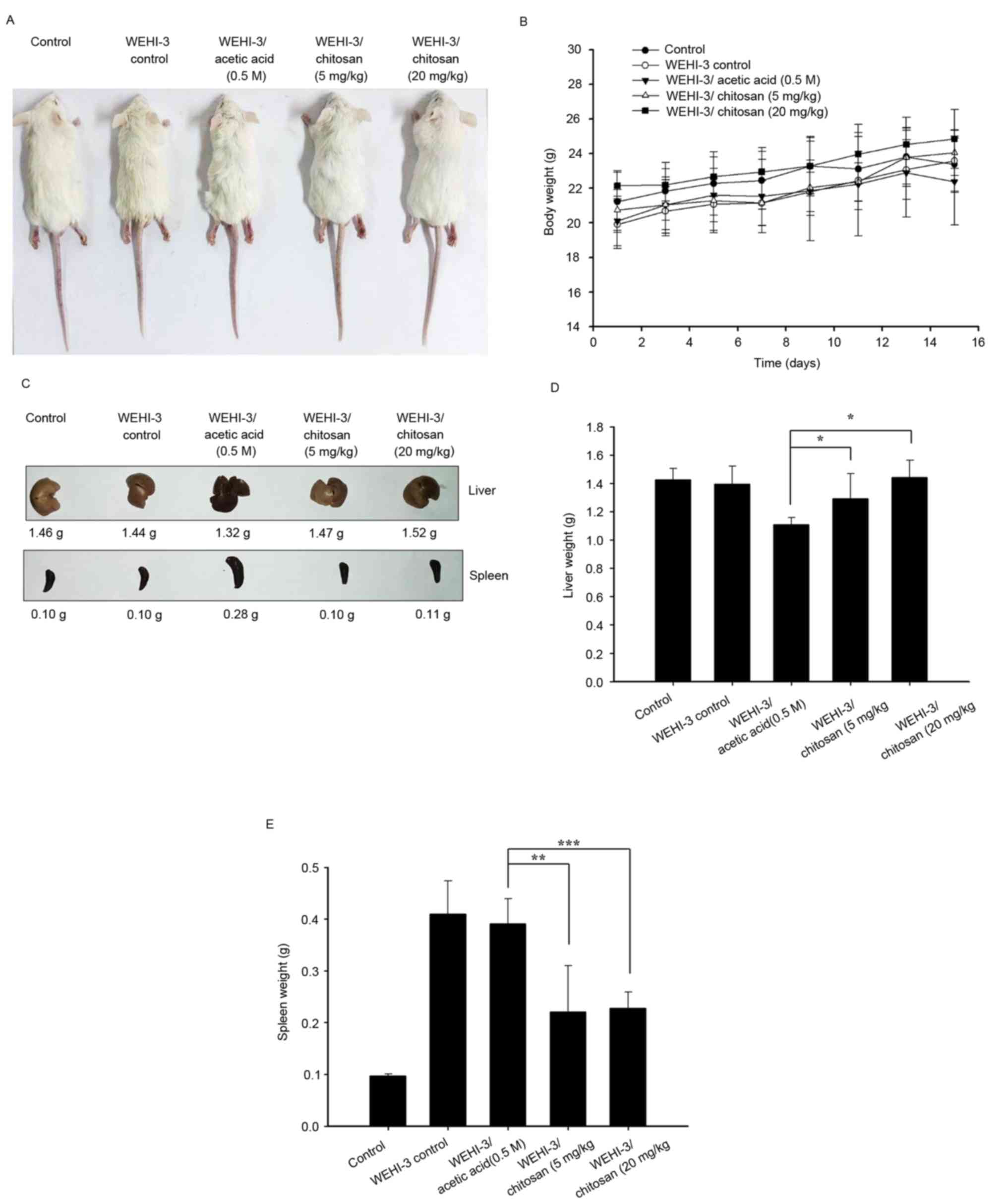
Chitosan affects the weights of body,
liver and spleen in leukemia BALB/c mice
WEHI-3 cells generated leukemia mice that were
divided into 4 experimental groups: One positive control, one
treated with acetic acid (vehicle) and the other two treated with
chitosan at 5 and 20 mg/kg per 2 days for 15 days (total 7 times).
Control mice (group I) were treated with a normal diet. The
representative animals, body weights, liver and spleen weights are
presented in Fig. 1, respectively.
These results indicated that chitosan did not significantly affect
the body weights and spleen weights of the animals, but it
significantly increased liver weights when compared with the
vehicle (acetic acid) treated group (Fig. 1D).
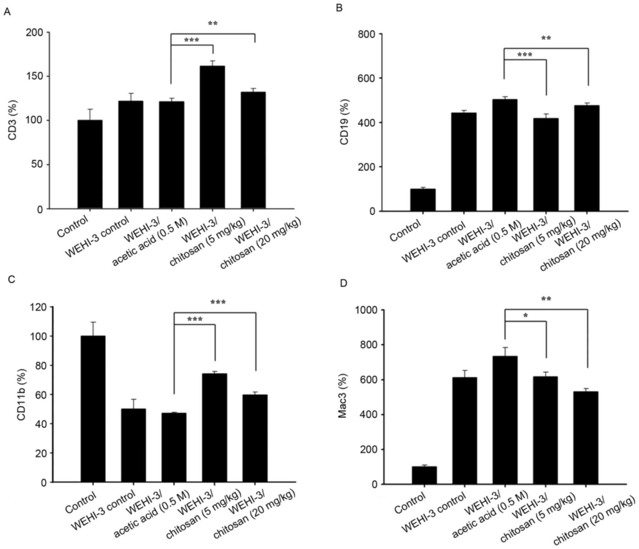
Chitosan affects cells markers of WBCs
in leukemia BALB/c mice
After animals were divided into 5 groups and were
treated with or without chitosan, blood samples were collected from
each mouse per group. The levels of cell markers CD3, CD19, CD11b
and Mac-3 from isolated cells were measured and the results are
presented in Fig. 2, respectively.
The results indicated that chitosan promoted CD3 (Fig. 2A) and CD19 (Fig. 2B) at both dose of treatment,
compared with acetic acid treatment only. However, chitosan
decreased the levels of CD11b at 5 mg/kg (Fig. 2C) and decreased the levels of Mac-3
at both doses (Fig. 2D) compared
with the acetic acid-treated groups.
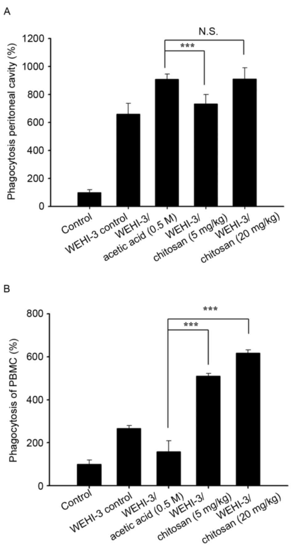
Chitosan affects macrophage
phagocytosis in PBMCs and the peritoneal cavity of leukemia BALB/c
mice
After treatment, cells were isolated from PBMCs and
the peritoneal cavity of each animal, and the percentage of
macrophages phagocytosis was measured. Chitosan treatment at both
doses (5 and 20 mg/kg) to leukemia mice did not significantly
induce macrophage phagocytosis in the peritoneal cavity (Fig. 3A), but significantly increased it
in PBMCs at both doses of treatment (Fig. 3B).
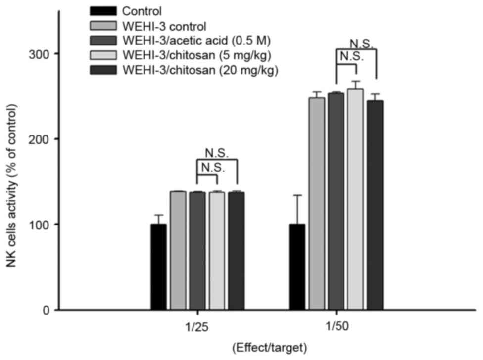
Chitosan affects the cytotoxic
activity of NK cells in leukemia BALB/c mice
YAC-1 cells were selected as targets for NK cells
isolated from splenocytes, and the activities of NK cells were
assayed by flow cytometry. The results indicated that chitosan did
not significantly affect NK cells cytotoxic activity at both doses
of treatment when compared with acetic acid treated groups
(Fig. 4).
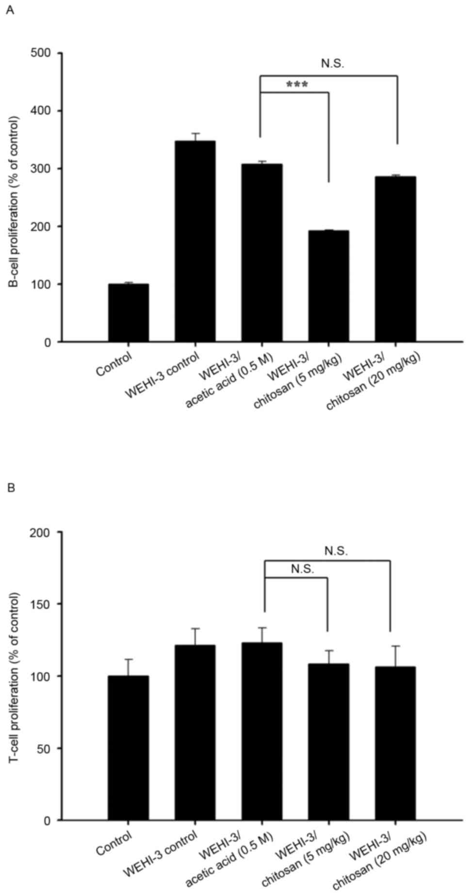
Chitosan affects T and B cell
proliferation in leukemia BALB/c mice
Splenocytes were isolated from each mouse per group
after treatment with or without chitosan, and were assessed by flow
cytometry for T- and B-cell proliferation after stimulating with
Con A or LPS, respectively. The lower dose of chitosan reduced
B-cell proliferation (Fig. 5A);
however, neither dose had any effect on T-cell proliferation
compared with the acetic acid treated group (Fig. 5B).
Chitosan affects WBC number, and the
activity of GOT, GPT and LDH in leukemia BALB/c mice
Blood samples were individually collected from
animals of each group. Chitosan at both doses of treatment
significantly decreased total WBC numbers compared with the acetic
acid group; however, 20 mg/kg chitosan resulted in a greater
decrease in cell numbers compared with 5 mg/kg (Fig. 6A). There was a similar trend in GOT
(Fig. 6B) and GPT (Fig. 6C) activity-both doses significantly
increased their activities, and the greatest effect was observed at
5 mg/kg. High doses of chitosan induced lower LDH activities
compared with low dose treatment (Fig.
6D). Both doses of chitosan treatment were reduced compared
with acetic acid treatment only.
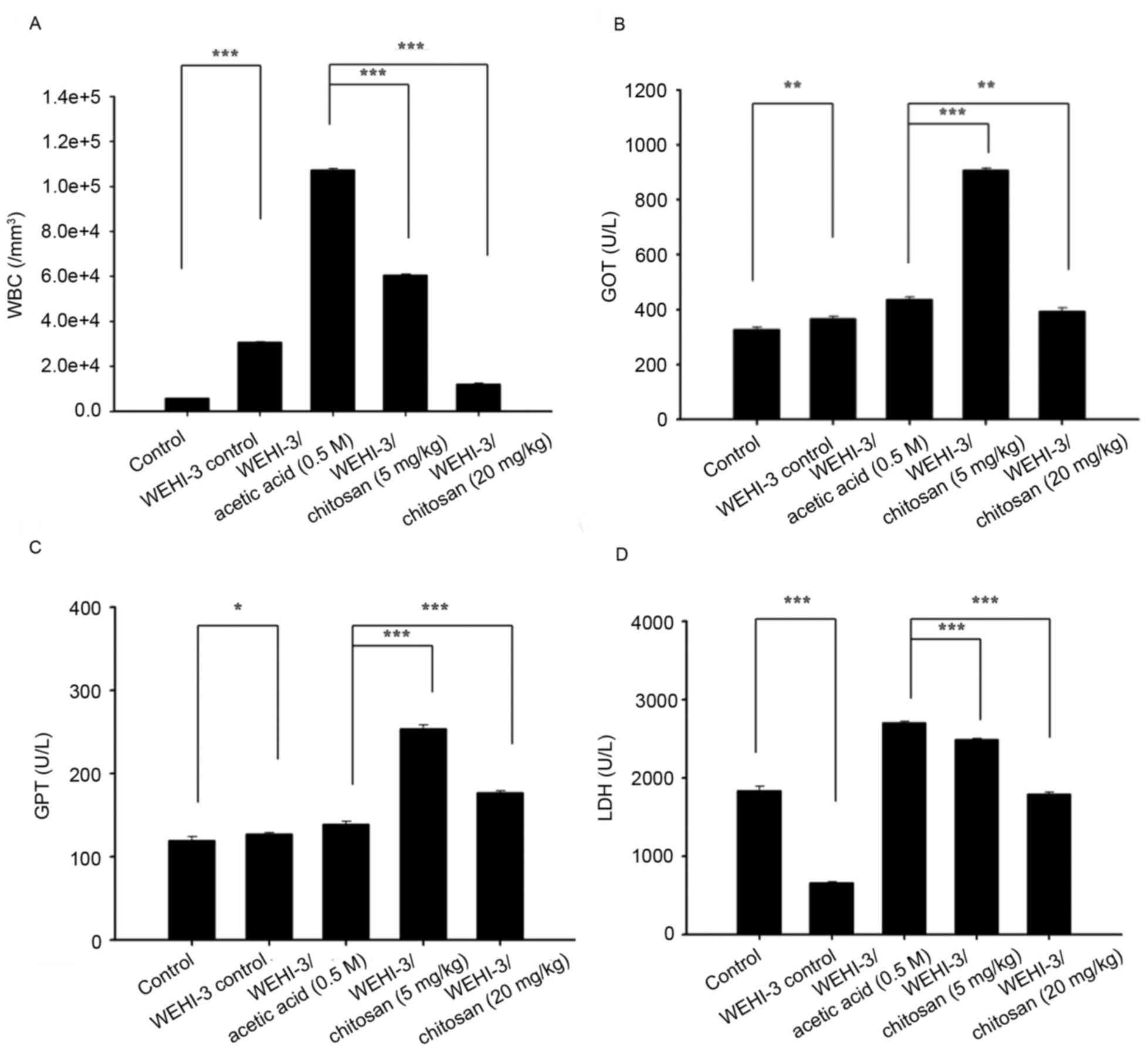 | Figure 6.Measurement of total WBCs, GOT, GPT
and LDH activity of leukemia BALB/c mice after exposure to
chitosan. (A) WBC numbers, and (B) GOT, (C) GPT and (D) LDH levels.
Data are expressed as the mean ± standard deviation of three
independent experiments (n=10/group). *P<0.05, **P<0.01,
***P<0.001. GOT, glutamic oxaloacetic transaminase; LDH, lactate
dehydrogenase; GPT, glutamic pyruvic transaminase; WBC, white blood
cell. |
Discussion
Previous studies have demonstrated that the
hydrolyzed products of chitosan (chitosan oligosaccharide) have
biological activities, but the majority of these studies are in
vitro or treated in animals via intravenous or intraperitoneal
administration (26–28). In our primary studies, chitosan was
demonstrated to have hypolipidemic effects which partly involved
the suppression of intestinal lipid absorption and hepatic acyl
coenzyme A:cholesterol acyltransferase-2 expression (29), and chitosan slowed down the rate of
tumor growth; however, it did not inhibit tumor formation (29). So far, there is no available
information on if chitosan affects immune responses in leukemia
mice. Therefore, in the present study, WEHI-3 mouse leukemia cells
were used to generate murine leukemia in BALB/c mice, and mice were
then randomly divided into 4 groups, including mice with a normal
diet, and others treated with acetic acid (vehicle) or oral
treatment of chitosan at 5 and 20 mg/kg. Each animal was weighed
throughout treatment. All blood samples and liver and spleen
tissues were collected under anesthesia for further measuring
levels of CD cell markers, macrophage phagocytosis, NK cell
activities and T and B cell proliferation. Blood samples also
measured the total WBC number, and the activity of blood GOT, GPT
and LDH.
These results indicated that chitosan did not
significantly affect the body weights and spleen weights of the
animals. Liver weights, however, were affected. After assessing
cell markers, it was demonstrated that chitosan increased the
population of surface markers such as CD3 and CD19, but decreased
the levels of CD11b at the 5 mg/kg dose level, and decreased the
levels of Mac-3 at both doses. These observations indicated that
chitosan may affect cell populations, such as T and B cells, and
monocytes and macrophages. Therefore, the present study also
examined T and B cell proliferation after stimulation with Con A or
LPS, respectively, from isolated splenocytes from each group of
mice. The results indicated that chitosan at 5 and 20 mg/kg did not
significantly affect T-cell proliferation; however, at low doses of
treatment, decreased B-cell proliferation. Furthermore, chitosan
treatment at both doses significantly decreased WBC numbers
compared to acetic acid treatment only. In human immune responses
for against invading foreign antigens, T and B cells, monocytes and
macrophages serve critical roles, and macrophages are responsible
for phagocytosis to destroy antigens (30), and serve important roles in innate
immunity (31,32). Thus, it is well known that agents
increase immune responses, and one hallmark is increased macrophage
phagocytosis.
After treatment of each group mice, cells were
isolated from PBMCs and the peritoneal cavity of each animal, and
macrophage phagocytosis was subsequently measured. Chitosan
treatment at 5 and 20 mg/kg significantly increased macrophage
phagocytosis, but did not significantly affect macrophage
phagocytosis in the peritoneal cavity. It was reported that
macrophage lineage is heterogeneous (33), and the location and inflammatory
environment can affect their function and activation (34). Chitosan downregulates expression of
pro-inflammatory markers (CD86 and major histocompatibility complex
II) from macrophages, decreases pro-inflammatory cytokines (TNF-α),
but increases anti-inflammatory cytokines (IL-10 and transforming
growth factor-β1) (35,36). Chitosan may be useful in the
prevention or treatment of periodontal inflammation (37). NK cells also serve an important
role in innate immunity especially during the early phase of immune
responses against certain viruses, parasites and microbial
pathogens (38). The results of
the present study indicated that chitosan did not significantly
affect NK cells cytotoxic activity at both doses of treatment, when
compared with acetic acid treated groups. Therefore, further
investigations are required to confirm associated immune responses
from chitosan.
The levels of GOT, GTP and LDH were measured from
blood sample of each animal from each group, and the results
indicated that chitosan at 5 and 20 mg/kg treatment significantly
increased GOT and GPT levels; however, 5 mg/kg increased higher
levels than 20 mg/kg treatment. In serum, high levels of GPT and
GOT which reflect the developments of hepatic cell destruction are
closely associated with hepatitis (39). Both doses of chitosan did not
significantly increase the levels of LDH activities, which were
lower than acetic acid-treated mice only. In patients with
abdominal trauma, abnormal hepatic transaminase and LDH levels are
associated with liver injury (10). The degrees of deacetylation,
molecular weight, viscosity, and pKa may be involved in the great
variability observed in chitosan treatment (10). Thus, further investigations are
required in the future.
In conclusion, the present study demonstrated that
chitosan modulates immune responses, potentially via T and B cell
populations and increase of macrophage phagocytosis in the blood
that may offer evidence for the function of chitosan in leukemia
patients in the future.
Acknowledgements
The present study was supported by Cheng Hsin
General Hospital (grant no 104-01) and Shin Kong Wu Ho-Su Memorial
Hospital (grant no SKH-8302-103-NDR-0).
References
|
1
|
Bennett JM, Catovsky D, Daniel MT,
Flandrin G, Galton DA, Gralnick HR and Sultan C: Proposals for the
classification of the acute leukaemias. French-American-British
(FAB) co-operative group. Br J Haematol. 33:451–458. 1976.
View Article : Google Scholar : PubMed/NCBI
|
|
2
|
American Cancer Society, . Global Cancer
Facts & Figures. 3rd. American Cancer Society; 2015
|
|
3
|
Bain BJ: Acute leukemia cytology,
cytochemistry and the FAB classificationLeukemia Diagnosis.
Blackwell Science; Oxford, UK: pp. 1–52. 1999
|
|
4
|
Bray F, Jemal A, Grey N, Ferlay J and
Forman D: Global cancer transitions according to the human
development index (2008–2030): A population-based study. Lancet
Oncol. 13:790–801. 2012. View Article : Google Scholar : PubMed/NCBI
|
|
5
|
American Cancer Society, . Cancer Facts
& Figures 201s. 2014. American Cancer Society. 2014.
|
|
6
|
Morton LM, Wang SS, Devesa SS, Hartge P,
Weisenburger DD and Linet MS: Lymphoma incidence patterns by WHO
subtype in the United States, 1992–2001. Blood. 107:265–276. 2006.
View Article : Google Scholar : PubMed/NCBI
|
|
7
|
Watson L, Wyld P and Catovsky D: Disease
burden of chronic lymphocytic leukaemia within the European Union.
Eur J Haematol. 81:253–258. 2008. View Article : Google Scholar : PubMed/NCBI
|
|
8
|
Azuma K, Ifuku S, Osaki T, Okamoto Y and
Minami S: Preparation and biomedical applications of chitin and
chitosan nanofibers. J Biomed Nanotechnol. 10:2891–2920. 2014.
View Article : Google Scholar : PubMed/NCBI
|
|
9
|
Azuma K, Osaki T, Minami S and Okamoto Y:
Anticancer and anti-inflammatory properties of chitin and chitosan
oligosaccharides. J Funct Biomater. 6:33–49. 2015. View Article : Google Scholar : PubMed/NCBI
|
|
10
|
Domard A: A perspective on 30 years
research on chitin and chitosan. Carbohydr Polym. 84:696–703. 2011.
View Article : Google Scholar
|
|
11
|
Aam BB, Heggset EB, Norberg AL, Sørlie M,
Vårum KM and Eijsink VG: Production of chitooligosaccharides and
their potential applications in medicine. Mar Drugs. 8:1482–1517.
2010. View Article : Google Scholar : PubMed/NCBI
|
|
12
|
Lee E, Kim H, Lee IH and Jon S: In vivo
antitumor effects of chitosan-conjugated docetaxel after oral
administration. J Control Release. 140:79–85. 2009. View Article : Google Scholar : PubMed/NCBI
|
|
13
|
Yuan WP, Liu B, Liu CH, Wang XJ, Zhang MS,
Meng XM and Xia XK: Antioxidant activity of chito-oligosaccharides
on pancreatic islet cells in streptozotocin-induced diabetes in
rats. World J Gastroenterol. 15:1339–1345. 2009. View Article : Google Scholar : PubMed/NCBI
|
|
14
|
No HK, Park NY, Lee SH and Meyers SP:
Antibacterial activity of chitosans and chitosan oligomers with
different molecular weights. Int J Food Microbiol. 74:65–72. 2002.
View Article : Google Scholar : PubMed/NCBI
|
|
15
|
Francesko A and Tzanov T: Chitin, chitosan
and derivatives for wound healing and tissue engineering. Adv
Biochem Eng Biotechnol. 125:1–27. 2011.PubMed/NCBI
|
|
16
|
Yeh MY, Wu MF, Shang HS, Chang JB, Shih
YL, Chen YL, Hung HF, Lu HF, Yeh C, Wood WG, et al: Effects of
chitosan on xenograft models of melanoma in C57BL/6 mice and
hepatoma formation in SCID mice. Anticancer Res. 33:4867–4873.
2013.PubMed/NCBI
|
|
17
|
Kim MS, Sung MJ, Seo SB, Yoo SJ, Lim WK
and Kim HM: Water-soluble chitosan inhibits the production of
pro-inflammatory cytokine in human astrocytoma cells activated by
amyloid beta peptide and interleukin-1beta. Neurosci Lett.
321:105–109. 2002. View Article : Google Scholar : PubMed/NCBI
|
|
18
|
Jeong EJ, Choi M, Lee J, Rhim T and Lee
KY: The spacer arm length in cell-penetrating peptides influences
chitosan/siRNA nanoparticle delivery for pulmonary inflammation
treatment. Nanoscale. 7:20095–20104. 2015. View Article : Google Scholar : PubMed/NCBI
|
|
19
|
Boyles MS, Kristl T, Andosch A, Zimmermann
M, Tran N, Casals E, Himly M, Puntes V, Huber CG, Lütz-Meindl U and
Duschl A: Chitosan functionalisation of gold nanoparticles
encourages particle uptake and induces cytotoxicity and
pro-inflammatory conditions in phagocytic cells, as well as
enhancing particle interactions with serum components. J
Nanobiotechnology. 13:842015. View Article : Google Scholar : PubMed/NCBI
|
|
20
|
Yeh MY, Shih YL, Chung HY, Chou J, Lu HF,
Liu CH, Liu JY, Huang WW, Peng SF, Wu LY and Chung JG: Chitosan
promotes immune responses, ameliorates glutamic oxaloacetic
transaminase and glutamic pyruvic transaminase, but enhances
lactate dehydrogenase levels in normal mice in vivo. Exp Ther Med.
11:1300–1306. 2016.PubMed/NCBI
|
|
21
|
Hung FM, Shang HS, Tang NY, Lin JJ, Lu KW,
Lin JP, Ko YC, Yu CC, Wang HL, Liao JC, et al: Effects of diallyl
trisulfide on induction of apoptotic death in murine leukemia
WEHI-3 cells in vitro and alterations of the immune responses in
normal and leukemic mice in vivo. Environ Toxicol. 30:1343–1353.
2015. View Article : Google Scholar : PubMed/NCBI
|
|
22
|
Chang YC, Lai TY, Yu CS, Chen HY, Yang JS,
Chueh FS, Lu CC, Chiang JH, Huang WW, Ma CY and Chung JG: Emodin
induces apoptotic death in murine myelomonocytic leukemia WEHI-3
cells in vitro and enhances phagocytosis in leukemia mice in vivo.
Evid Based Complement Alternat Med. 2011:5235962011. View Article : Google Scholar : PubMed/NCBI
|
|
23
|
Wang S, Zhu J and Liu Y: A novel
anti-adhesion peptide (β3) inhibits hepatocellular carcinoma
activity in vitro and in vivo. Oncol Lett. 12:4744–4748.
2016.PubMed/NCBI
|
|
24
|
Recommendations of the german society for
clinical chemistry. Standardization of methods for the
determination of enzyme activities in biological fluids. Z Klin
Chem Klin Biochem. 8:658–660. 1970.PubMed/NCBI
|
|
25
|
Nagamatsu Y, Yamamoto J, Fukuda A, Ohta M,
Tsuda Y and Okada Y: Determination of leukocyte elastase
concentration in plasma and serum by a simple method using a
specific synthetic substrate. Haemostasis. 21:338–345.
1991.PubMed/NCBI
|
|
26
|
Wei P, Ma P, Xu QS, Bai QH, Gu JG, Xi H,
Du YG and Yu C: Chitosan oligosaccharides suppress production of
nitric oxide in lipopolysaccharide-induced N9 murine microglial
cells in vitro. Glycoconj J. 29:285–295. 2012. View Article : Google Scholar : PubMed/NCBI
|
|
27
|
Madhuprakash J, El Gueddari NE,
Moerschbacher BM and Podile AR: Production of bioactive chitosan
oligosaccharides using the hypertransglycosylating chitinase-D from
Serratia proteamaculans. Bioresour Technol. 198:503–509. 2015.
View Article : Google Scholar : PubMed/NCBI
|
|
28
|
Liu B, Liu WS, Han BQ and Sun YY:
Antidiabetic effects of chitooligosaccharides on pancreatic islet
cells in streptozotocin-induced diabetic rats. World J
Gastroenterol. 13:725–731. 2007. View Article : Google Scholar : PubMed/NCBI
|
|
29
|
Wu CC, Lin SY, Chen CT, Chang YP, Huang
YS, Lii CK, Yu CC, Hsieh SL and Chung JG: Differential blood
lipid-lowering effects of alkylsulfonated chitosan of different
molecular weights in Syrian hamsters in vivo. Mol Med Rep.
5:688–694. 2012.PubMed/NCBI
|
|
30
|
Arpinati M and Curti A: Immunotherapy in
acute myeloid leukemia. Immunotherapy. 6:95–106. 2014. View Article : Google Scholar : PubMed/NCBI
|
|
31
|
Kim KH, Kim TS, Lee JG, Park JK, Yang M,
Kim JM, Jo EK and Yuk JM: Characterization of proinflammatory
responses and innate signaling activation in macrophages infected
with mycobacterium scrofulaceum. Immune Netw. 14:307–320. 2014.
View Article : Google Scholar : PubMed/NCBI
|
|
32
|
Gordon S, Plüddemann A and Mukhopadhyay S:
Sinusoidal immunity: Macrophages at the lymphohematopoietic
interface. Cold Spring Harb Perspect Biol. 7:a0163782015.
View Article : Google Scholar :
|
|
33
|
Gordon S and Taylor PR: Monocyte and
macrophage heterogeneity. Nat Rev Immunol. 5:953–964. 2005.
View Article : Google Scholar : PubMed/NCBI
|
|
34
|
Davies LC, Jenkins SJ, Allen JE and Taylor
PR: Tissue-resident macrophages. Nat Immunol. 14:986–995. 2013.
View Article : Google Scholar : PubMed/NCBI
|
|
35
|
Chou TC, Fu E and Shen EC: Chitosan
inhibits prostaglandin E2 formation and cyclooxygenase-2 induction
in lipopolysaccharide-treated RAW 264.7 macrophages. Biochem
Biophys Res Commun. 308:403–407. 2003. View Article : Google Scholar : PubMed/NCBI
|
|
36
|
Yoon HJ, Moon ME, Park HS, Im SY and Kim
YH: Chitosan oligosaccharide (COS) inhibits LPS-induced
inflammatory effects in RAW 264.7 macrophage cells. Biochem Biophys
Res Commun. 358:954–959. 2007. View Article : Google Scholar : PubMed/NCBI
|
|
37
|
Arancibia R, Maturana C, Silva D, Tobar N,
Tapia C, Salazar JC, Martinez J and Smith PC: Effects of chitosan
particles in periodontal pathogens and gingival fibroblasts. J Dent
Res. 92:740–745. 2013. View Article : Google Scholar : PubMed/NCBI
|
|
38
|
Santoni A, Zingoni A, Cerboni C and
Gismondi A: Natural killer (NK) cells from killers to regulators:
Distinct features between peripheral blood and decidual NK cells.
Am J Reprod Immunol. 58:280–288. 2007. View Article : Google Scholar : PubMed/NCBI
|
|
39
|
Yamashita T, Ohshima H, Asanuma T, Inukai
N, Miyoshi I, Kasai N, Kon Y, Watanabe T, Sato F and Kuwabara M:
The effects of alpha-phenyl-tert-butyl nitrone (PBN) on
copper-induced rat fulminant hepatitis with jaundice. Free Radic
Biol Med. 21:755–761. 1996. View Article : Google Scholar : PubMed/NCBI
|