Introduction
Varicocele, which is a dilation of the veins of the
pampiniform plexus draining the testes, is present in ~15% of all
males and 19–41% of males diagnosed with primary infertility
(1). The pathophysiological
mechanism of varicocele remains to be elucidated but is possibly
caused by blood flow occlusion in the spermatic vein, or
hypofunction of its valves, causing blood stasis and making the
spermatic vein coil and expand (2).
Previous studies (3,4) have
indicated that, in addition to decreasing sperm count and motility
and causing DNA damage, varicocele may also disturb the development
of the testes. Although the exact mechanism by which varicocele
causes abnormal spermatogenesis remains to be elucidated, oxidative
stress is regarded as one of the primary etiologic causes (5–7).
Oxidative stress has been reported to affect the activity of the
spermatozoa, to injure DNA structure and to accelerate apoptosis,
all of which would lead to decreased sperm numbers and motility,
abnormal development of morphology and impaired function of sperm
in the testes (8). Enzymatic
antioxidants in the testes, including glutathione peroxidase
(GSH-Px), heme oxygenase-1 (HO-1) and NAD(P)H:quinone
oxido-reductase-1, can scavenge free radicals to tackle oxidative
stress (9,10). Hence, upregulation of these
enzymatic antioxidants can prevent the damage to the testis and
spermatozoa caused by oxidative stress (11).
Nuclear factor E2-related factor 2 (Nrf2)
coordinately modulates the expression of >200 genes in humans
and other animals, acting as an endogenous antioxidant mechanism
(12). The
Nrf2-antioxidant-response element (ARE) signaling pathway is
hypothesized to act as one of the major defense systems, which may
inhibit oxidative stress via promoting the expression of HO-1 and
other downstream antioxidative genes (13). Furthermore, Nrf2 combines with
Kelch-like ECH-associated protein 1 (KEAP1) and remains in the
cytoplasm without oxidative stimulation (14). KEAP1 can promote the ubiquitylation
of Nrf2 to repress its function. However, when excess reactive
oxygen species (ROS) are present, KEAP1 becomes inactivated
(15) and Nrf2 translocates into
the nucleus and binds to the ARE to initiate the transcription of
downstream genes (11,14).
Grape seed proanthocyanidin extract (GSPE) consists
of a group of polyphenolic bioflavonoids that possess a broad
spectrum of biological, pharmacological and therapeutic properties
that combat oxidative stress (16). GSPE has been reported (17) to demonstrate more powerful
antioxidative activity than vitamin C or E. Previous studies
(18–20) have demonstrated that GSPE can
activate the Nrf2 defense pathway to exert its protective function
in various situations, including diabetes mellitus, nickel sulfate
poisoning and arsenic poisoning.
The present study aimed to investigate whether GSPE
can alleviate varicocele-induced oxidative injury by activating the
Nrf2 defense pathway in rat testes.
Materials and methods
Reagents
GSPE (lot. no. G050412) was purchased from Tianjin
Jianfeng, Inc. (Tianjin, China). Rabbit anti-Nrf2 antibody (cat.
no. ab137550), mouse anti-HO-1 antibody (cat. no. ab13243), rabbit
anti-H2A histone family member X (H2A.X) antibody (cat. no.
ab11175) and rabbit anti-B-cell lymphoma 2 (Bcl-2)-like protein 4
(Bax) antibody (cat. no. ab32503) were all purchased from Abcam
(Cambridge, UK); rabbit anti-Bcl-2 antibody (cat. no. sc-783),
mouse anti-actin antibody (cat. no. sc-58673), goat-anti-rabbit
immunoglobulin G (IgG)-horseradish peroxidase (HRP) (cat. no.
sc-2004) and goat-anti-mouse IgG-HRP (cat. no. sc-2005) were all
purchased from Santa Cruz Biotechnology, Inc. (Dallas, TX, USA);
rabbit anti-cleaved-caspase-3 (cat. no. 9661) was purchased from
Cell Signaling Technology, Inc. (Danvers, MA, USA). The total
superoxide dismutase (SOD) assay, GSH-Px assay andmalondialdehyde
(MDA) assay kits were obtained from Nanjing Jiancheng
Bioengineering Institute (Nanjing, China). The nuclear and
cytoplasmic protein extraction, bicinchoninic acid (BCA) assay and
terminal deoxynucleotidyl transferase dUTP nick end labeling
(TUNEL) apoptosis assay kits were all purchased from Beyotime
Institute of Biotechnology (Beijing, China).
Animals and groups
Adult (6–8 weeks old) male Wistar rats weighing
280–320 g were supplied by the Animal Center of Shandong University
(license no. SCXK20130007). The animals were supplied with food and
water and were maintained on a standard 12 h light:dark cycle with
60% relative humidity and a temperature of 25°C. Animal care and
management was approved by the Ethics Committee of Qilu Hospital of
Shandong University (approval no. DWLL-20B-025; Jinan, China).
Anesthesia was induced by an intraperitoneal injection of 10%
chloral hydrate and animals were sacrificed using cervical
dislocation.
The rats were randomly divided into six groups (10
rats/group) following 7 days of acclimatization: i) Control; ii)
control + GSPE; iii) sham; iv) sham + GSPE; v) varicocele (V); and
vi) varicocele + GSPE.
Establishment of varicocele model
The procedure for establishing a varicocele model
was previously reported by Tuner (21). The rats in the varicocele and
varicocele + GSPE groups were anesthetized by intraperitoneal
injection of 10% chloral hydrate (3 ml/kg). A midline laparotomy
incision was made and the abdominal contents were gently pushed to
the right side to expose the left renal vein, inferior vena cava
and left spermatic vein. The adipose tissue surrounding the left
renal vein was carefully dissected and a 4/0 silk suture was
loosely placed around it, just distal to the insertion of the left
spermatic vein. A rigid guide wire (0.8 mm in diameter) was placed
over the left renal vein and the suture was tied around the vein
over it. The guide wire was then carefully removed. Congestion of
the left renal vein and spermatic vein in each animal was
immediately apparent. The incision was closed in two layers. The
rats in the sham and sham + GSPE groups underwent the same
procedure, however the sutures were not tied down. The rats were
fasted for 24 h prior to and following surgery. The varicocele
model was considered to be successfully established when the
diameter of the left spermatic vein reached more than twice its
previous size.
GSPE administration
At 4 weeks after surgery, the rats in the control +
GSPE, sham + GSPE and varicocele + GSPE groups received an
intragastric dose of 250 mg/kg GSPE daily for another 4 weeks,
while the rats in the other three groups received the same volume
of normal saline, also intragastrically.
Histological analysis
All the animals were euthanized following 4 weeks of
GSPE or normal saline administration. The left testis was removed
using a low abdominal incision. Half of the testis was put into
liquid nitrogen and then stored at −80°C. The other half was fixed
in 4% paraformaldehyde, dehydrated sequentially with ethanol and
embedded in paraffin wax. The wax-embedded tissue was sliced into 5
µm thick sections. Five sections per rat were stained with
hematoxylin and eosin (H&E) for morphological analysis.
Immunohistochemistry of Nrf2
The sections were placed in 0.01 M sodium citrate
buffer and incubated between 92–98°C to perform antigen retrieval.
The sections were then incubated in 3% H2O2
for 30 min and then blocked with 10% normal goat serum (OriGene
Technologies, Inc., Rockville, MD, USA) for 30 min at 37°C. The
sections were then incubated with rabbit polyclonal antibody
specific for Nrf2 (dilution 1:200) overnight at 4°C. The sections
were then incubated with goat polyclonal secondary antibody (1:500)
to rabbit for 30 min. Finally, the sections were stained using a
3,3′-diaminobenzidine kit and then stained with hematoxylin and
observed with a light microscope.
Sperm counts and motility
The left cauda epididymis tissue was chopped into
small pieces and incubated in 3 ml normal saline in a 37°C water
bath for 2 min. Approximately 10 µl of sperm suspension was dropped
onto a blood cell counting plate. The sperm concentration and rate
of sperm motility (progressive + non-progressive) were measured by
computer-assisted sperm analysis (Microptic, Barcelona, Spain).
Western blotting
As described above, half of the testis was stored at
−80°C. The nuclear and cytoplasmic proteins of the testis were
extracted according to the protocol of the nuclear and cytoplasmic
protein extraction kit (cat. no. P0028; Beyotime Institute of
Biotechnology). The concentration of the proteins was measured by
the BCA method. Then proteins (50 µg) were fractionated by 10%
SDS-PAGE and transferred onto PVDF membranes. The membranes were
blocked in 5% milk for 2 h and then incubated with primary
antibodies specific for Nrf2 (1:500), HO-1 (1:500), H2A.X (1:500),
β-actin (1:1,000), cleaved caspase-3 (1:500), Bcl-2 (1:1,000) or
Bax (1:1,000) at 4°C overnight. Then the membrane was incubated
with secondary antibodies (1:5,000) at room temperature for 2 h and
visualized using ECL reagent (Beyotime Institute of Biotechnology).
The intensity of the blotted bands was analyzed by Quantity One
software (version 4.2; Bio-Rad Laboratories, Inc., Hercules, CA,
USA). H2A.X and β-actin were used as loading controls for nuclear
and total protein, respectively.
Measurement of oxidative stress
Oxidative stress in the testes was evaluated by
assessing the concentration of MDA, SOD and GSH-Px. The
concentrations of MDA, SOD and GSH-Px were tested according to the
protocols of the lipid peroxidation MDA assay, total SOD assay kit
with WST-8 and cellular GSH-Px assay kits (Beyotime Institute of
Biotechnology).
TUNEL assay
TUNEL assays were conducted to evaluate the
apoptotic level of the testes using the One-Step TUNEL apoptosis
assay kit (Beyotime Institute of Biotechnology). After being
incubated with the TUNEL reagent in humid and dark conditions for
60 min, the sections were stained with DAPI (Sigma-Aldrich; Merck
KGaA, Darmstadt, Germany) for 10 min and observed under a
fluorescence microscope. The apoptotic cells exhibited red
fluorescence when excited by light of 550 nm, while the nucleus
exhibited blue fluorescence when excited by light of 358 nm.
TUNEL-positive cells were counted per 103 cells.
Statistical analysis
All the measured data were presented as the mean ±
standard deviation (n≥5 in each group). One-way analysis of
variance was used to make comparisons between different groups
followed by Dunnett's test to perform comparisons between two
groups. P<0.05 was considered to indicate a statistically
significant difference. All of the data were analyzed using SPSS
version 19.0 (IBM SPSS, Armonk, NY, USA).
Results
Effects of GSPE on morphology and
function of testes
As demonstrated in Fig.
1A and B, sperm concentration and motility (progressive +
nonprogressive) tended to be significantly lower in the varicocele
compared with the control group (P=0.000 and P=0.001,
respectively). However, compared with the varicocele group, GSPE
ameliorated the sperm concentration and motility in the varicocele
+ GSPE group (P=0.013 and P=0.033, respectively).
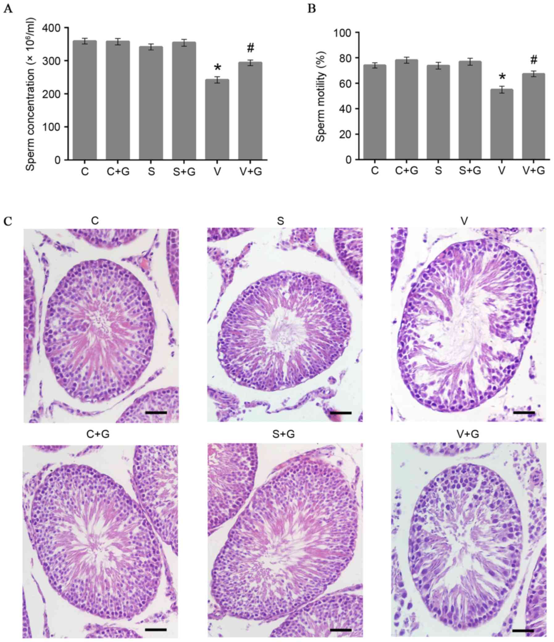 | Figure 1.Morphological and functional
evaluations of the testis. (A) Statistical results of testicular
sperm concentration. *n=9, P=0.000 vs. control group;
#n=9, P=0.013 vs. varicocele group. (B) Statistical
results of testicular sperm motility. *n=9, P=0.001 vs. control
group; #n=9, P=0.033 vs. varicocele group. (C)
Histological sections of the left testis with hematoxylin and eosin
staining exhibit fewer and looser testicular epithelial cells with
structural damage in the varicocele group compared with the control
group, whereas GSPE can constrain the varicocele-triggered
morphological changes. Scale bar, 50 µm. C, control group; C+G,
control + GSPE group; S, sham group; S+G, sham + GSPE group; V,
varicocele group; V+G, varicocele + GSPE group; GSPE, grape seed
proanthocyanidin extract. |
H&E staining demonstrated that varicocele caused
damaging effects to the compared with the control and sham groups
(Fig. 1C). The number testicular
epithelial cells in the varicocele group were observably fewer and
were looser than those in the control and sham groups. Abnormal
spermatogenic cell types in damaged seminiferous tubules were also
observed in the testicular sections of the varicocele group.
However, the varicocele + GSPE group exhibited limited histological
changes, which indicated that GSPE may alleviate the damage
triggered by varicocele.
GSPE attenuated the oxidative stress
in varicocele testes
SOD, GSH-Px and MDA activity were measured to
evaluate the oxidative stress occurring in the left rat testis. The
concentrations of SOD and GSH-Px were significantly lower in the
varicocele group compared with the control or sham groups
(P<0.05). By contrast, administration of GSPE markedly restored
SOD and GSH-Px concentrations (P=0.018 and P=0.020, respectively;
Fig. 2B and C). Compared with the
control and sham groups, the level of MDA was much higher due to
the effect of varicocele (P=0.000). GSPE can decrease the level of
MDA in the testes of varicocele rats significantly (P=0.007;
Fig. 2A). In addition, according
to the results of the control + GSPE and sham + GSPE groups, no
evident effects of GSPE were observed on the levels of MDA, SOD or
GSH-Px (P>0.05) in the control or sham groups.
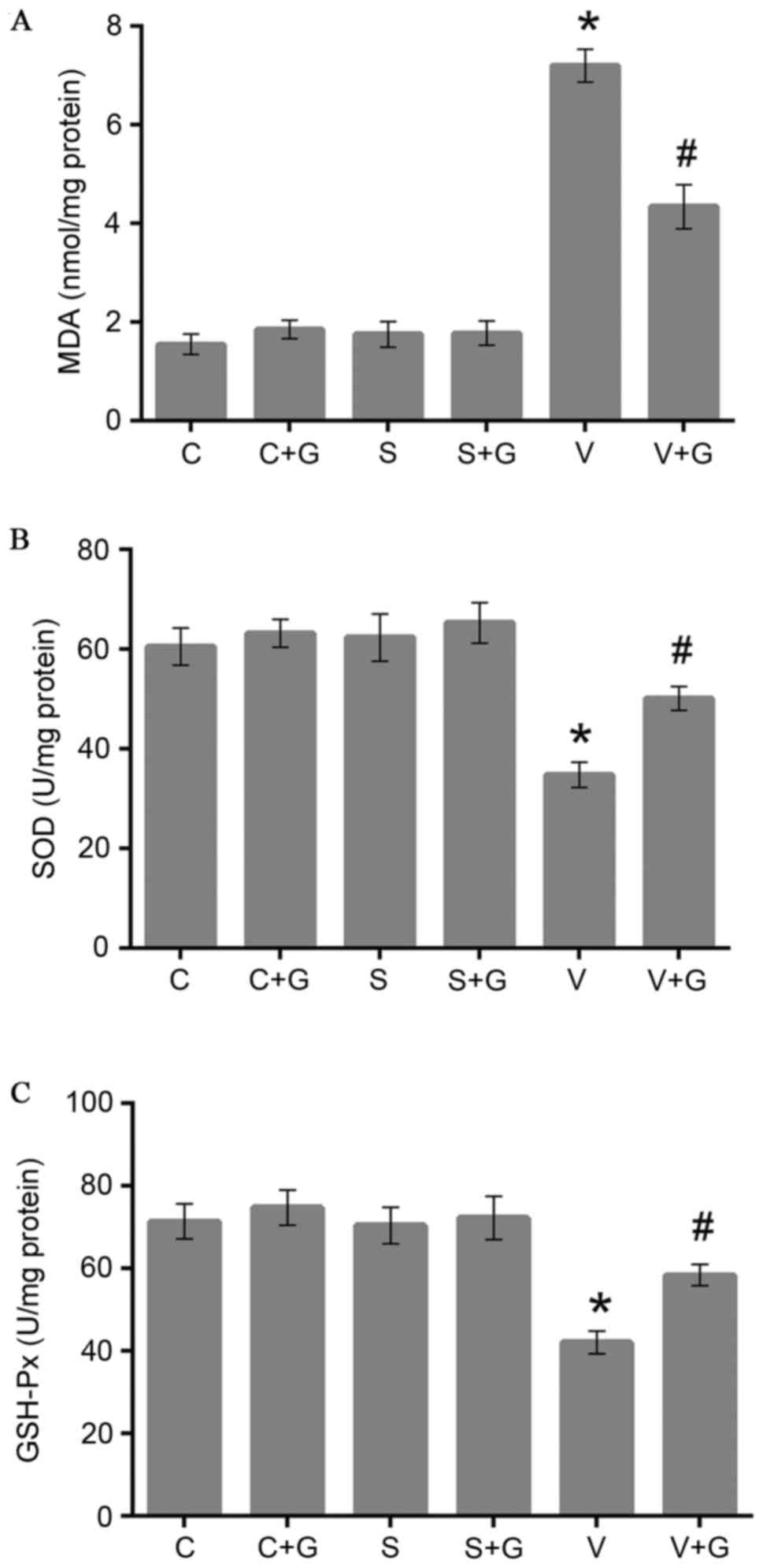 | Figure 2.Effect of varicocele and GSPE on
oxidative stress in the testis. (A) MDA level in the testis. *n=6,
P=0.000 vs. control group; #n=6, P=0.007 vs. varicocele
group. (B) SOD activity in the testis. *n=6, P=0.004 vs. control
group; #n=6, P=0.018 vs. varicocele group. (C) GSH-Px
activity in the testis. *n=6, P=0.004 vs. control group;
#n=6, P=0.020 vs. varicocele group. GSPE, grape seed
proanthocyanidin extract; MDA, malondialdehyde; C, control group;
C+G, control + GSPE group; S, sham group; S+G, sham + GSPE group;
V, varicocele group; V+G, varicocele + GSPE group; SOD, superoxide
dismutase; GSH-Px, glutathione peroxidase. |
Protective effects of GSPE on
apoptosis in varicocele testis
Several studies (8,22)
have demonstrated that varicocele can increase the apoptosis level
in the testis due to oxidative stress. The present study examined
whether GSPE had any protective effects on the apoptosis level of
varicocele testes by TUNEL staining. The number of TUNEL-positive
cells in the varicocele group was markedly increased compared with
the control group (P=0.004), whereas GSPE significantly decreased
TUNEL-positive cell numbers in the varicocele rats (P=0.026;
Fig. 3A and B). In addition,
western blotting demonstrated that the expression of cleaved
caspase-3 in the testis of the varicocele group increased
significantly compared with the control group (P=0.006), whereas
treatment of varicocele with GSPE decreased the expression of
cleaved caspase-3 significantly (P=0.028; Fig. 3C).
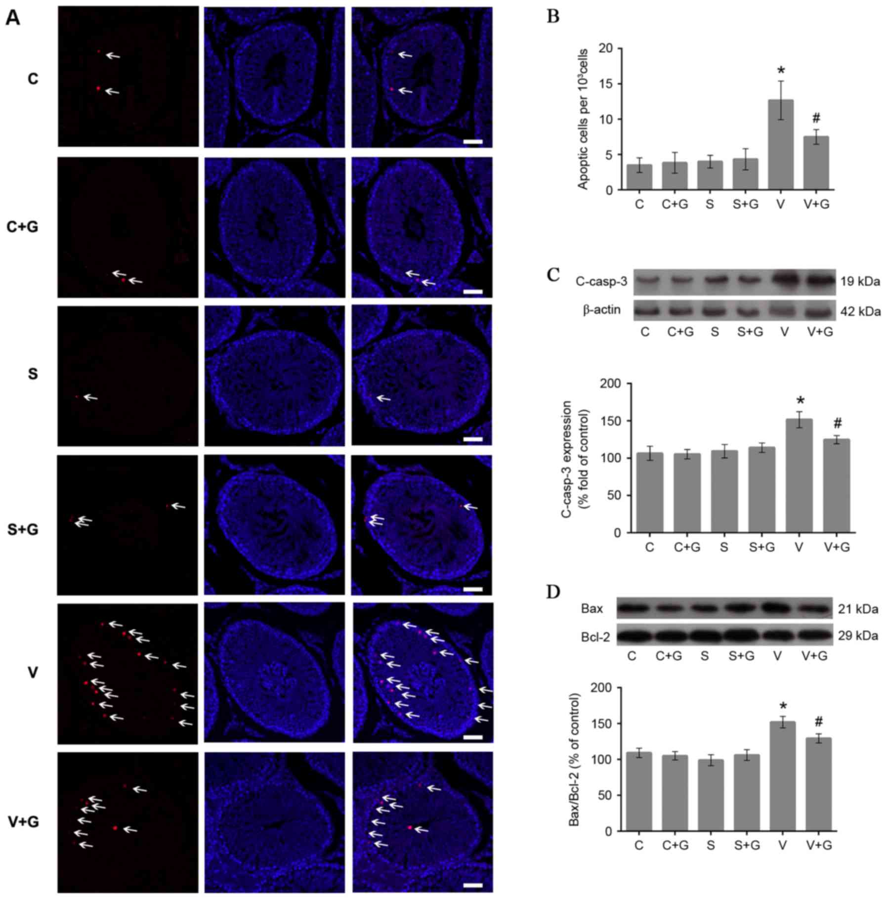 | Figure 3.Effect of GSPE on apoptosis in the
testis. (A) TUNEL staining showing cell apoptosis in the testis.
The arrows indicate TUNEL-positive cells (red), the nuclei stained
with DAPI (blue). Scale bar, 50 µm. (B) Statistical results of
TUNEL staining, *n=6, P=0.004 vs. control group; #n=6,
P=0.026 vs. varicocele group. (C) The expression of c-casp-3
protein in the testis. *n=5, P=0.006 vs. the control group;
#n=5, P=0.028 vs. the varicocele group. (D) The Bax to
Bcl-2 protein expression ratio in the testis. *n=5, P=0.000 vs.
control group; #n=5, P=0.015 vs. varicocele group. GSPE,
grape seed proanthocyanidin extract; TUNEL, terminal
deoxynucleotidyl transferase dUTP nick end labeling; C, control
group; C+G, control + GSPE group; S, sham group; S+G, sham + GSPE
group; V, varicocele group; V+G, varicocele + GSPE group; Bax,
Bcl-2-like protein 4; Bcl-2, B-cell lymphoma 2; c-casp-3, cleaved
caspase-3. |
The mitochondrial apoptotic pathway has been
reported (23) to be involved in
varicocele testes, therefore whether GSPE can avert the activation
of the mitochondrial apoptotic pathway in varicocele testes was
investigated. Western blotting demonstrated that the Bax to Bcl-2
expression ratio was significantly higher in the varicocele group
compared with the control group (P=0.000). GSPE significantly
alleviated the increase of the Bax to Bcl-2 expression ratio in the
testes of the varicocele rats (P=0.015; Fig. 3D).
GSPE exerted protective effects
through activation of the Nrf2 pathway
The transcription factor Nrf2 has an important role
in regulating the cellular antioxidant system. The effect of GSPE
on the expression of Nrf2 was investigated and immunohistochemical
results demonstrated that varicocele can slightly increase the
expression of Nrf2 in interstitial cells compared with the control
group, while administration of GSPE markedly increased the
expression of Nrf2 in interstitial cells in the three groups that
were administered GSPE (Fig. 4A).
The expression of Nrf2 was predominantly located in the nucleus of
the interstitial cells. Western blotting demonstrated that the
varicocele group expressed a significantly higher level of Nrf2
than the control group (P=0.044; Fig.
4B), and the varicocele + GSPE group demonstrated a
significantly higher level of Nrf2 expression compared with the
varicocele group (P=0.006; Fig.
4B). The groups administered with GSPE all demonstrated
significantly higher expression of Nrf2 compared with the control
group (P<0.01 in three groups; Fig.
4B).
 | Figure 4.Effect of GSPE on the Nrf2 pathway in
the testis. (A) Immunohistochemical staining results for Nrf2 in
the testis. The arrow shows Nrf2 positive staining predominantly
located in the nucleus of the interstitial cells. Scale bar: 50 µm.
(B) The protein expression of total Nrf2 in the testis. *n=5, C+G
(P=0.004), S+G (P=0.002), V (P=0.044) vs. control group;
#n=5, P=0.006 vs. varicocele group. (C) The protein
expression of n-Nrf2 in the testis. *n=5, C+G (P=0.0024), S+G
(P<0.001), V (P=0.014) vs. control group; #n=5,
P=0.034 vs. varicocele group. (D) The protein expression of HO-1 in
the testis. *n=5, C+G (P<0.001), S+G (P<0.001), V (P=0.039)
vs. control group; #n=5, P=0.022 vs. varicocele group.
GSPE, grape seed proanthocyanidin extract; C, control group; C+G,
control + GSPE group; S, sham group; S+G, sham + GSPE group; V,
varicocele group; V+G, varicocele + GSPE group; n, nuclear; Nrf2,
nuclear factor (erythroid-derived 2)-like 2; H2A.X, H2A histone
family member X; HO-1, heme oxygenase-1. |
Previous studies (24,25)
have demonstrated that Nrf2, as a transcription factor, can only
exert its function by entering the nucleus. Therefore, the nuclear
expression of Nrf2 was measured. As demonstrated in Fig. 4C, the varicocele group expressed a
higher level of Nrf2 than the control group (P=0.014). The
varicocele + GSPE group expressed a significantly increased level
of Nrf2 than the varicocele group (P=0.034). The groups treated
with GSPE all demonstrated a higher level of nuclear Nrf2
expression than the control group (P<0.01).
The expression of HO-1, a gene downstream of Nrf2,
was next measured to evaluate the antioxidative effect of Nrf2. As
demonstrated in Fig. 4D, the
varicocele + GSPE group demonstrated higher expression of HO-1 than
the varicocele group (P=0.022). However, no significant differences
were demonstrated when comparing the expression of total Nrf2,
nuclear Nrf2 or HO-1 between the three groups that were given GSPE
(Fig. 4).
Discussion
Varicocele is an abnormal tortuosity of the
pampiniform plexus of the spermatic cord, which has incidence rates
of 4.4–22.6% in the male population, particularly in males with
primary or secondary infertility. Many associated factors are
considered relevant in the pathophysiology of varicocele. By using
a well-established rat model of varicocele (21), the injured spermatogenic function
and pathologic alterations of the testes in the varicocele group
was demonstrated. In addition, the sperm concentration and motility
(based on computer assisted semen analysis) and the histological
results of H&E staining were clearly ameliorated following 4
weeks of GSPE administration, which illustrated that GSPE can
attenuate the functional and morphologic damage to rat testes and
epididymis caused by varicocele.
GSPE, chemically a mixture of pycnogenol and
flavonoid including oligomeric proanthocyanidins, is a potent
antioxidant (19). The
biologically active compounds possess protective effects against
oxidative stress induced by free radicals and ROS. Studies
(19,26,27)
have demonstrated that the administration of GSPE can ameliorate
oxidative damage from various causes. First, in accordance with
previous findings (28), the
present study demonstrated that varicocele can result in
deterioration of oxidative stress markers. Additionally, in the
varicocele + GSPE group, the testicular activity of GSH-Px and SOD
were higher than those in the varicocele group. MDA concentrations
were lower in the varicocele + GSPE group. These results proved
that GSPE can partially relieve the oxidative stress in testes
induced by varicocele.
Apoptosis is one of the dominant mechanisms of
testicular dysfunction resulting from varicocele (29–32).
Varicocele has been reported (33–35)
to increase sperm DNA damage and pachytene spermatocyte programmed
death. The TUNEL assay results also identified an increased
apoptosis level in the spermatogonia of the varicocele group
compared with the control group. This finding was compatible with
the protein expression of Bcl-2, Bax and cleaved caspase-3
(Fig. 3C and D). Conversely, GSPE
has been demonstrated (20,36,37)
to protect cells from the apoptosis induced by oxidative stress.
The number of apoptotic nuclei per 103 cells revealed
less cell DNA damage in the varicocele + GSPE group compared with
the varicocele group. Furthermore, the ratio of Bax to Bcl-2 and
activated caspase-3 expression fell in the varicocele + GSPE group.
Caspase-3 is considered to be a major executioner protease and the
ratio of Bax to Bcl-2 is also a good indicator of apoptosis levels
(38). Consequently, it was
hypothesized that the administration of GSPE for 4 weeks was able
to alleviate the apoptosis in the testicular tissues of rats with
varicocele.
However, the exact mechanism of the elevated
oxidative stress and apoptosis in male reproductive organs has yet
to be fully elucidated. Nrf2 is a transcription factor that
improves the expression of antioxidant proteins and can function
against oxidative damage (39).
Nrf2 is known to be associated with the testicular apoptosis
induced by diabetes (40).
Similarly, the results of the present study revealed higher
expression of Nrf2 and its downstream gene HO-1 in the varicocele
group compared with the control group due to the oxidative
stimulation caused by varicocele (Fig.
4). Several studies (18,41)
have demonstrated that GSPE can activate the Nrf2 pathway to exert
its antioxidative function. However, few reports have focused on
the correlation between GSPE and the Nrf2 signaling pathway in the
testis. The present study revealed that Nrf2 and HO-1 were
significantly upregulated due to the administration of GSPE
compared to the varicocele and control groups. In addition, the
immunohistochemical staining results for Nrf2 were in agreement
with the western blot analysis. Thus, GSPE may exert a sustained
effect on Nrf2 expression, which can further attenuate the
oxidative stress and apoptosis triggered by varicocele in rat
testes. However, the results demonstrated that the control + GSPE,
sham + GSPE and varicocele + GSPE groups all had significantly
higher expression of Nrf2 and its downstream gene HO-1, which
suggests that GSPE activates the Nrf2 pathway in normal and
varicocele rat testis.
Admittedly, there may also be several limitations in
the present study. The rat model of varicocele cannot be used to
interpret the equivalent situation in human patients due to the
anatomical structure differences of the left spermatic vein between
rats and humans and different standards for semen analysis. In
addition, the present study did not adopt different concentration
gradients or time courses of GSPE treatment. It is possible that
employing a different dose or period of gavage would reinforce the
functions of GSPE. Future studies may compare the different effects
of GSPE with internal spermatic vein ligation, which is the main
surgical treatment for varicocele in clinics (42).
In summary, the data from the present study
indicated that varicocele leads to oxidative and mitochondrial
apoptotic injuryand GSPE upreg ulates Nrf2 and its downstream genes
to mitigate such damage and resume normal spermatogenic organ
function. Thus, GSPE appears to be an effective protective agent
that may be used in the treatment of male varicocele in the
future.
GSPE, which consists of a group of polyphenolic
bioflavonoids, possesses potent antioxidative activity. Oxidative
stress is one of the major pathogenic mechanisms in varicocele, and
can cause morphological and functional damage to the testis and
epididymis. The present study confirmed that GSPE can activate the
Nrf2-antioxidant system to attenuate varicocele induced testicular
injury caused by oxidative stress.
Acknowledgements
This work was supported by the National Natural
Science Foundation of China (grant nos. 81470987 and 81670687 to B.
Shi), the Tai Shan Scholar Foundation to B. Shi and the Natural
Science Foundation of Shandong Province (grant no. ZR2014HQ062 to
Y. Zhu). The authors thank Dr Yongxin Zou for providing associated
experimental equipment and reagents.
References
|
1
|
Pastuszak AW and Wang R: Varicocele and
testicular function. Asian J Androl. 17:659–667. 2015. View Article : Google Scholar :
|
|
2
|
Liang M, Wen J, Dong Q, Zhao LG and Shi
BK: Testicular hypofunction caused by activating p53 expression
induced by reactive oxygen species in varicocele rats. Andrologia.
47:1175–1182. 2015. View Article : Google Scholar
|
|
3
|
Razi M and Malekinejad H:
Varicocele-induced infertility in animal models. Int J Fertil
Steril. 9:141–149. 2015.
|
|
4
|
Shiraishi K, Takihara H and Matsuyama H:
Effects of grade 1 varicocele detected in the pediatric age-group
on testicular development. J Pediatr Surg. 44:1995–1998. 2009.
View Article : Google Scholar
|
|
5
|
Sohrabipour S, Jafari A, Kamalinejad M,
Sarrafnejd A, Shahrestany T and Sadeghipour HR: The role of
flaxseed and vitamin E on oxidative stress in prepubertal rats with
experimental varicocele: An experimental study. Iran J Reprod Med.
11:459–466. 2013.
|
|
6
|
Razi M, Sadrkhanloo RA, Malekinejad H and
Sarrafzadeh-Rezaei F: Testicular biohistochemical alterations
following experimental varicocele in rats. Iran J Reprod Med.
10:209–218. 2012.
|
|
7
|
Altunoluk B, Efe E, Kurutas EB, Gul AB,
Atalay F and Eren M: Elevation of both reactive oxygen species and
antioxidant enzymes in vein tissue of infertile men with
varicocele. Urol Int. 88:102–106. 2012. View Article : Google Scholar
|
|
8
|
Walczak-Jedrzejowska R, Wolski JK and
Slowikowska-Hilczer J: The role of oxidative stress and
antioxidants in male fertility. Cent European J Urol. 66:60–67.
2013. View Article : Google Scholar :
|
|
9
|
Hayes JD and McLellan LI: Glutathione and
glutathione-dependent enzymes represent a co-ordinately regulated
defence against oxidative stress. Free Radic Res. 31:273–300. 1999.
View Article : Google Scholar
|
|
10
|
Hong CC, Ambrosone CB, Ahn J, Choi JY,
McCullough ML, Stevens VL, Rodriguez C, Thun MJ and Calle EE:
Genetic variability in iron-related oxidative stress pathways
(Nrf2, NQ01, NOS3, and HO-1), iron intake, and risk of
postmenopausal breast cancer. Cancer Epidemiol Biomarkers Prev.
16:1784–1794. 2007. View Article : Google Scholar
|
|
11
|
Li Y, Cao Y, Wang F, Pu S, Zhang Y and Li
C: Tert-butylhydroquinone attenuates scrotal heat-induced damage by
regulating Nrf2-antioxidant system in the mouse testis. Gen Comp
Endocrinol. 208:12–20. 2014. View Article : Google Scholar
|
|
12
|
Hayes JD and McMahon M: NRF2 and KEAP1
mutations: Permanent activation of an adaptive response in cancer.
Trends Biochem Sci. 34:176–188. 2009. View Article : Google Scholar
|
|
13
|
Wang R, Paul VJ and Luesch H: Seaweed
extracts and unsaturated fatty acid constituents from the green
alga Ulva lactuca as activators of the cytoprotective Nrf2-ARE
pathway. Free Radic Biol Med. 57:141–153. 2013. View Article : Google Scholar :
|
|
14
|
Kobayashi A, Kang MI, Okawa H, Ohtsuji M,
Zenke Y, Chiba T, Igarashi K and Yamamoto M: Oxidative stress
sensor Keap1 functions as an adaptor for Cul3-based E3 ligase to
regulate proteasomal degradation of Nrf2. Mol Cell Biol.
24:7130–7139. 2004. View Article : Google Scholar :
|
|
15
|
McMahon M, Lamont DJ, Beattie KA and Hayes
JD: Keap1 perceives stress via three sensors for the endogenous
signaling molecules nitric oxide, zinc, and alkenals. Proc Natl
Acad Sci USA. 107:18838–18843. 2010. View Article : Google Scholar :
|
|
16
|
Zhang Z, Zheng L, Zhao Z, Shi J, Wang X
and Huang J: Grape seed proanthocyanidins inhibit
H2O2-induced osteoblastic MC3T3-E1 cell
apoptosis via ameliorating H2O2-induced
mitochondrial dysfunction. J Toxicol Sci. 39:803–813. 2014.
View Article : Google Scholar
|
|
17
|
Bagchi D, Garg A, Krohn RL, Bagchi M, Tran
MX and Stohs SJ: Oxygen free radical scavenging abilities of
vitamins C and E, and a grape seed proanthocyanidin extract in
vitro. Res Commun Mol Pathol Pharmacol. 95:179–189. 1997.
|
|
18
|
Chen S, Zhu Y, Liu Z, Gao Z, Li B, Zhang
D, Zhang Z, Jiang X, Liu Z, Meng L, et al: Grape seed
proanthocyanidin extract ameliorates diabetic bladder dysfunction
via the activation of the Nrf2 pathway. PLoS One. 10:e01264572015.
View Article : Google Scholar :
|
|
19
|
Li SG, Ding YS, Niu Q, Xu SZ, Pang LJ, Ma
RL, Jing MX, Feng GL, Liu JM and Guo SX: Grape seed
proanthocyanidin extract alleviates arsenic-induced oxidative
reproductive toxicity in male mice. Biomed Environ Sci. 28:272–280.
2015.
|
|
20
|
Su L, Deng Y, Zhang Y, Li C, Zhang R, Sun
Y, Zhang K, Li J and Yao S: Protective effects of grape seed
procyanidin extract against nickel sulfate-induced apoptosis and
oxidative stress in rat testes. Toxicol Mech Methods. 21:487–494.
2011. View Article : Google Scholar
|
|
21
|
Turner TT: The study of varicocele through
the use of animal models. Hum Reprod Update. 7:78–84. 2001.
View Article : Google Scholar
|
|
22
|
Cam K, Simsek F, Yuksel M, Turkeri L,
Haklar G, Yalcin S and Akdas A: The role of reactive oxygen species
and apoptosis in the pathogenesis of varicocele in a rat model and
efficiency of vitamin E treatment. Int J Androl. 27:228–233. 2004.
View Article : Google Scholar
|
|
23
|
Mostafa T, Rashed L, Nabil N and Amin R:
Seminal BAX and BCL2 gene and protein expressions in infertile men
with varicocele. Urology. 84:590–595. 2014. View Article : Google Scholar
|
|
24
|
Alfadda AA and Sallam RM: Reactive oxygen
species in health and disease. J Biomed Biotechnol.
2012:9364862012. View Article : Google Scholar :
|
|
25
|
Bryan HK, Olayanju A, Goldring CE and Park
BK: The Nrf2 cell defence pathway: Keap1-dependent and -independent
mechanisms of regulation. Biochem Pharmacol. 85:705–717. 2013.
View Article : Google Scholar
|
|
26
|
Nazima B, Manoharan V and Miltonprabu S:
Oxidative stress induced by cadmium in the plasma, erythrocytes and
lymphocytes of rats: Attenuation by grape seed proanthocyanidins.
Hum Exp Toxicol. 35:428–447. 2016. View Article : Google Scholar
|
|
27
|
Lin KN, Lin ML and Wei EQ: Protective
effect of grape seed proanthocyanidin on cultured RGC-5 cells
against CoCl2-induced hypoxic injury. Zhejiang Da Xue
Xue Bao Yi Xue Ban. 44:24–29. 2015.(In Chinese).
|
|
28
|
Khosravanian N, Razi M, Farokhi F and
Khosravanian H: Testosterone and vitamin E administration
up-regulated varicocele-reduced Hsp70-2 protein expression and
ameliorated biochemical alterations. J Assist Reprod Genet.
31:341–354. 2014. View Article : Google Scholar :
|
|
29
|
Lee JD, Lee TH, Cheng WH and Jeng SY:
Involved intrinsic apoptotic pathway of testicular tissues in
varicocele-induced rats. World J Urol. 27:527–532. 2009. View Article : Google Scholar
|
|
30
|
Barqawi A, Caruso A and Meacham RB:
Experimental varicocele induces testicular germ cell apoptosis in
the rat. J Urol. 171:501–503. 2004. View Article : Google Scholar
|
|
31
|
Zhang K, Wang Z, Wang H, Fu Q, Zhang H and
Cao Q: Hypoxia-induced apoptosis and mechanism of epididymal
dysfunction in rats with left-side varicocele. Andrologia.
48:318–324. 2016. View Article : Google Scholar
|
|
32
|
Du J and Dianjun G: Cell cycle specificity
of spermatogenic cell apoptosis in rats with experimental
varicocele. Clin Lab. 59:851–859. 2013.
|
|
33
|
Naughton CK, Nangia AK and Agarwal A:
Pathophysiology of varicoceles in male infertility. Hum Reprod
Update. 7:473–481. 2001. View Article : Google Scholar
|
|
34
|
Ricci JE, Muñoz-Pinedo C, Fitzgerald P,
Bailly-Maitre B, Perkins GA, Yadava N, Scheffler IE, Ellisman MH
and Green DR: Disruption of mitochondrial function during apoptosis
is mediated by caspase cleavage of the p75 subunit of complex I of
the electron transport chain. Cell. 117:773–786. 2004. View Article : Google Scholar
|
|
35
|
Sharma RK, Pasqualotto FF, Nelson DR,
Thomas AJ Jr and Agarwal A: The reactive oxygen species-total
antioxidant capacity score is a new measure of oxidative stress to
predict male infertility. Hum Reprod. 14:2801–2807. 1999.
View Article : Google Scholar
|
|
36
|
Sato M, Bagchi D, Tosaki A and Das DK:
Grape seed proanthocyanidin reduces cardiomyocyte apoptosis by
inhibiting ischemia/reperfusion-induced activation of JNK-1 and
C-JUN. Free Radic Biol Med. 31:729–737. 2001. View Article : Google Scholar
|
|
37
|
Song Q, Shi Z, Bi W, Liu R, Zhang C, Wang
K and Dang X: Beneficial effect of grape seed proanthocyanidin
extract in rabbits with steroid-induced osteonecrosis via
protecting against oxidative stress and apoptosis. J Orthop Sci.
20:196–204. 2015. View Article : Google Scholar
|
|
38
|
Said TM, Paasch U, Glander HJ and Agarwal
A: Role of caspases in male infertility. Hum Reprod Update.
10:39–51. 2004. View Article : Google Scholar
|
|
39
|
Palanisamy K, Krishnaswamy R, Paramasivan
P, Chih-Yang H and Vishwanadha VP: Eicosapentaenoic acid prevents
TCDD-induced oxidative stress and inflammatory response by
modulating MAP kinases and redox-sensitive transcription factors.
Br J Pharmacol. 172:4726–4740. 2015. View Article : Google Scholar :
|
|
40
|
Jiang X, Bai Y, Zhang Z, Xin Y and Cai L:
Protection by sulforaphane from type 1 diabetes-induced testicular
apoptosis is associated with the up-regulation of Nrf2 expression
and function. Toxicol Appl Pharmacol. 279:198–210. 2014. View Article : Google Scholar
|
|
41
|
Güçlü A, Yonguç N, Dodurga Y, Gündoğdu G,
Güçlü Z, Yonguç T, Adıgüzel E and Turkmen K: The effects of grape
seed on apoptosis-related gene expression and oxidative stress in
streptozotocin-induced diabetic rats. Ren Fail. 37:192–197. 2015.
View Article : Google Scholar
|
|
42
|
Akdemir S, Gurocak S, Konac E, Ure I, Onen
HI, Gonul II, Sozen S and Menevse A: Different surgical techniques
and L-carnitine supplementation in an experimental varicocele
model. Andrologia. 46:910–916. 2014. View Article : Google Scholar
|


