Introduction
Vascular endothelial dysfunction is generally caused
by pressure or volume overload, such as hypertension, myocardial
infarction, valve stenosis or regurgitation (1), as well as the increased secretion and
release of inflammatory cytokines, such as interleukin-lβ (IL-1β),
IL-6, tumor necrosis factor-α (TNF-α), monocyte chemoattractant
protein-1 (MCP-1), and angiotensin II (Ang II). Vascular
endothelial dysfunction induces or exacerbates cardiac disease,
which will eventually result in heart failure (HF) (2–4).
Although previous studies have implicated nuclear transcription
factor-κB (NF-κB) (5), p38
mitogen-activated protein kinase (MAPK) (6) and phosphatidylinositol 3-hydroxy
kinase (PI3K) (7) as potential
targets in vascular endothelial dysfunction, the precise mechanisms
are still incompletely understood. A human umbilical vein
endothelial cells (HUVECs) have been widely used to establish
experimental models for vascular inflammation in vitro, and
the lipopolysaccharide (LPS)-induced HUVEC injury is commonly used
as an in vitro model (4),
we used LPS-stimulated HUVECs as the model for our study.
Baicalein (5,6,7-trihydroxy-2-phenyl-4H-1-benzop
yran-4-one, BAI) is a flavonoid extracted from the root of
Scutellaria baicalensis (3). Many evidences have suggested that BAI
possesses many pharmacological activities. It is reported that BAI
inhibits agonist-induced and tumor cell-induced platelet
aggregation (8), and could
suppress tumor growth and metastasis (9). BAI also combines synergistically with
cefotaxime to prevent Klebsiella pneumoniae through the
inhibition of CTX-M-1 gene expression (10). Tsai et al (11) proved that BAI protects against
oxLDL-induced oxidative stress and inflammation via the modulation
of AMPK-α. BAI reduced angiogenesis in the inflammatory
microenvironment through the inhibition of the expression of AP-1
(12); and according to Zong et
al (13), BAI exerts
protection against cardiac hypertrophy and fibrosis through the
suppression of mitogen-activated protein kinase (MEK)/extracellular
signal-regulated kinase 1/2 (ERK1/2) signaling.
In the present study, we found that BAI protects
HUVECs from LPS-induced injury associated with the Toll-like
receptor 4 (TLR4), phosphorylation of TGF-β-activated kinase 1
(p-TAK1), phosphorylation of TANK-binding kinase 1 (p-TBK1) and
phosphorylation of nuclear transcription factor-κB (NF-κB) p65
(p-p65) signaling, which inhibits inflammation and apoptosis in
HUVECs. Further, using MRT67307, a IKKε and TBK1 inhibitor, we
verified that BAI targets the expression of p-TBK1 to suppress
TLR4/NF-κB signaling. These results suggested that BAI could
protect against vascular endothelial injury and exert potential
therapeutic effects for the prevention of HF.
Materials and methods
Cell culture
HUVECs (8000; ScienCell Research Laboratories,
Carlsbad, CA, USA) were grown in Dulbecco's modified Eagle's medium
(DMEM/F-12) supplemented with 10% fetal bovine serum (FBS) (both
from Gibco, Grand Island, NY, USA) and endothelial cell growth
supplement (ECGS, 1052; ScienCell Research Laboratories) under
standard conditions (5% CO2; temperature, 37°C). The
cells were passaged when 70–80% confluent and starved in serum-free
DMEM for 12 h before LPS stimulation. LPS (L2630) and BAI (≥98%,
CAS 491-67-8) were purchased from Sigma-Aldrich; Merck KGaA
(Darmstadt, Germany). MRT67307 (S7948) was bought from Selleck
Chemicals (Houston, TX, USA).
Determination of cytotoxicity and
efficacy
To determine the cytotoxicity, the cell viability of
HUVECs was tested after treatment with different concentrations of
BAI (0, 3.125, 6.25, 12.5, 25, 50, 100, 200, or 400 µM) for a
specific period of time. The same process was repeated in HUVECs
stimulated by LPS (10 µM) and simultaneously treated by different
concentrations of BAI (0, 3.125, 6.25, 12.5, 25, or 50 µM) to
determine the efficacy of BAI. Both the cytotoxicity and efficacy
of BAI were determined by the cell counting kit assay (Enhanced
Cell Counting Kit-8, C0042; Beyotime Institute of Biotechnology,
Haimen, China). The absorbance at 450 nm was measured using the
Bio-Tek Synergy HT Multi-Detection Microplate Reader (BioTek
Instruments, Inc., Winooski, VT, USA).
Detection of apoptosis through TUNEL
assay
The HUVECs were divided into three groups: i)
Control: Cells treated with neither LPS nor BAI; ii) LPS: Cells
treated with LPS (10 µM); iii) LPS + BAI: Cells treated with LPS
(10 µM) and BAI (6.25 µM). Apoptosis was identified through the DNA
fragmentation in the terminal deoxynucleotidyl transferase dUTP
nick end labeling (TUNEL) assay (14). The TUNEL assay works through the
incorporation of modified dUTPs at the 3′-OH ends of fragmented DNA
by the enzyme terminal deoxynucleotidyl transferase (TdT), which
subsequently tags cells with damaged DNA (15). After the TUNEL assay treatment,
damaged DNA is stained green, whereas intact DNA is not stained.
HUVECs were detected using the Apo-Direct TUNEL Assay kit (EMD
Millipore, Billerica, MA, USA). All steps were conducted in strict
accordance with the specifications and photographed using a
fluorescent microscope.
Flow cytometric detection of HUVEC
apoptosis
The cells were resuspended, rinsed and processed
with Annexin V-FITC/PI Apoptosis Detection kit (KGA108; Nanjing
KeyGen Biotech Co., Ltd., Nanjing, China). Annexin V was considered
as a sensitive index of early stage apoptotic cells, while PI was
the indicator of advanced stage apopotic cells. Briefly, the cells
were re-suspended in 500 µl binding buffers and stained
consecutively by 5 µl Annexin V-FITC and 5 µl PI. We then analyzed
HUVECs by flow cytometry (FACSCalibur; BD Biosciences, Franklin
Lakes, NJ, USA) to differentiate apoptotic cells from necrotic
cells. Both HUVECs stained Annexin+/PI− and
Annexin+/PI+ in the flow cytometric analyses
were considered as apoptotic cells.
Reverse transcription-quantitative
polymerase chain reaction (RT-qPCR)
The mRNA expression levels of IL-1β, IL-6, TNF-α,
MCP-1 and GAPDH (glyceraldehyde-3-phosphate dehydrogenase) were
analyzed by RT-qPCR. Total RNA was extracted from HUVECs using
TRIzol reagent in accordance with the manufacturer's instructions
(Invitrogen Life Technologies, Carlsbad, CA, USA), and the purity
of the RNA was evaluated based on the OD260/OD280 ratios detected
using the SmartSpec Plus Spectrophotometer (Bio-Rad Laboratories,
Inc., Hercules, CA, USA). The mRNA was reverse transcribed into
cDNA using oligo(dT) primers and the Transcriptor First Strand cDNA
Synthesis kit (Roche, Basel, Switzerland). The following primer
sequences were used: IL-1β forward, 5′-ATTTGAGTCTGCCCAGTTCCC−3′ and
reverse, 5′-CCAGGAAGACGGGCATGTTT-3′; IL-6 forward,
5′-CAATGAGGAGACTTGCCTGG-3′ and reverse, 5′-GGCATTTGTGGTTGGGTCAG-3′;
TNF-α forward, 5′-TCTGGGCAGGTCTACTTTGG-3′ and reverse,
5′-GGTTGAGGGTGTCTGAAGGA-3′; MCP-1 forward,
5′-AATCAATGCCCCAGTCACCT-3′ and reverse,
5′-CTTCTTTGGGACACTTGCTGC−3′. The transcripts were mixed with
LightCycler 480 SYBR-Green I Master Mix and quantified in the
LightCycler® 480 Real-Time Quantitative PCR System (both
from Roche) using the double standard curve method. The results
were normalized to the expression of GAPDH.
Inflammatory cytokines expression
measured by enzyme-linked immunosorbent assay (ELISA)
We detected the expressions of IL-1β, IL-6, and
TNF-α in HUVECs with Human Quantikine ELISA kits (R&D Systems,
Inc., Minneapolis, MN, USA). HUVECs were divided into four groups:
i) Control: Cells treated with neither LPS nor BAI; ii) LPS: Cells
stimulated by LPS (10 µM); iii) LPS + BAI: Cells stimulated by LPS
(10 µM) and treated with BAI (6.25 µM); and iv) BAI: Cells treated
with BAI (6.25 µM). We collected the culture supernatant and
measured the inflammatory cytokine expression in strict accordance
with the manufacturer's specifications and read the absorbance of
each well at 450 nm using a microplate reader; 550 nm was used as
the reference wavelength. The values of the samples were calculated
through a comparison of the absorbance values with prediluted
inflammatory cytokines standards (R&D Systems, Inc.).
Immunofluorescent staining of p65
The HUVECs were fixed, contained, blocked, and
incubated with a dilution of the p65 antibody, and then incubated
with green fluorescent secondary antibody in the dark. The nuclei
were stained with DAPI. The cells were photographed using a
fluorescent microscope; green fluorescence represents p65, whereas
the nuclei are shown in blue. Overlapping areas of green and blue
indicate the occurrence of the nuclei translocation of NF-κB.
Western blotting
HUVECs were lysed with lysis buffer RIPA (G2002;
Wuhan Goodbio Biotechnology Co., Ltd., Wuhan, China). Protein
quantification was conducted using the Pierce BCA protein assay kit
(Thermo Fisher Scientific, Inc., Waltham, MA, USA). The following
primary antibodies were used: Cleaved-caspase-3 (9664), caspase-3
(9665), B-cell lymphoma 2-associated X protein (Bax; 2722), and
Bcl-2 (2870) (all from Cell Signaling Technology, Inc., Danvers,
MA, USA), TLR4 (sc-10741; Santa Cruz Biotechnology, Inc., Dallas,
TX, USA), p-TAK1 (4508), p-TBK1 (5483), p-p65 (3033), TAK1 (5206),
TBK1 (3013), p65 (8242), and GAPDH (2118) (all from Cell Signaling
Technology, Inc.). The corresponding peroxidase-conjugated
secondary antibodies used were IRdye 800CW-conjugated goat
anti-mouse immunoglobulin G (926–32210) and IRDye 800CW-conjugated
goat anti-rabbit immunoglobulin G (926–32211) (both from LI-COR
Biosciences, Lincoln, NE, USA). The blots were scanned using a
two-color infrared imaging system (Odyssey; LI-COR Biosciences) and
the expression of target proteins were normalized to the expression
of GAPDH.
Statistical analysis
All statistical analyses were computed using the
SPSS software (version 17.0; SPSS, Inc., Chicago, IL, USA) and the
results are presented as the mean ± SD. For measurement data, a
one-sample Kolmogorov-Smirnov test was utilized for normal
distribution detection, all data detected being normal distribution
(P>0.05). Then a homogeneity variance test followed. If the data
with homoscedasticity (P>0.05), they were analyzed with one-way
ANOVA and Tukey HSD test. If the data with heteroscedasticity
(P<0.05), a Tamhane's T2 test was appropriate. Besides,
enumeration data were compared with a Chi-square test. A value of
P<0.05 was considered to indicate a statistically significant
difference.
Results
Effects of BAI on HUVEC cell
survival
The characterization of the cytotoxicity of BAI
(3.125–400 µM) in HUVECs revealed that high concentrations of BAI
(400 µM) caused a reduction in cell viability by approximately 30%,
but there was no significant difference of cell viability
(P>0.05) occurred in HUVECs. Thus, BAI concentrations of
3.125–400 µM (Fig. 1A) were
considered not to be cytotoxic to HUVECs. The cell viability of
LPS-stimulated HUVECs (10 µM) was reduced by more than 30% compared
with that of the control cells; this decrease was rescued by the
addition of 3.125–50 µM BAI (Fig.
1B). We selected 6.25 µM BAI for further experiments, as it
exerted the clearest rescue effect on cell viability. We also
studied cells stimulated for 3, 6, 12, 24 and 48 h, finding out
that HUVECs stimulated for 12 h started to have significant change
of cell viability.
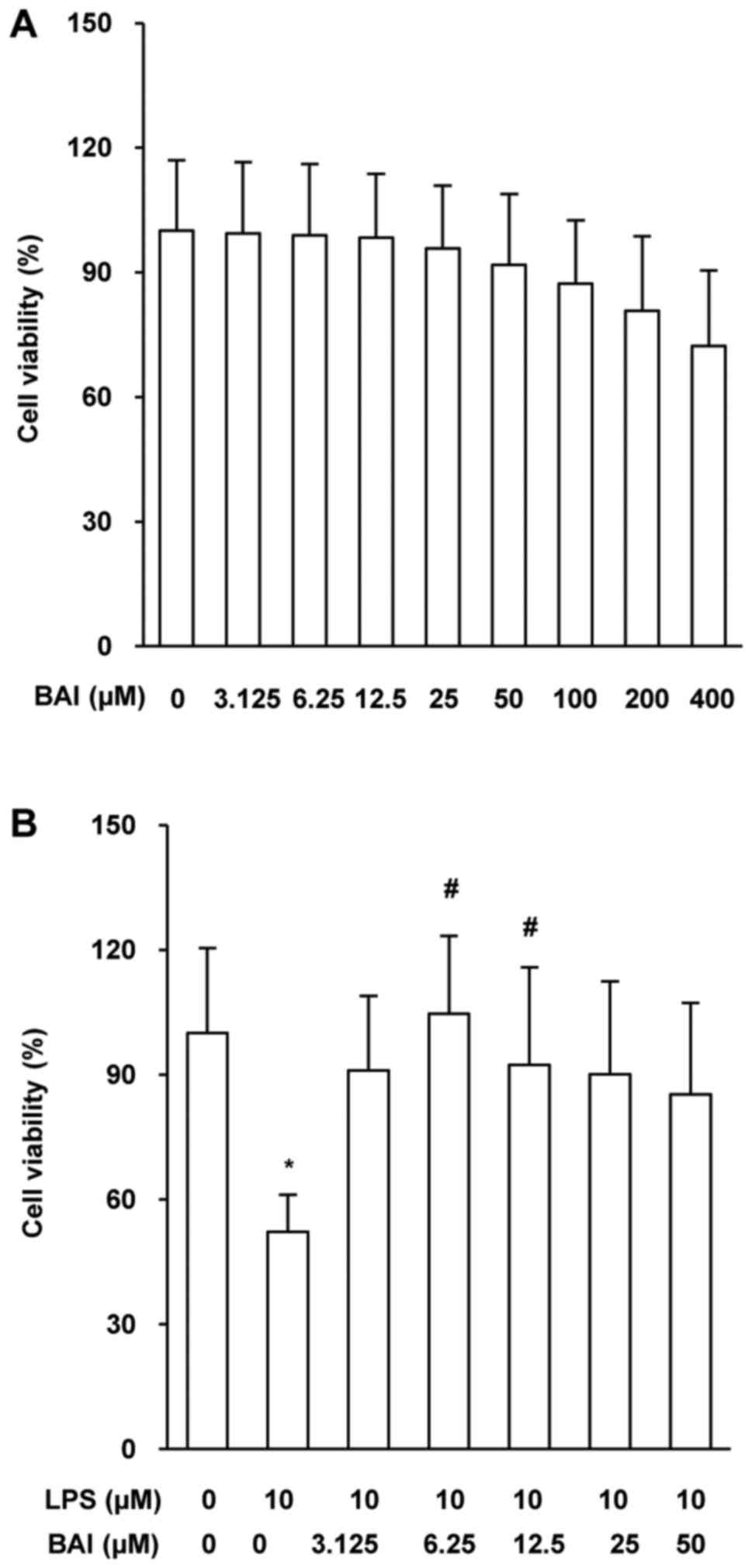 | Figure 1.BAI protected against LPS-induced
injury in HUVECs. HUVECs were stimulated by LPS or treated with BAI
for 12 h. Cell viability was evaluated in HUVECs treated with (A)
different concentrations of BAI (0, 3.125, 6.25, 12.5, 25, 50, 100,
200 and 400 µM) alone or (B) in combination (BAI, 0, 3.125, 6.25,
12.5, 25 or 50 µM) with 0 or 10 µM LPS. Cell viability was measured
by Cell Counting Kit-8 assays and was calculated using the optical
density values. The data are expressed as the mean ± standard
deviation. *P<0.05 vs. control (0 µM BAI/LPS);
#P<0.05 vs. LPS group (10 µM LPS only). BAI,
baicalein; LPS, lipopolysaccharide; HUVECs, human umbilical vein
endothelial cells. |
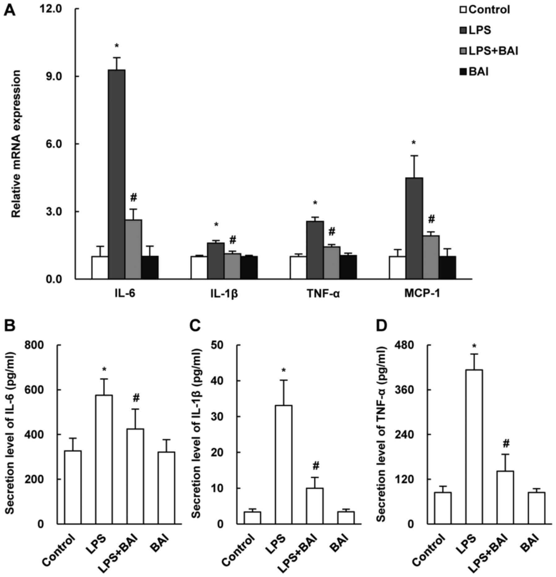
BAI attenuated LPS-induced
inflammation in HUVECs
LPS-induced injury in HUVECs is related to increased
inflammation. To determine the protective effect of BAI on
inflammation, we measured the mRNA expression levels of IL-1β,
IL-6, TNF-α and MCP-1 by quantitative PCR and the protein levels by
ELISA. Our results showed that the expressions of the inflammatory
markers was markedly suppressed in BAI-treated HUVECs (Fig. 2B-D). Besides, the changes of
inflammatory cytokines mRNA level in HUVECs treated for 24 h, which
were not shown in the article, were detected to be similar to cells
treated for 12 h.
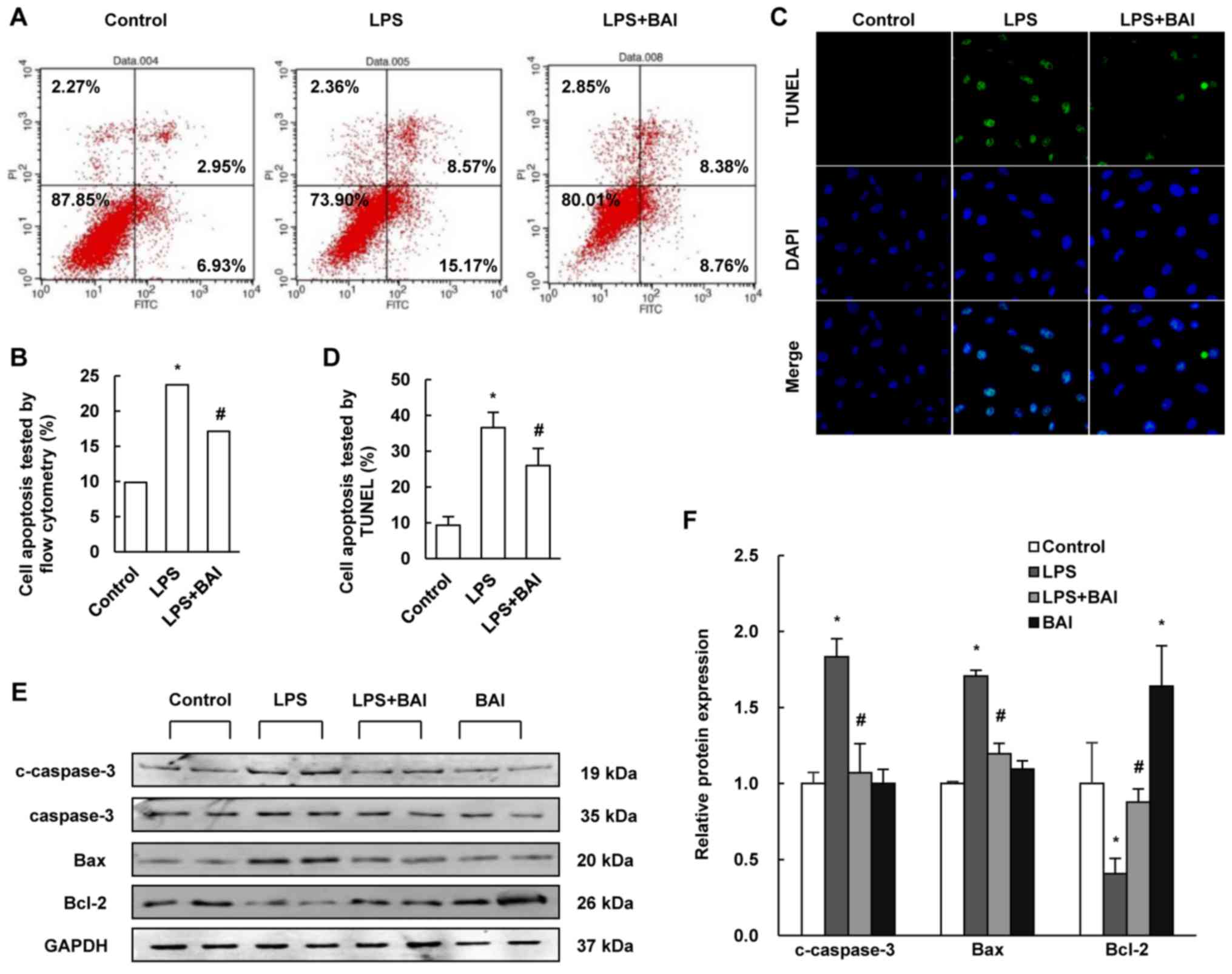
BAI inhibited LPS-induced apoptosis in
HUVECs
To examine the protective effect of BAI in the
regulation of endothelial cell injury, flow cytometry was used to
differentiate apoptotic cells and the TUNEL assay was performed to
determine DNA fragmentation. Compared with the control group,
treatment with BAI profoundly reduced apoptosis in LPS-stimulated
cells (Fig. 3A-D).
To further clarify the effect of BAI on LPS-induced
injury, we measured the relative expressions of Bcl-2, Bax and
cleaved caspase-3 by using western blotting. The data revealed that
BAI increased the level of Bcl-2, but reduced the level of Bax and
cleaved caspase-3, which contrasted with the changes induced by LPS
(Fig. 3E and F).
BAI reduced LPS-induced HUVECs injury
via TLR4/NF-κB signaling
The above results suggested that BAI might attenuate
LPS induced endothelial cell injury. However, the molecular
mechanism by which BAI exerted these protective effects was
unclear. As the TLR4/NF-κB cascade signaling pathway is proven to
play an important part in the development of endothelial
inflammation and apoptosis, we examined whether BAI affected the
LPS-induced activation of the TLR4/NF-κB signaling pathway. In
comparison with the LPS group, the expression of TLR4 and the
LPS-induced phosphorylation of TAK1, TBK1, and NF-κB p65 were
reduced by BAI (Fig. 4A and B).
MRT67307 removed the protective effects of BAI on the
phosphorylation of TBK1 (Fig. 4C and
D). In addition, the immunofluorescent staining of p65 revealed
that nuclear translocation in HUVECs was inhibited by BAI treatment
(Fig. 4E and F).
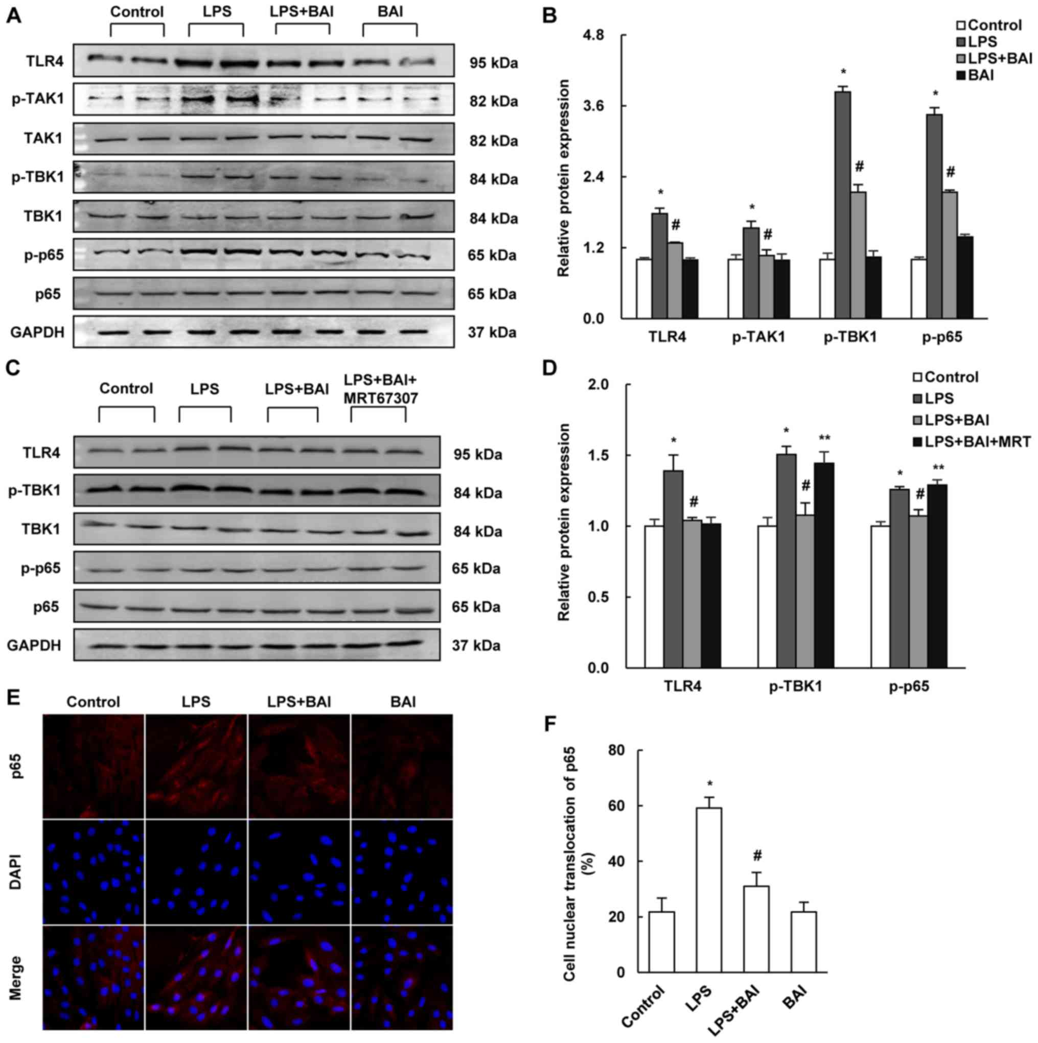 | Figure 4.BAI reduced LPS-induced damage via
TLR4/NF-κB signaling. The protein expressions of TLR4/NF-κB
signaling were established by (A-D) western blotting. (A) Blots for
LPS stimulation demonstrated that (B) TLR4, p-TAK1, p-TBK1 and
p-p65 were upregulated by LPS, then downregulated by BAI treatment.
(C) MRT67307 treatment and (D) inhibited the effect of BAI on
p-TBK1. (E) The immunofluorescent staining of p65 (magnification,
×200) revealed that (F) there was more cell nuclear translocation
with LPS stimulation and less with BAI treatment. The data are
expressed as the mean ± standard deviation. *P<0.05 vs. control
(0 µM BAI/LPS); #P<0.05 vs. LPS group (10 µM LPS
only); **P<0.05 vs. LPS+BAI (10 µM LPS, 6.25 µM BAI). BAI,
baicalein; LPS, lipopolysaccharide; HUVECs, human umbilical vein
endothelial cells; TLR4, Toll-like receptor 4; p, phosphorylated;
TAK1, transforming growth factor β-activated kinase 1; TBK1, tumor
necrosis factor receptor-associated family member associated
nuclear factor-κB activator-binding kinase 1. |
Discussion
In this study, we found that BAI protected against
the LPS-induced injury in HUVECs. BAI reduced the activation of
TLR4/NF-κB signaling in blunt inflammation and apoptosis. This
discovery points to an important role for BAI in the regulation of
vascular endothelial dysfunction.
Endothelial cells are reported to have a critical
role in the process of HF (16).
Due to their advantages of plasticity, secretion ability and repair
function, endothelial cells have been confirmed as an important
cardiac moiety (1). Traditionally,
inflammatory cytokines are considered to be derived from immune
cells, such as monocytes, but recent studies have indicated that
endothelial cells could also be the source of inflammatory
cytokines during inflammatory responses (17). Endothelial cells are reported to be
involved in the adhesion and migration of immunocytes (18).
To elucidate the mechanisms of BAI in HUVECs, we
detected the expression of certain proteins in HUVECs. This helped
in revealing a reduction in the expression of TLR4 and in the
phosphorylation of NF-κB p65 (p-p65) in BAI-treated HUVECs compared
with those of the control group. Previous studies have also
indicated that BAI reduced p-p65, including the BAI-induced
prevention of cisplatin-induced acute kidney injury through the
downregulation of the NF-κB pathway (19) and the BAI-induced suppression of
p-p65 expression in the IL-1β-induced proliferation of human
rheumatoid arthritis fibroblast-like synoviocytes (20). Wang et al (21) also reported, decreased expressions
of TLR4 and p-p65 in their experiment, which investigated the
ameliorative effects of BAI in early brain injury in rats induced
by experimental subarachnoid hemorrhage. Our data showed that BAI
downregulated the expression of TLR4 and p-p65 in LPS-stimulated
HUVECs. We also found the reduced activation of TBK1 and TAK1 after
BAI treatment.
MRT67307 (Formula
C26H36N6O2·xHCl) is a
potent and dual IKKε and TBK1 inhibitor. According to the MRT67307
specification from Selleck Chemicals, MRT67307 prevents the
phosphorylation of IRF3 and the production of IFNβ in macrophages;
enhances the IL-1-stimulated phosphorylation of p105 and RelA, as
well as enhances IL-1-stimulated activation of NF-κB-dependent gene
transcription in wild-type MEFs; increases IL-10 production and
suppresses proinflammatory cytokine production via a cAMP response
element-binding protein (CREB)-regulated transcriptional
coactivator (CRTC) 3 dependent mechanism; inhibit ULK and block
autophagy in MEF cells. Furthermore, MRT67307 was reported to
transiently enhance the protein level of p-TBK1 in 1 h, such as
enhancing the LPS-stimulated p-TBK1 (22). In this study, we found that the
protective effects of BAI were negated by MRT67307.
The present study has revealed the protective effect
of BAI on endothelial cells injury and provided insights into the
mechanism underlying the protective effect of BAI. BAI decreased
LPS-induced cell apoptosis through the inactivation of
TLR4/TBK/NF-κB signaling. Our observations provided further
insights into the pathogenesis of vascular endothelial injury and
HF and demonstrated that BAI may contribute to the inhibition of
endothelial dysfunction and in the prevention of HF. However, we
focused on only one reported signaling pathway of BAI; the study is
also limited by the absence of research into the in vivo
effects and mechanisms of BAI. Additional studies are required to
conduct further exploration into the impact of BAI on LPS-induced
injury in HUVECs.
Acknowledgements
This study was supported by National Natural Science
Foundation of China (grant nos. 81270303, 81470516, 81530012); and
Fundamental Research Funds of the Central Universities (grant no.
2042017kf0145).
Glossary
Abbreviations
Abbreviations:
|
BAI
|
baicalein
|
|
LPS
|
lipopolysaccharide
|
|
HUVECs
|
human umbilical vein endothelial
cells
|
References
|
1
|
McCarron JG, Lee MD and Wilson C: The
endothelium solves problems that endothelial cells do not know
exist. Trends Pharmacol Sci. 38:322–338. 2017. View Article : Google Scholar : PubMed/NCBI
|
|
2
|
Fraisl P, Mazzone M, Schmidt T and
Carmeliet P: Regulation of angiogenesis by oxygen and metabolism.
Dev Cell. 16:167–179. 2009. View Article : Google Scholar : PubMed/NCBI
|
|
3
|
Wei F, Liu S, Luo L, Gu N, Zeng Y, Chen X,
Xu S and Zhang D: Anti-inflammatory mechanism of ulinastatin:
Inhibiting the hyperpermeability of vascular endothelial cells
induced by TNF-α via the RhoA/ROCK signal pathway. Int
Immunopharmacol. 46:220–227. 2017. View Article : Google Scholar : PubMed/NCBI
|
|
4
|
Jang J, Jung Y, Kim Y, Jho EH and Yoon Y:
LPS-induced inflammatory response is suppressed by Wnt inhibitors,
Dickkopf-1 and LGK974. Sci Rep. 7:416122017. View Article : Google Scholar : PubMed/NCBI
|
|
5
|
Yu X, Lan P, Hou X, Han Q, Lu N, Li T,
Jiao C, Zhang J, Zhang C and Tian Z: HBV inhibits LPS-induced NLRP3
inflammasome activation and IL-1β production via suppressing the
NF-κB pathway and ROS production. J Hepatol. 66:693–702. 2017.
View Article : Google Scholar : PubMed/NCBI
|
|
6
|
Peng T, Lu X, Lei M, Moe GW and Feng Q:
Inhibition of p38 MAPK decreases myocardial TNF-alpha expression
and improves myocardial function and survival in endotoxemia.
Cardiovasc Res. 59:893–900. 2003. View Article : Google Scholar : PubMed/NCBI
|
|
7
|
Ives A, Nomura J, Martinon F, Roger T,
LeRoy D, Miner JN, Simon G, Busso N and So A: Xanthine
oxidoreductase regulates macrophage IL-1β secretion upon NLRP3
inflammasome activation. Nat Commun. 6:65552015. View Article : Google Scholar : PubMed/NCBI
|
|
8
|
Kim SD, Lee YJ, Baik JS, Han JY, Lee CG,
Heo K, Park YS, Kim JS, Ji HD, Park SI, et al: Baicalein inhibits
agonist- and tumor cell-induced platelet aggregation while
suppressing pulmonary tumor metastasis via cAMP-mediated VASP
phosphorylation along with impaired MAPKs and PI3K-Akt activation.
Biochem Pharmacol. 92:251–265. 2014. View Article : Google Scholar : PubMed/NCBI
|
|
9
|
Wang Y, Han E, Xing Q, Yan J, Arrington A,
Wang C, Tully D, Kowolik CM, Lu DM, Frankel PH, et al: Baicalein
upregulates DDIT4 expression which mediates mTOR inhibition and
growth inhibition in cancer cells. Cancer Lett. 358:170–179. 2015.
View Article : Google Scholar : PubMed/NCBI
|
|
10
|
Cai W, Fu Y, Zhang W, Chen X, Zhao J, Song
W, Li Y, Huang Y, Wu Z, Sun R, et al: Synergistic effects of
baicalein with cefotaxime against Klebsiella pneumoniae through
inhibiting CTX-M-1 gene expression. BMC Microbiol. 16:1812016.
View Article : Google Scholar : PubMed/NCBI
|
|
11
|
Tsai KL, Hung CH, Chan SH, Shih JY, Cheng
YH, Tsai YJ, Lin HC and Chu PM: Baicalein protects against
oxLDL-caused oxidative stress and inflammation by modulation of
AMPK-alpha. Oncotarget. 7:72458–72468. 2016.PubMed/NCBI
|
|
12
|
Huang Y, Miao Z, Hu Y, Yuan Y, Zhou Y, Wei
L, Zhao K, Guo Q and Lu N: Baicalein reduces angiogenesis in the
inflammatory microenvironment via inhibiting the expression of
AP-1. Oncotarget. 8:883–899. 2017.PubMed/NCBI
|
|
13
|
Zong J, Zhang DP, Zhou H, Bian ZY, Deng W,
Dai J, Yuan Y, Gan HW, Guo HP and Tang QZ: Baicalein protects
against cardiac hypertrophy through blocking MEK-ERK1/2 signaling.
J Cell Biochem. 114:1058–1065. 2013. View Article : Google Scholar : PubMed/NCBI
|
|
14
|
Zheng K, Setyawati MI, Lim TP, Leong DT
and Xie J: Antimicrobial cluster bombs: Silver nanoclusters packed
with daptomycin. ACS Nano. 10:7934–7942. 2016. View Article : Google Scholar : PubMed/NCBI
|
|
15
|
Grasl-Kraupp B, Ruttkay-Nedecky B,
Koudelka H, Bukowska K, Bursch W and Schulte-Hermann R: In situ
detection of fragmented DNA (TUNEL assay) fails to discriminate
among apoptosis, necrosis, and autolytic cell death: A cautionary
note. Hepatology. 21:1465–1468. 1995. View Article : Google Scholar : PubMed/NCBI
|
|
16
|
Burchfield JS, Xie M and Hill JA:
Pathological ventricular remodeling: Mechanisms: Part 1 of 2.
Circulation. 128:388–400. 2013. View Article : Google Scholar : PubMed/NCBI
|
|
17
|
Heim A, Zeuke S, Weiss S, Ruschewski W and
Grumbach IM: Transient induction of cytokine production in human
myocardial fibroblasts by coxsackievirus B3. Circ Res. 86:753–759.
2000. View Article : Google Scholar : PubMed/NCBI
|
|
18
|
Pober JS and Sessa WC: Evolving functions
of endothelial cells in inflammation. Nat Rev Immunol. 7:803–815.
2007. View
Article : Google Scholar : PubMed/NCBI
|
|
19
|
Sahu BD, Mahesh Kumar J and Sistla R:
Baicalein, a bioflavonoid, prevents cisplatin-induced acute kidney
injury by up-regulating antioxidant defenses and down-regulating
the MAPKs and NF-κB pathways. PLoS One. 10:e1341392015. View Article : Google Scholar
|
|
20
|
Chen S, Yang Y, Feng H, Wang H, Zhao R and
Liu H: Baicalein inhibits interleukin-1-induced proliferation of
human rheumatoid arthritis fibroblast-like synoviocytes.
Inflammation. 37:163–169. 2014. View Article : Google Scholar : PubMed/NCBI
|
|
21
|
Wang CX, Xie GB, Zhou CH, Zhang XS, Li T,
Xu JG, Li N, Ding K, Hang CH, Shi JX and Zhou ML: Baincalein
alleviates early brain injury after experimental subarachnoid
hemorrhage in rats: Possible involvement of TLR4/NF-κB-mediated
inflammatory pathway. Brain Res. 1594:245–255. 2015. View Article : Google Scholar : PubMed/NCBI
|
|
22
|
Clark K, Peggie M, Plater L, Sorcek RJ,
Young ER, Madwed JB, Hough J, McIver EG and Cohen P: Novel
cross-talk within the IKK family controls innate immunity. Biochem
J. 434:93–104. 2011. View Article : Google Scholar : PubMed/NCBI
|