Introduction
Vanadium is a soft, silvery-grey metal, which exists
in a number of different oxidation states (−1, 0, +2, +3, +4 and
+5); vanadium pentoxide (V2O5) is the most
common commercial form, and most of the studies on toxicity have
been conducted on vanadium pentoxide, as it is the primary form
found in industrial exposure situations (1). All vanadium compounds are considered
toxic. The Occupational Safety and Health Administration (Bellevue,
WA, USA) have set an exposure limit for the workplace (considering
an 8 h workday, and a 40 h work week), of 0.05 mg/m3 for
V2O5 dust and 0.1 mg/m3 for
V2O5 fumes (2).
The exposure dose of vanadium that is considered
life-threatening is 35 mg/m3 [as determined by the
National Institute for Occupational Safety and Health (NIOSH;
Washington, DC, USA)], which could cause serious and perpetuating
health issues, including death (2). The respiratory system is the most
vulnerable to vanadium toxicity, while the effect on the
gastrointestinal system is minimal due to the low gut absorption
rate (3–5). However, quantitative data are not
sufficient to obtain a chronic or subchronic inhalation reference
dose.
In rat models, the effects resulting from an inhaled
or oral vanadium were evaluated in the sera (6,7),
nervous tissue (8), liver
(9) and other types of tissue
(kidney, gut, lungs) development (10). In vanadium workers (NIOSH 1983)
increases in skin rashes and atopic dermatitis have been recorded.
To the best of our knowledge, no prior in vivo or in
vitro studies have been conducted to evaluate the effect of
vanadium exposure on dermal fibroblasts. Here, we evaluated the
effect of V2O5 on the proliferation and
chemokine secretion profiles of dermal fibroblasts.
Materials and methods
Fibroblast cell cultures
Dermal fibroblasts were obtained from 6 patients who
underwent surgery for thyroid nodular goiter (discarded dermal
material was used). The local Ethics Committee of the University of
Pisa approved the study protocol, and all subjects provided
informed consent.
As previously described, tissue explants from the
derma were minced and placed in culture dishes, to allow the
fibroblasts to proliferate (11).
Fibroblasts were propagated in Medium 199 containing 20% FBS
(Gibco; Invitrogen, Ltd., Paisley, UK), gentamycin (20 µg/ml) and
penicillin (100 U/ml), in a 37°C humidified incubator with 5% of
CO2. Cells were subsequently maintained in medium 199
containing 10% FBS (and antibiotics) (12).
Cell viability and proliferation
assay
The evaluation of cell proliferation and viability
was conducted using a WST-1 assay (Roche Diagnostics, Almere, The
Netherlands), which uses MTT (13,14).
Fibroblasts were seeded (35,000 cells/ml, in a final volume of 100
µl) into 96-well plates. The effect V2O5 on
fibroblast proliferation was determined following exposure of the
cells for 24 h to increasing concentrations of
V2O5 (1, 10 and 100 nM). Cells were then
plated and treated for 24 h with V2O5, or
with its vehicle alone; all experiments were performed in
triplicate for each cell preparation.
Proliferation assay: cell
counting
The cell counting assay was also used to assess
fibroblast proliferation (13,14).
Chemokines secretion assays
Chemokine (C-X-C motif) ligand (CXCL)9 and CXCL10
secretion assays were performed by seeding 30,000 cells/ml into
96-well plates, with a final volume of 100 µl/well, in growth
medium that was removed after 24 h. Cells were subsequently washed
in PBS, then incubated (24 h) in phenol red and serum-free medium
with interferon (IFN)-γ (R&D Systems, Minneapolis, MN, USA;
500; 1,000; 5,000; 10,000 IU/ml), and/or 10 ng/ml tumor necrosis
factor (TNF)-α (R&D Systems) (11). Preliminary experiments were
conducted to select the TNF-α concentration, in order to obtain the
highest secretion rate. The supernatants were collected after 24 h,
then frozen at −20°C until use in the chemokine assay.
To understand the effect of
V2O5 on the chemokine secretion induced by
IFN-γ, cells were treated for 24 h with increasing concentrations
of V2O5 (1, 10 and 100 nM), in the presence
or absence of IFN-γ (1,000 IU/ml), and/or TNF-α (10 ng/ml). An
ELISA was used to measure the CXCL9 and CXCL10 levels in the
supernatants. The experiments were performed three times for each
different cell preparation.
ELISA for CXCL9 and CXCL10
CXCL9 and CXCL10 were assessed in the supernatants
obtained from cell cultures, using commercially available kits
(R&D Systems). The minimum (mean) detectable doses were 1.5 and
1.2 pg/ml for CXCL9 and CXCL10, respectively. The intra- and
inter-assay coefficients of variation were 3.5 and 6.4%
respectively, for CXCL9, and 4.5 and 7.3% respectively, for CXCL10.
Quality control pools of normal, low and high concentrations were
also included in each assay.
Data analysis
For normally distributed variables, values are given
as the mean ± SD in text, and in figures, otherwise as the median
and interquartile range. Mean group values were compared using
one-way analysis of variance (ANOVA) for variables normally
distributed variables, or by using the Kruskal-Wallis test or
Mann-Whitney U test. Proportions were compared using the
Chi-square test. In addition, the Bonferroni-Dunn test was used for
the post hoc comparison of normally distributed variables.
Results
Cell proliferation
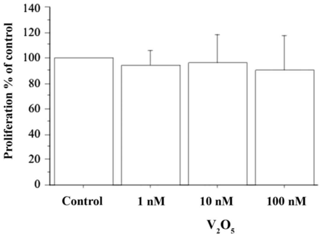
The WST-1 cell viability and proliferation assay
showed that V2O5 (1, 10 and 100 nM) did not
alter the viability or proliferation of dermal fibroblasts
(Fig. 1). These results were
confirmed by a cell counting assay (data not presented).
CXCL9
CXCL9 was not detectable in the supernatants
gathered from primary fibroblast samples, whereas its concentration
was elevated following IFN-γ dose-dependent induction (0, 75±31,
141±29, 210±35 and 297±74 pg/ml for IFN-γ 0; 500; 1,000; 5,000 and
10,000 IU/ml, respectively; P<0.001, ANOVA). TNF-α alone had no
significant impact on CXCL9, which remained undetectable, whereas
IFN-γ plus TNF-α exhibited a synergistic effect on the CXCL9
release (CXCL9, 11,154±1,673 vs. 151±42 pg/ml with IFN-γ alone;
P<0.0001, ANOVA).
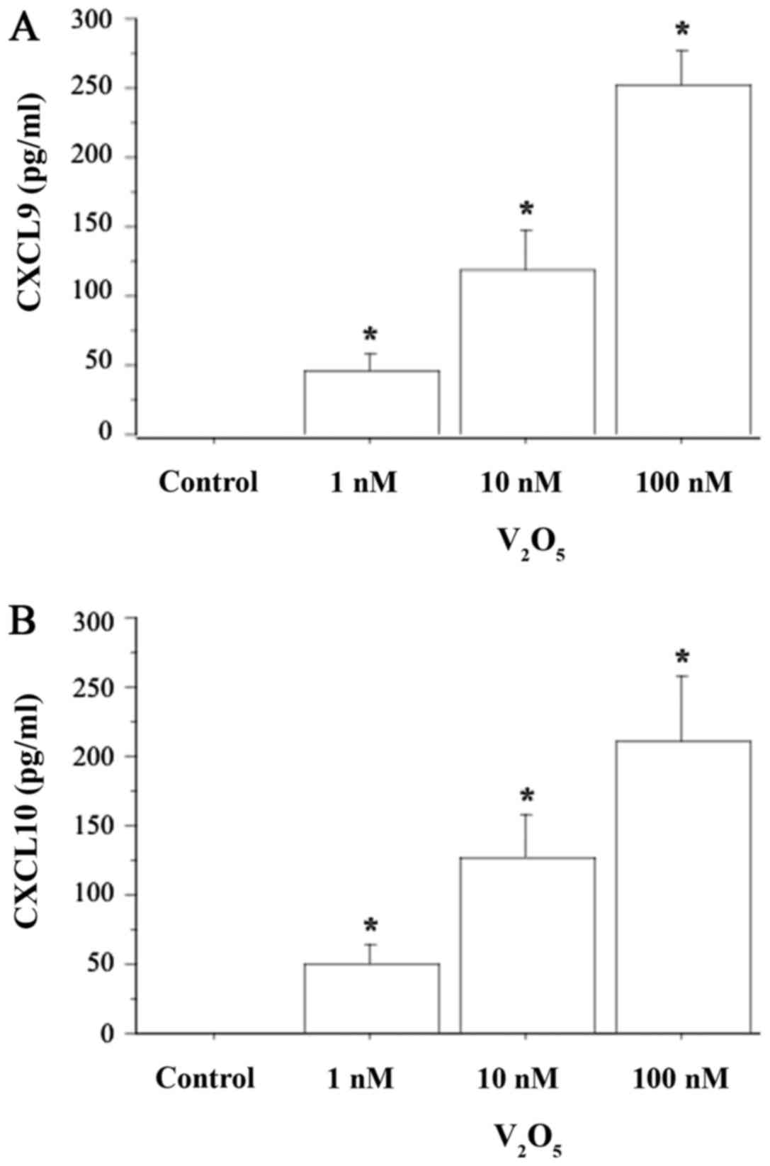
CXCL9 release was dose-dependently stimulated
(P<0.0001, ANOVA) when fibroblasts were treated with increasing
V2O5 concentrations (1, 10 and 100 nM)
(Fig. 2A). Following the treatment
of fibroblasts with V2O5 (1, 10 and 100 nM),
together with TNF-α, CXCL9 secretion was not significantly changed
with respect to V2O5 alone (data not
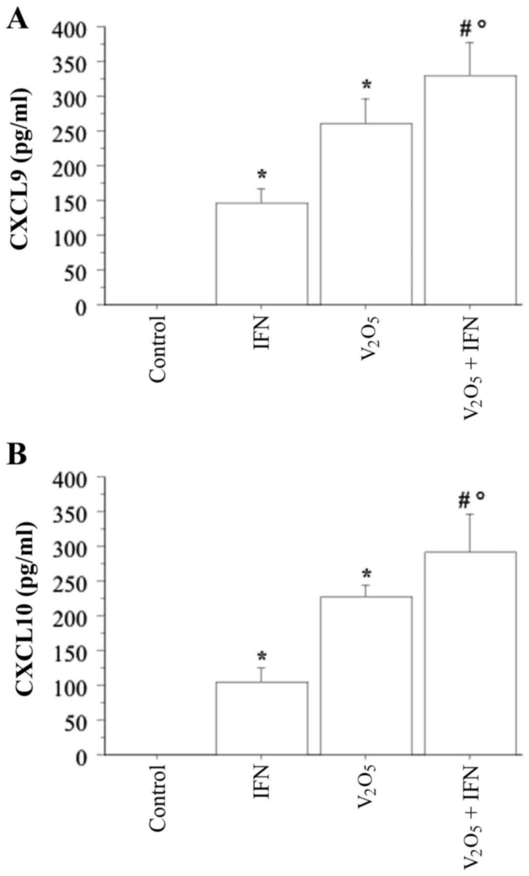
presented). Treating fibroblasts with 100 nM
V2O5 plus IFN-γ induced a synergistic
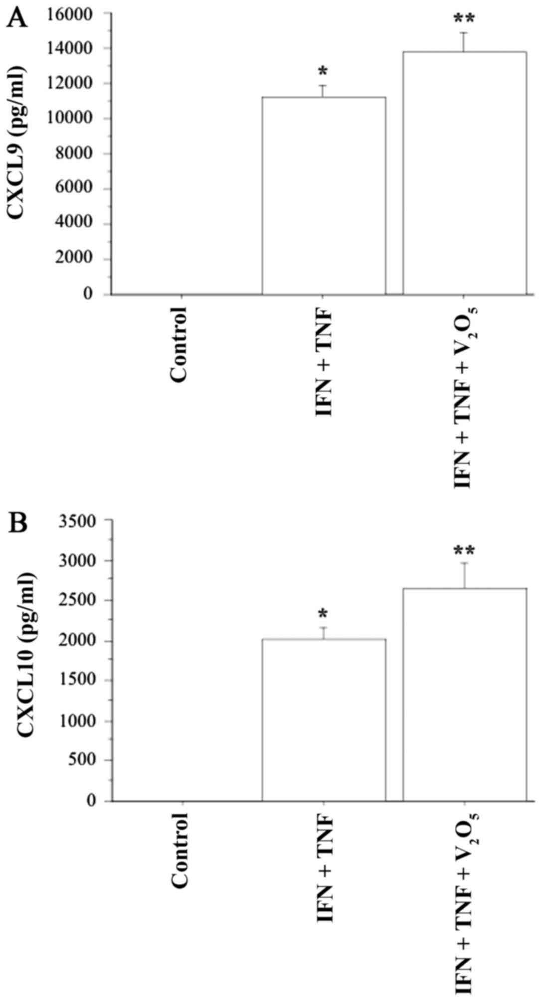
increase in CXCL9 release (P<0.0001, ANOVA) (Fig. 3A). When fibroblasts were treated
with V2O5 (100 nM), together with IFN-γ and
TNF-α stimulation, CXCL9 release was synergistically increased
(P<0.0001, ANOVA) (Fig.
4A).
CXCL10
CXCL10 was also not detectable in the supernatants
obtained from primary fibroblast cultures under basal conditions.
IFN-γ induced CXCL10 secretion dose-dependently (0, 34±18, 107±42,
187±32 and 272±76 pg/ml, respectively, for IFN-γ 0; 500; 1,000;
5,000; 10,000 IU/ml; ANOVA, P<0.001). TNF-α alone did not have a
significant impact on CXCL10 secretion, whereas IFN-γ plus
TNF-α exhibited a synergistic effect on CXCL10 secretion (3,043±234
vs. 117±27 pg/ml with IFN-γ alone; P<0.0001, ANOVA).
CXCL10 release was dose-dependently stimulated
(P<0.0001, ANOVA) when fibroblasts were treated with increasing
V2O5 concentrations (1, 10 and 100 nM)
(Fig. 2B). Following the treatment
of fibroblasts with V2O5 (1, 10 and 100 nM),
and together with TNF-α, CXCL10 secretion was not significantly
changed with respect to V2O5 alone (data not
presented).
Treating fibroblasts with 100 nM
V2O5 plus IFN-γ caused a synergistic
increase in CXCL10 release (P<0.0001, ANOVA) (Fig. 3B). When fibroblasts were treated
with V2O5 (100 nM) together with IFN-γ and
TNF-α stimulation, CXCL10 release was also synergistically
increased (P<0.0001, ANOVA) (Fig.
4B).
Discussion
The results of the present study demonstrated that
V2O5 could promote IFN-γ-dependent chemokine
secretion in dermal fibroblasts, without altering their cell
proliferation and viability. In addition, our results confirmed
that IFN-γ and TNF-α stimulate CXCL9 and CXCL10 secretion, as
hypothesized (11,15). It is notable that
V2O5 was able to synergize with IFN-γ and
TNF-α, further increasing chemokines secretion.
These results are concordant with the hypothesis
that V2O5 is able to induce and perpetuate
inflammation in the dermal tissues, evolving from a predominant
T-helper (Th)1 immune response (13). IFN-γ-inducible C-X-C chemokines are
secreted by several types of mammalian cells, including
fibroblasts, thyrocytes, islet cells, colon epithelial cells and
endothelial cells, among others (11,13–21).
These cell types are unable to produce these chemokines under basal
conditions; they are induced following stimulation by IFN-γ (alone
or in combination with TNF-α), a cytokine that is produced by
Th1-activated lymphocytes in several autoimmune diseases, including
in the thyroid in Graves' disease, and in autoimmune thyroiditis.
It has been hypothesized that this process can be involved in the
initiation and/or the perpetuation of various autoimmune disorders
(11,13–21),
and that it may also be applied to the thyroid.
Our results are concordant with those of other
studies in different cell types. V2O5
exposure is a cause of occupational bronchitis; an in vitro
study was conducted to evaluate the gene expression profiles of
human lung fibroblasts following V2O5
exposure, in order to identify genes that might play a role in the
bronchial inflammation, repair and fibrosis during the pathogenesis
of bronchitis. A dozen genes are overexpressed by
V2O5, including CXCL9 and
CXCL10 (1). A further study
reported that fibroblasts responded to vanadium oxidative stress by
producing IFN-β and activating STAT-1, which lead to increased
CXCL10 levels (22), thus serving
a role in the innate immune response.
It is notable that vanadium is able to increase
chemokine secretion in the dose range of 1–100 nM. Since the normal
blood levels of vanadium range from 0.45–18.4 nM, 100 nM could be
noted as a dose that might mimic an abnormally high exposure
(23). Thus, we could hypothesize
that V2O5 in this concentration range is able
to induce an inflammatory reaction in dermal tissues, prompting the
appearance of skin rashes or atopic dermatitis.
Moreover, it has been shown that exposure of human
skin fibroblasts to vanadate causes DNA strand breaks at relevant
concentration of 1 µM (24). In
the present study we have considered lower concentrations (1, 10
and 100 nM), that did not alter the viability or proliferation of
dermal fibroblasts.
In conclusion, the results of our study showed that
V2O5 is able to induce Th1 chemokine
secretion in dermal tissues, and that it can synergize with
important Th1 cytokines (such as IFN-γ and TNF-α), leading to the
induction and perpetuation of inflammation in the dermis. Moreover,
different genes are overexpressed by V2O5,
including CXCL9 and CXCL10, that appear to have
important functions in inflammation, fibrosis and repair. To the
best of our knowledge, no prior study has evaluated the immune
modulatory effects of vanadium in dermal fibroblasts; therefore,
our results could be important for evaluating the pathogenesis of
clinical dermatological manifestations of vanadium exposure in
humans. The induction and perpetuation of inflammation in the
dermis and the variety of involved candidate genes could be at the
basis of V2O5-induced effects after
occupational and environmental exposures. Additional studies are
required to assess dermal integrity, as well as the manifestations
of toxicity in subjects who are occupationally exposed, or are
living in polluted areas.
Acknowledgements
Not applicable.
Funding
No funding was received.
Availability of data and materials
All data generated or analyzed during this study are
included in this published article.
Authors' contributions
PF, RF, AC, AA and SMF made substantial
contributions to the conception and design of the study and to the
acquisition of the data. GE, FR, AP, GG, GF and SB analysed the
data. PF, AA and SMF. have been involved in drafting the
manuscript. AA critically revised the manuscript for important
intellectual content.
Ethics approval and consent to
participate
Not applicable.
Consent for publication
Not applicable.
Competing interests
The authors confirm that they have no competing
interests.
References
|
1
|
Ingram JL, Antao-Menezes A, Turpin EA,
Wallace DG, Mangum JB, Pluta LJ, Thomas RS and Bonner JC: Genomic
analysis of human lung fibroblasts exposed to vanadium pentoxide to
identify candidate genes for occupational bronchitis. Respir Res.
8:342007. View Article : Google Scholar : PubMed/NCBI
|
|
2
|
Occupational Safety and Health
Administration (OSHA): Occupational safety and health guideline for
vanadium pentoxide dust. OSHA; Washington, DC: 2007
|
|
3
|
Sax NI: Dangerous Properties of Industrial
Materials. 6th. Van Nostrand Reinhold Company; New York, NY: pp.
2717–2720. 1984
|
|
4
|
Ress NB, Chou BJ, Renne RA, Dill JA,
Miller RA, Roycroft JH, Hailey JR, Haseman JK and Bucher JR:
Carcinogenicity of inhaled vanadium pentoxide in F344/N rats and
B6C3F1 mice. Toxicol Sci. 74:287–296. 2003. View Article : Google Scholar : PubMed/NCBI
|
|
5
|
Wörle-Knirsch JM, Kern K, Schleh C,
Adelhelm C, Feldmann C and Krug HF: Nanoparticulate vanadium oxide
potentiated vanadium toxicity in human lung cells. Environ Sci
Technol. 41:331–336. 2007. View Article : Google Scholar : PubMed/NCBI
|
|
6
|
Scibior A, Zaporowska H and Ostrowski J:
Selected haematological and biochemical parameters of blood in rats
after subchronic administration of vanadium and/or magnesium in
drinking water. Arch Environ Contam Toxicol. 51:287–295. 2006.
View Article : Google Scholar : PubMed/NCBI
|
|
7
|
González-Villalva A, Fortoul T,
Avila-Costa MR, Piñón-Zarate G, Rodriguez-Laraa V, Martínez-Levy G,
Rojas-Lemus M, Bizarro-Nevarez P, Díaz-Bech P, Mussali-Galante P
and Colin-Barenque L: Thrombocytosis induced in mice after subacute
and subchronic V2O5 inhalation. Toxicol Ind
Health. 22:113–116. 2006. View Article : Google Scholar : PubMed/NCBI
|
|
8
|
Soazo M and Garcia GB: Vanadium exposure
through lactation produces behavioral alterations and CNS myelin
deficit in neonatal rats. Neurotoxicol Teratol. 29:503–510. 2007.
View Article : Google Scholar : PubMed/NCBI
|
|
9
|
Kobayashi K, Himeno S, Satoh M, Kuroda J,
Shibata N, Seko Y and Hasegawa T: Pentavalent vanadium induces
hepatic metallothionein through interleukin-6-dependent and
-independent mechanisms. Toxicology. 228:162–170. 2006. View Article : Google Scholar : PubMed/NCBI
|
|
10
|
Barceloux DG: Vanadium. J Toxicol Clin
Toxicol. 37:265–278. 1999. View Article : Google Scholar : PubMed/NCBI
|
|
11
|
Antonelli A, Ferri C, Fallahi P, Ferrari
SM, Frascerra S, Sebastiani M, Franzoni F, Galetta F and Ferrannini
E: High values of CXCL10 serum levels in patients with hepatitis C
associated mixed cryoglobulinemia in presence or absence of
autoimmune thyroiditis. Cytokine. 42:137–143. 2008. View Article : Google Scholar : PubMed/NCBI
|
|
12
|
Valyasevi RW, Harteneck DA, Dutton CM and
Bahn RS: Stimulation of adipogenesis, peroxisome
proliferator-activated receptor-gamma (PPARgamma), and thyrotropin
receptor by PPARgamma agonist in human orbital preadipocyte
fibroblasts. J Clin Endocrinol Metab. 87:2352–2358. 2002.
View Article : Google Scholar : PubMed/NCBI
|
|
13
|
Antonelli A, Rotondi M, Fallahi P,
Romagnani P, Ferrari SM, Buonamano A, Ferrannini E and Serio M:
High levels of circulating CXC chemokine ligand 10 are associated
with chronic autoimmune thyroiditis and hypothyroidism. J Clin
Endocrinol Metab. 89:5496–5499. 2004. View Article : Google Scholar : PubMed/NCBI
|
|
14
|
Kemp EH, Metcalfe RA, Smith KA, Woodroofe
MN, Watson PF and Weetman AP: Detection and localization of
chemokine gene expression in autoimmune thyroid disease. Clin
Endocrinol (Oxf). 59:207–213. 2003. View Article : Google Scholar : PubMed/NCBI
|
|
15
|
Antonelli A, Ferrari SM, Fallahi P,
Frascerra S, Santini E, Franceschini SS and Ferrannini E: Monokine
induced by interferon gamma (IFNgamma) (CXCL9) and IFNgamma
inducible T-cell alpha-chemoattractant (CXCL11) involvement in
Graves' disease and ophthalmopathy: Modulation by peroxisome
proliferator-activated receptor-gamma agonists. J Clin Endocrinol
Metab. 94:1803–1809. 2009. View Article : Google Scholar : PubMed/NCBI
|
|
16
|
Antonelli A, Ferrari SM, Frascerra S,
Pupilli C, Mancusi C, Metelli MR, Orlando C, Ferrannini E and
Fallahi P: CXCL9 and CXCL11 chemokines modulation by peroxisome
proliferator-activated receptor-alpha agonists secretion in Graves'
and normal thyrocytes. J Clin Endocrinol Metab. 95:E413–E420. 2010.
View Article : Google Scholar : PubMed/NCBI
|
|
17
|
Garcià-Lòpez MA, Sancho D, Sànchez-Madrid
F and Marazuela M: Thyrocytes from autoimmune thyroid disorders
produce the chemokines IP-10 and Mig and attract CXCR3v
lymphocytes. J Clin Endocrinol Metab. 86:5008–5016. 2001.
View Article : Google Scholar : PubMed/NCBI
|
|
18
|
Antonelli A, Ferrari SM, Corrado A,
Ferrannini E and Fallahi P: CXCR3, CXCL10 and type 1 diabetes.
Cytokine Growth Factor Rev. 25:57–65. 2014. View Article : Google Scholar : PubMed/NCBI
|
|
19
|
Antonelli A, Ferrari SM, Giuggioli D,
Ferrannini E, Ferri C and Fallahi P: Chemokine (C-X-C motif) ligand
(CXCL)10 in autoimmune diseases. Autoimmun Rev. 13:272–280. 2014.
View Article : Google Scholar : PubMed/NCBI
|
|
20
|
Antonelli A, Fallahi P, Delle Sedie A,
Ferrari SM, Maccheroni M, Bombardieri S, Riente L and Ferrannini E:
High values of Th1 (CXCL10) and Th2 (CCL2) chemokines in patients
with psoriatic arthtritis. Clin Exp Rheumatol. 27:22–27.
2009.PubMed/NCBI
|
|
21
|
Fallahi P, Ferrari SM, Ruffilli I, Elia G,
Biricotti M, Vita R, Benvenga S and Antonelli A: The association of
other autoimmune diseases in patients with autoimmune thyroiditis:
Review of the literature and report of a large series of patients.
Autoimmun Rev. 15:1125–1128. 2016. View Article : Google Scholar : PubMed/NCBI
|
|
22
|
Antao-Menezes A, Turpin EA, Bost PC,
Ryman-Rasmussen JP and Bonner JC: STAT-1 signaling in human lung
fibroblasts is induced by vanadium pentoxide through an IFN-beta
autocrine loop. J Immunol. 180:4200–4207. 2008. View Article : Google Scholar : PubMed/NCBI
|
|
23
|
Sabbioni E, Kuèera J, Pietra R and
Vesterberg O: A critical review on normal concentrations of
vanadium in human blood, serum, and urine. Sci Total Environ.
188:49–58. 1996. View Article : Google Scholar : PubMed/NCBI
|
|
24
|
Ivancsits S, Pilger A, Diem E, Schaffer A
and Rüdiger HW: Vanadate induces DNA strand breaks in cultured
human fibroblasts at doses relevant to occupational exposure. Mutat
Res. 519:25–35. 2002. View Article : Google Scholar : PubMed/NCBI
|