Introduction
Ovarian cancer (OC) is a principal cause of cancer
mortality in women, with an estimated 14,080 cases of mortality in
the USA (1) every year, and 22,500
cases of mortality annually in China (2). Although patients with OC are
initially responsive to standard therapy (generally debulking
surgery followed by platinum-centered chemotherapy) (3), ~70% of them develop recurrent disease
(4). The 5-year overall survival
(OS) is very poor as the relapsed disease is frequently incurable
(5), with little improvement over
the last 30 years (6). The
emergence of a drug-resistant disease is thus a primary obstacle in
the clinical management of OC. To overcome these unsatisfactory
treatment outcomes, understanding the molecular mechanisms that
contribute to drug resistance and identifying predictive biomarkers
are critical (7).
Accumulating evidence indicates that ion channels
serve crucial roles in cancer biology (8,9) and
mediate numerous aspects of cancer pathology, including apoptosis,
angiogenesis, cell growth, migration, invasion and metastasis
(10). Among the ion channels,
potassium channels are the most diverse and ubiquitous, and
represent easily accessible cancer biomarkers and targets for
therapy (11,12). In OC, potassium channels have been
demonstrated to be closely associated with cancer progression and
outcomes. Potassium two pore domain channel subfamily K member 9 is
involved in the oncogenesis of OC, although its prominent
expression is paradoxically associated with better survival
(13); potassium voltage-gated
channel subfamily H (KCNH) member 1 and KCNH member 2 (KCNH2)
expression is associated with poor prognosis (14,15),
and KCNH2 channel activity is associated with tumor drug resistance
(16).
Potassium calcium-activated channel subfamily N
member 3 (KCNN3), a potassium channel of the small conductance
Ca2+-activated potassium channel family (17), contributes to the development and
progression of numerous solid tumors. In melanoma cells,
upregulation of KCNN3 enhances cell motility by hyperpolarizing the
cell membrane potential (18); in
breast cancer cells, KCNN3 is a mediator of cell migration
(19), and together with P2X
purinoceptor 7, contributes to cysteine cathepsin-dependent cell
invasiveness (20); and in colon
cancer, KCNN3, together with short transient receptor potential
channel 1 (TRPC1) and calcium release-activated calcium channel
protein 1 (orai-1), regulates store operated calcium entry
(SOCE)-dependent cell migration (21). Furthermore, KCNN3 is upregulated by
a 16-fold change in bortezomib-resistant BN myeloma cells (22), which suggests that its expression
is associated with drug resistance. However, KCNN3 has not been
widely studied with respect to cancer, and research on its role in
OC is rare. To the best of the authors' knowledge, the present
study, which used public data, bioinformatics and
immunohistochemistry analyses, is the first to report the
association of KCNN3 with prognosis and drug resistance in OC.
Materials and methods
Data acquisition
Data regarding gene expression determined using Log2
median-centered intensity in OC and normal controls were retrieved
from microarrays downloaded from The Cancer Genome Atlas (TCGA)
Ovarian cohort, including 586 ovarian serous adenocarcinoma and 8
normal control samples; and the Yoshihara Ovarian cohort, including
43 ovarian serous adenocarcinoma and 10 normal control samples,
deposited in Oncomine (https://www.oncomine.org/resource/main.html) (23,24))
(Tables I and II). The mRNA expression values of 489
tissues from a total of 586 patient samples with ovarian serous
adenocarcinoma were determined via Agilent microarray analysis
(Agilent Technologies, Inc., Santa Clara, CA, USA) (25), and these data were downloaded from
the cBioportal (http://www.cbioportal.org/) (26,27).
Corresponding data of microRNA (miR), DNA methylation and clinical
data of the 489 OC tissues were also downloaded from cBioportal
(http://www.cbioportal.org/). Among the
489 tissues, 197 were platinum-sensitive and 90 were resistant
tissues. KCNN3 mRNA expression (probe: 205903_s_at) data and
survival information of the 1,656 patients with OC [including 395
patients with OC who had low cancer antigen (CA) 125 expression
levels] were downloaded from the KM Plotter (http://kmplot.com/), which is a collection of 14
independent microarrays (data set nos. GSE14764, GSE15622,
GSE18520, GSE19829, GSE23554, GSE26193, GSE26712, GSE27651,
GSE30161, GSE3149, GSE51373, GSE63885, GSE65986 and GSE9891) from
the Gene Expression Omnibus (GEO) profiles and TCGA ovarian cohort
(28). No alterations were made to
any of the aforementioned data used in the analysis.
 | Table I.Association between KCNN3 expression
and clinical factors of patients with ovarian cancer in TCGA cohort
(489 patients) and lab collection (51 patients). |
Table I.
Association between KCNN3 expression
and clinical factors of patients with ovarian cancer in TCGA cohort
(489 patients) and lab collection (51 patients).
|
| TCGA cohort, 489
patients | Lab collection, 51
patients |
|---|
|
|
|
|
|---|
|
|
| KCNN3 mRNA
expression |
|
| KCNN3 protein
expression |
|
|---|
|
|
|
|
|
|
|
|
|---|
| Clinical
factors | No. of patients
(Percentage of total cohort) | High (%) | Low (%) |
P-valuea | No. of patients
(Percentage of total cohort) | High (%) | Low (%) |
P-valuea |
|---|
| Drug
resistance | 287 |
|
| 0.003 | 51 |
|
| 0.011 |
|
Resistance | 90 (31.4%) | 33 (36.7) | 57 (63.3) |
| 24 (47.1) | 6 (25.0) | 18 (75.0) |
|
|
Sensitive | 197 (68.6) | 110 (55.8) | 87 (44.2) |
| 27 (52.9) | 17 (63.0) | 10 (37.0) |
|
| Grade | 477 |
|
| 1.000 | 51 |
|
| 0.002 |
|
I–II | 57 (11.9) | 29 (50.9) | 28 (49.1) |
| 15 (29.4) | 12 (20.0) | 3 (80.0) |
|
|
III | 420 (88.1) | 210 (50.0) | 210 (50.0) |
| 36 (70.6) | 11 (30.6) | 25 (69.4) |
|
| Stage | 484 |
|
| 1.000 | 51 |
|
| 1.000 |
|
I–II | 24 (5.0) | 12 (50.0) | 12 (50.0) |
| 11 (21.6) | 5 (45.5) | 6 (54.5) |
|
|
III–IV | 460 (95.0) | 229 (49.8) | 231 (50.2) |
| 40 (78.4) | 18 (45.0) | 22 (55.0) |
|
| Primary therapy
outcome success |
|
|
| 0.098 |
|
|
|
|
| Stable
and progressive disease | 62 (15.7) | 24 (38.7) | 38 (61.3) |
|
|
|
|
|
|
Complete response and partial
response | 333 (84.3) | 168 (50.5) | 165 (49.5) |
|
|
|
|
|
| Serum CA 125,
U/ml |
|
|
|
| 51 |
|
| 0.012 |
|
<400 |
|
|
|
| 23 (45.1) | 15 (65.2) | 8 (34.8) |
|
|
≥400 |
|
|
|
| 28 (54.9) | 8 (28.6) | 20 (71.4) |
|
| Table II.On the basis of the microarray data
retrieved from Oncomine, KCNN3 is differentially expressed and
downregulated in the majority of tumor types. |
Table II.
On the basis of the microarray data
retrieved from Oncomine, KCNN3 is differentially expressed and
downregulated in the majority of tumor types.
|
|
Datasetsa |
|---|
|
|
|
|---|
| Cancer type | All | KCNN3
upregulated | KCNN3
downregulated |
|---|
| Bladder | 6 |
| 2 |
| Brain and central
nervous system | 13 | 3 | 1 |
| Breast | 10 |
| 1 |
| Cervical | 6 | – | – |
| Colorectal | 11 |
| 1 |
| Esophageal | 7 | – | – |
| Gastric | 6 |
| 1 |
| Head and neck
cancer | 18 | – | – |
| Kidney | 7 |
| 1 |
| Leukemia | 14 |
| 1 |
| Liver | 8 | – | – |
| Lung | 13 | – | – |
| Lymphoma | 11 |
| 1 |
| Melanoma | 5 | – | – |
| Myeloma | 4 | 1 |
|
| Ovarian | 8 |
| 2 |
| Pancreatic | 9 | 1 | 2 |
| Prostate | 15 | – | – |
| Sarcoma | 6 |
| 2 |
| Other | 14 |
| 2 |
| Sum | 191 | 5 | 17 |
Samples
OC specimens were collected from adult patients
(aged 18–87 years old) with ovarian serous adenocarcinoma who were
treated at The Affiliated Tumor Hospital of Guangxi Medical
University (Nanning, China) between April 2005 and December 2012.
All patients underwent optimal cytoreductive surgeries (residual
<2 cm), and at least six cycles of platinum-paclitaxel
chemotherapy following surgery. The classification of response to
chemotherapy was defined as sensitive (complete remission and
relapse >6 months following stopping chemotherapy; n=27) or
resistant (complete remission and relapse <6 months following
stopping chemotherapy; n=24) to primary chemotherapy. Specimens
were fixed in 10% formalin for 48 h at room temperature and then
embedded in paraffin. Paraffin-embedded sections (4 µm) from 51
patients (aged 26–71 years old; median age, 49 years old) were
subsequently stained with 0.5% hematoxylin for 8 min at room
temperature and 0.5% eosin for 1 min at room temperature (H&E),
and the stained sections were evaluated by two independent
pathologists. The present study was approved by The Ethics
Committee of Guangxi Medical University and was performed in
accordance with The Declaration of Helsinki. Informed consent was
obtained from all individual participants included in the present
study. Furthermore, commercially available adult human normal
tissue arrays were purchased from Cybrdi, Inc. (Rockville, MD, USA;
cat. no. OV241c), consisting of six OC tissues and six adjacent
normal ovarian tissues. H&E staining on these tissues was
performed prior to purchase by Cybridi, Inc. In total, six normal
ovarian tissues and 57 OC tissues were included in the present
study.
Immunohistochemistry
The primary antibody used in the present study was
rabbit monoclonal antibody against human KCNN3 (1:1,000; Abcam,
Cambridge, UK; cat. no. ab192515), and the secondary antibody was
goat anti-rabbit immunoglobulin G heavy and light chains
(horseradish peroxidase; 1:2,000; Abcam; cat. no. ab97051). The
sections was incubated with 3% peroxidase blocking solution (SPlink
Detection kits, OriGene Technologies, Inc., Beijing, China; cat.
no. SP-9000) for 15 min at room temperature, and then incubated
with 10% goat serum (SPlink Detection kits; OriGene Technologies,
Inc.; cat. no. SP-9000) for 15 min at room temperature.
Subsequently, the sections were incubated with primary antibody in
0.01 M PBS for 12 h at 4°C, and then incubated with secondary
antibody for 1 h at room temperature. The slides were imaged using
an EVOS FL Auto Imaging System (Life Technologies; Thermo Fisher
Scientific, Inc., Waltham, MA, USA; magnification, ×10 and ×40).
All slides were evaluated independently by two pathologists. Slide
immunostaining was scored based on the percentage and intensity of
the stained tumor cells (29). The
intensity of immunostaining was graded as following: 0, negative
staining; 1+, weak staining; 2+, moderate staining; and 3+, strong
staining. The staining percentage was graded as following: 0,
stained tumor cells <25%; 1+, stained tumor cells 25–50%; 2+,
stained tumor cells 50–75%; and 3+, stained tumor cells >75%.
The final immunostaining score was calculated by multiplying the
staining intensity score by the staining percentage score. Final
values ranged between 0 and 9. Scores <5 were considered ‘low
expression’ and scores ≥5 were considered ‘high expression’.
Bioinformatics analysis
Biological process annotation was performed using
Coremine Medical (http://www.coremine.com/medical/) (30). A protein-gene interaction network
was generated using GeneMania (http://www.genemania.org/) (31,32).
miR-mRNA predictions used miRsystem, which has seven prediction
tools, including TARGETSCAN, RNA22, PICTAR, DIANA, MIRANDA,
MIRBRIDGE and PITA (http://mirsystem.cgm.ntu.edu.tw/) (33).
Statistical analysis
The data were analyzed using SPSS 20.0 software (IBM
Corp., Armonk, NY, USA). Gene mRNA expression levels are presented
as the mean ± standard deviation. Homogeneity of variance was
analyzed using the Student's t-test. Correlations between
gene-protein expression and clinicopathological factors were
evaluated using the Pearson's χ2 test and Spearman's
correlation (2-sided). The probability of survival and significance
was calculated using the Kaplan-Meier method. Gene expression
values were dichotomized into high and low expression, using the
median as a cut-off in all the above analyses (34). Correlations between miRs/DNA
methylation and gene expression were analyzed using bivariate
correlations. P<0.05 was considered to indicate a statistically
significant difference.
Results
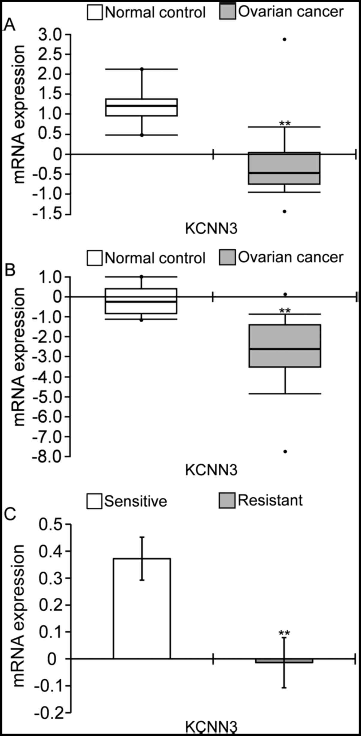
KCNN3 expression is decreased in OC
tissues and drug-resistant tissues compared with normal
tissues
KCNN3 expression was significantly decreased in OC
tissues compared with the normal control, according to the TCGA
Ovarian cohort and Yoshihara Ovarian cohort deposited in Oncomine.
(Fig. 1A and B). KCNN3 mRNA was
significantly lower in 586 samples ovarian serous
cystadenocarcinomas compared with eight normal ovaries, by
2.770-fold change obtained from the TCGA Ovarian cohort, as
determined via Log2 median-centered intensity (Fig. 1A); and was lower in 40 ovarian
serous cystadenocarcinomas compared with 10 normal peritoneal
samples, by 5.778-fold changes, according to the Yoshihara Ovarian
cohort (Fig. 1B). Furthermore,
KCNN3 was significantly lower in drug-resistant OC tissues compared
with sensitive tissues. KCNN3 mRNA expression in 90
platinum-resistant tissues was significantly lower compared with in
197 platinum-sensitive tissues in TCGA cohort (Fig. 1C; P<0.01; Table I).
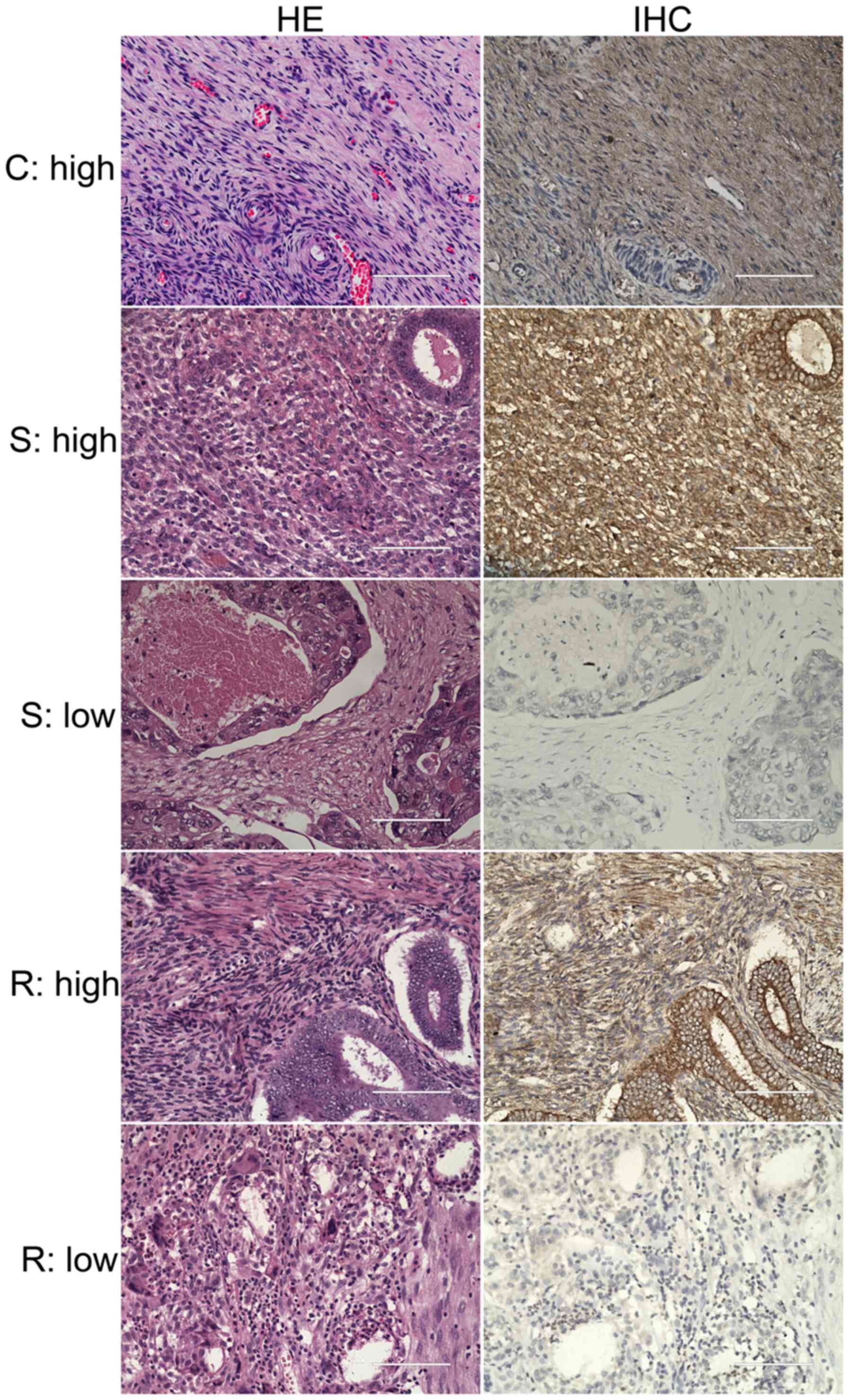
The morphology of 57 OC tissues and 6 normal ovarian
tissues were investigated via H&E staining (Fig. 2). The immunohistochemistry results
of these tissues demonstrated that the majority of OC tissues
revealed low expression levels of KCNN3 (33/57 cases); whereas, all
six normal controls revealed high expression levels of the protein
(Fig. 2). Statistical analysis
using the χ2 test revealed a significant low expression
of KCNN3 in OC tissues, as determined via imaging of
immunohistochemistry results (P=0.009). Furthermore, among the 51
OC specimens from the lab collection, KCNN3 protein expression was
significantly lower in 24 drug-resistant OC tissues compared with
27 sensitive tissues, as determined via imaging of
immunohistochemistry results (Fig.
2; Table I; P=0.011). The
percentage of drug-resistant tissues with low expression of KCNN3
was 75% (18/24 cases), whereas the low expression of the gene in
drug-sensitive tissues was only 37% (10/27 cases; P=0.011; Table I).
Low KCNN3 expression predicts shorter
disease-free survival (DFS) and OS in OC
KCNN3 expression was analyzed against OC clinical
factors, and identified to be associated with prognosis. Lower
KCNN3 mRNA expression was significantly associated with shorter DFS
in 489 patients with OC in TCGA ovarian cohort (low vs. high
groups; average values, 22.630±1.720 vs. 35.231±4.684; median
values, 15.410±1.049 vs. 18.040±1.253; P=0.016; Fig. 3A), although its association with OS
was not significant (low vs. high groups; average values,
49.126±3.051 vs. 59.070±4.489; median values, 43.400±3.858 vs.
43.890±2.449; P=0.153; data not shown). However, lower KCNN3 mRNA
expression was significantly associated with poor OS in a large
sample of 1,656 patients with OC from the KM Plotter (low vs. high
groups; average values, 61.517±2.608 vs. 77.324±4.214; median
values, 44.430±2.001 vs. 47.820±2.564; P=0.014; Fig. 3B), which included the above 489
patients in TCGA cohort (28).
These results were consistent with observations in specimens from
51 patients with OC, in which lower KCNN3 protein expression was
significantly associated with poor DFS (low vs. high groups,
average values; 16.920±4.230 vs. 38.559±5.764; median values.
6.000±0.400 vs. 47.000±10.441; P=0.026; Fig. 3C) and OS (low vs. high groups;
average values, 38.950±5.388 vs. 68.712±5.025; P=0.006; Fig. 3D). There is no median data for OS
as the mortality rate in this subgroup was <50%.
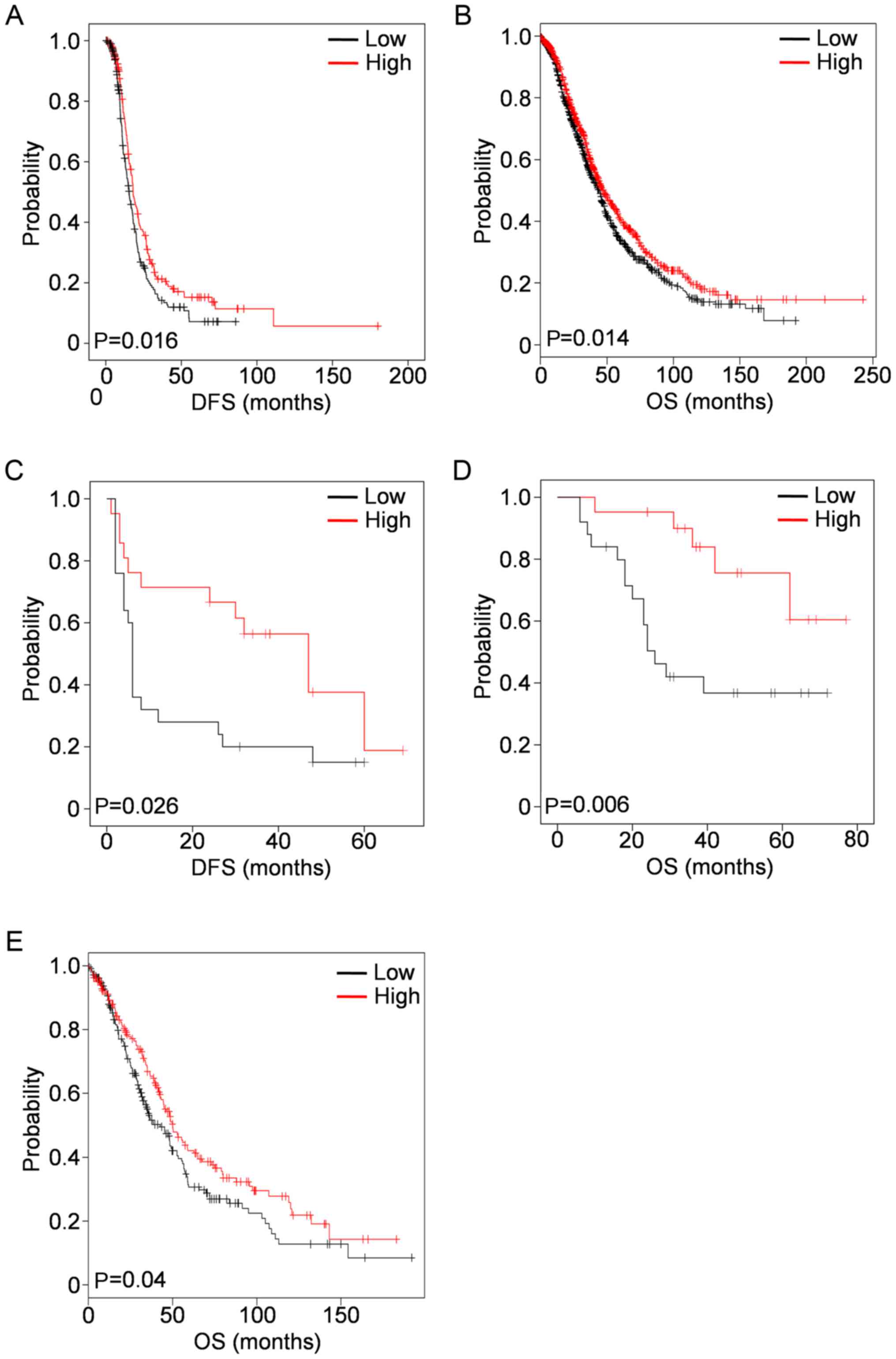 | Figure 3.Low KCNN3 expression is associated
with DFS and OS in OC, as determined using KM survival plots. (A)
Low KCNN3 mRNA expression was associated with shorter DFS in 489
patients with OC (TCGA ovarian cohort). (B) Low KCNN3 mRNA
expression (probe: 205903_s_at) was associated with shorter OS in
1,656 patients (data from KM Plotter). Low KCNN3 protein expression
was associated with (C) shorter OS and (D) DFS in 51 OC specimens.
(E) Low KCNN3 mRNA expression was associated with shorter OS in 395
patients with OC whose CA 125 expression levels were in the lowest
quartile (data from KM Plotter). mRNA expression values were
dichotomized into high and low, using the median as a cutoff.
Protein expression values were dichotomized into high and low,
according to slide immunostaining scores. KCNN3, potassium
calcium-activated channel subfamily N member 3; DFS, disease-free
survival; OS, overall survival; OC, ovarian cancer; KM,
Kaplan-Meier; CA 125, cancer antigen 125. |
A low expression level of KCNN3 protein in the
subgroup of patients with OC with high expression levels of
mucin-16 [cancer antigen (CA) 125] (≥400 µl/ml) was additionally
observed, compared with a higher KCNN3 expression level in patients
with low expression levels of CA 125 (<400 µl/ml; Table I). Of these 395 patients with OC
who had low CA 125 expression levels (in the lowest quartile), low
KCNN3 expression was notably associated with shorter OS in the KM
Plotter cohort (Fig. 3E). In
addition, low KCNN3 expression was significantly associated with
higher histological grade (Grade III) in the 51 OC specimens from
the lab collection, although no significant association was
detected between KCNN3 and tumor stage (Table I).
Bioinformatics analyses suggest that
KCNN3 mediates drug resistance
Bioinformatics approaches were performed, including
protein interaction and text mining, to predict the function of
KCNN3 in OC drug resistance. The search terms ‘KCNN3’, ‘OC’ and
‘drug resistance (DR)’ were significantly associated with 51 drugs
(e.g. cisplatin, oxaliplatin and doxorubicin), 47 genes and
proteins (including, TP53, JUN and CD34), 43 biological processes
(including, ‘apoptosis’, ‘DNA repair’ and ‘cell cycle’), nine
cellular components and four molecular functions (Fig. 4A). Possible networks of protein or
gene interactions with KCNN3 included 49 gene or gene products
associated with drug-resistance in OC, which further explained its
association with drug resistance. These genes/gene products
included 25 oncogenes (35); URI1,
STAT3, SRC, RSF1, PIK3CA, NOTCH3, NINL, NFKB1, MYC, MIEN1, MET,
KRAS, JUN, IKBKE, FOS, ERBB2, EGFR, DAXX, CUZD1, CLU, BCL2, BAX,
AKT2, AKT1 and ACTN4 and 15 tumor suppressors (36) including BRCA1, BRCA2, CHEK2,
FBXO32, MLH1, SULF1, IL24, CDKN2A, CDKN1A, TP53, TP73, PDCD4, PTEN,
RASSF1 and WWOX, as well as 9 other genes, including CCL21 and
SPARCL1 (37), GGNBP2 and RNASET2
(38), NEK2 (39), NEK11 (40), ALDH1A2 and ADH1B (41) and TRPC1 (42). KCNN3 directly interacted with 18 of
these genes or gene products, and exhibited indirect interactions
with the rest (Fig. 4B). As there
were associations between KCNN3 and a wide range of drugs,
genes/proteins, biological processes, cellular components and
molecular functions with known roles in OC and drug resistance
(Fig. 4), it was concluded that
KCNN3 expression possibly affects drug resistance in OC.
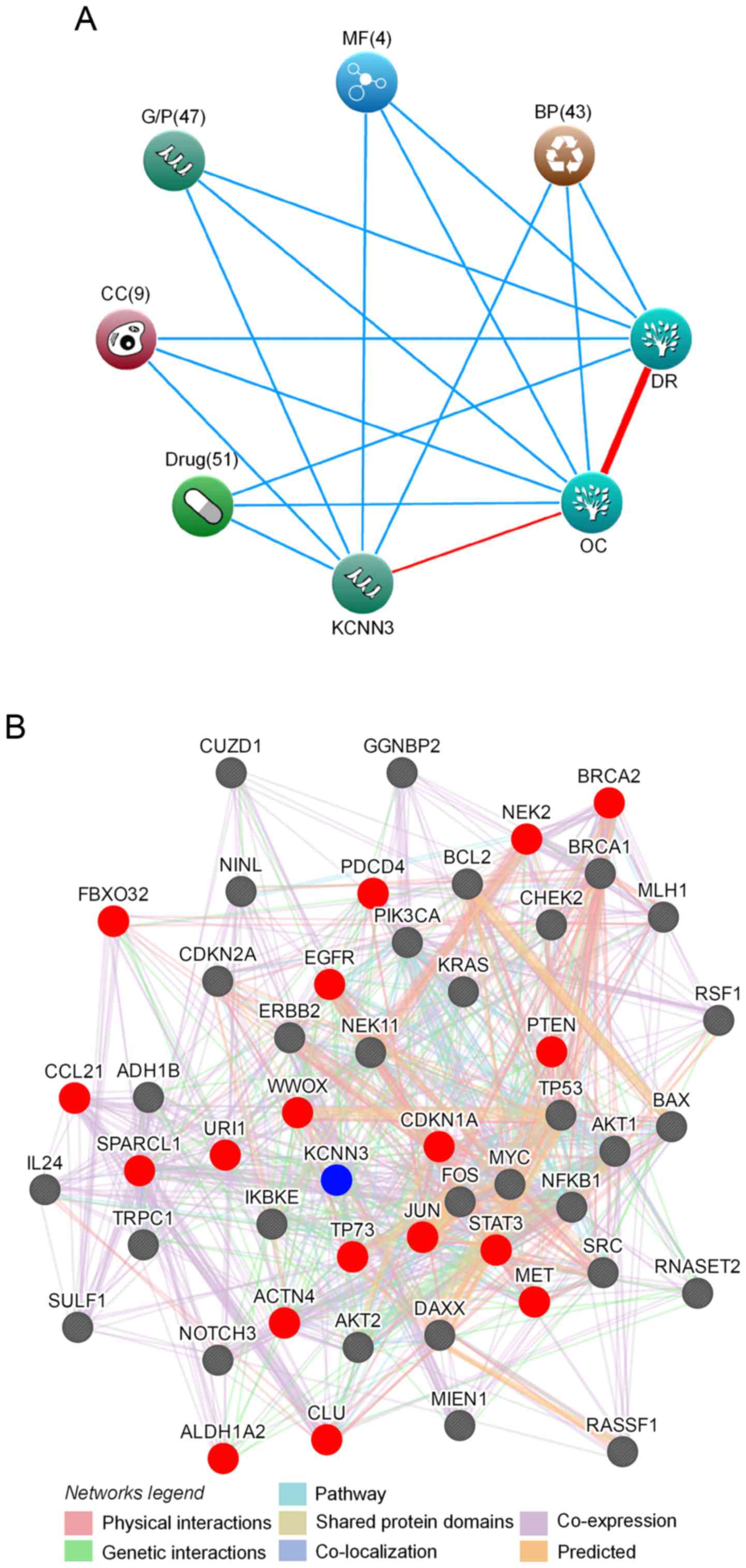 | Figure 4.Bioinformatics analyses of
associations between KCNN3 and drug resistance in OC. (A) KCNN3
associations with drug resistance and OC, as determined by text
mining using Coremine Medical. The input terms were ‘KCNN3’, ‘drug
resistance’ and ‘ovarian cancer’. (B) A protein interaction network
of KCNN3 with 49 drug resistance-associated proteins in OC, as
generated using GeneMANIA. Red and grey circles represent target
proteins that interact with KCNN3 directly and indirectly,
respectively. KCNN3, potassium calcium-activated channel subfamily
N member 3; OC, ovarian cancer; DR, drug resistance; BP, biological
process; MF, molecular function; G/P, genes and proteins; CC,
cellular component. |
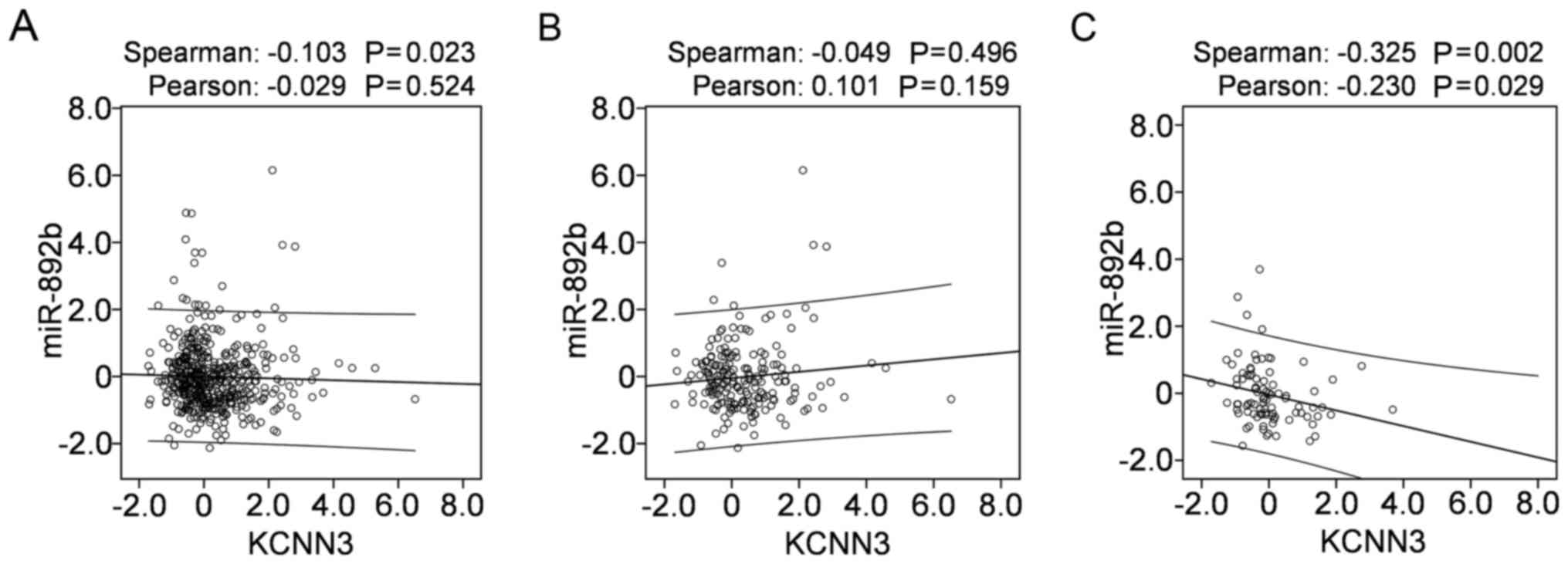
KCNN3 expression is potentially
regulated by miR-892b
To investigate the possible mechanism that mediates
KCNN3 expression in OC, mRNA-miR prediction was conducted using the
miRSystem, which predicted 35 miRNAs that potentially target KCNN3.
Of these, 24 with expression data available in TCGA were downloaded
from cBioportal, from which correlations were analyzed between
miRNA expression and KCNN3 mRNA expression in 489 OC tissues. Among
the 24 miRNAs, only the expression of miR-892b was negatively
correlated with KCNN3 mRNA expression in the 489 OC tissues when
determined using Spearman's correlation (Fig. 5A; P<0.05); however, not when
determined using Pearson's χ2 test (Fig. 5A; P>0.05). In 197
platinum-sensitive tissues of the 489 OC tissues, miR-892b was not
correlated with KCNN3 (Fig. 5B);
however, they were significantly and negatively correlated in 90
platinum-resistant tissues of the 489 OC tissues (Fig. 5C; P<0.05). The present results
support the possibility that miR-892b targets KCNN3 and contributes
to its downregulation in OC, particularly in platinum-resistant
tissues.
DNA methylation with KCNN3 mRNA expression levels in
489 OC tissues (including 197 sensitive tissues and 90 resistant
tissues) was additionally analyzed; however, no correlation was
observed, although DNA methylation of KCNN3 was negatively
correlated with mRNA expression (data not shown).
Discussion
Novel technologies have led to an increase in the
volume and diversity of large-scale public data (43), which are a vital pillar of open
science and a key enabler of reproducibility and novel discoveries
(44). Reuse of public data may
potentially answer questions beyond those originally envisioned
(45), and provide a systems-level
approach to predicting treatment response and disease progression,
and to developing precision therapies (43,46).
Computational approaches based on these public datasets may
additionally facilitate more rapid annotation of protein function
and guide laboratory experiments (47). In the present study, microarrays
and associated clinical data retrieved from Oncomine, TCGA and KM
Plotter were used to identify genes associated with prognosis and
drug resistance in OC.
It was identified that KCNN3 was significantly lower
in OC tissues compared with normal controls, in agreement with the
two independent microarrays, Yoshihara ovarian statistics and TCGA
ovarian cohort. This result was consistent with findings that KCNN3
was significantly downregulated in ≥10 tumor types and upregulated
in only three different tumors. Further analyses based on TCGA
cohort indicated significantly lower KCNN3 expression in
drug-resistant OC tissues, which was supported by experiments
conducted with 51 OC specimens. Low KCNN3 expression in OC,
particularly in drug-resistant tissues, appears to be regulated by
miR-892, which has been demonstrated to affect cancer growth,
migration, invasion, metastasis and angiogenesis (48,49).
Significantly lower expression of KCNN3 in OC and
drug-resistant OC suggests that KCNN3 mediates cancer progression
and drug resistance. This hypothesis is supported by bioinformatics
analyses, including text mining and protein interaction analyses,
and is consistent with a previous study, in which KCNN3 was
predicted to be one of 1,298 genes that contribute to drug
resistance in OC (50). A previous
study demonstrated that KCNN3 together with the TRPC1 and orai-1
complex regulates SOCE-dependent colon cancer cell migration
(21). Specifically, acquisition
of drug resistance in multiple myeloma is associated with the
suppression of inositol 1,4,5-triphosphate receptor type 1,
phospholipase C, transient receptor potential cation channel
subfamily M member 7 and TRPC1 expression, and reducing the
expression of TRPC1 markedly inhibits drug-induced cell death
(51). It was observed that
decreased expression of TRPC1 is associated with drug resistance in
OC (42), and the protein
interacts with KCNN3. Thus, it was concluded that low expression of
KCNN3 may contribute to drug resistance via interactions with
TRPC1, through inhibition of drug induced cell death.
Downregulation of KCNN3 predicted worse DFS and OS
in 51 patients with OC, and consistently predicted worse DFS and OS
in 489 and 1,656 patients, respectively, suggesting that it may be
a marker for prognosis in OC, in particular for OS. Low KCNN3
expression was notably associated with decreased OS in 395 patients
with OC with CA 125 expression levels in the lowest quartile. The
downregulation of KCNN3 was associated with higher serum CA 125
(≥400 µl/ml); whereas, KCNN3 upregulation was associated with lower
serum CA 125 (<400 µl/ml), thus it was concluded that the gene
may be used to predict OS among patients whose serum CA 125 is
<400 µl/ml.
In conclusion, the present study reported for the
first time, to the best of the authors' knowledge, the associations
between KCNN3 and drug resistance and prognosis in OC, which
indicate that KCNN3 is a potential therapeutic target and
prognostic marker in the treatment of OC. Further in vitro
and in vivo studies are required to validate and clarify the
present results.
Acknowledgements
The authors would like to thank Mrs Marla Brunker,
from Liwen Bianji, Edanz Group China www.liwenbianji.cn/ac), for editing the English text
of a draft of this manuscript.
Funding
The present study was supported by National Natural
Science Foundation of China (grant nos. 81460397, 81660606 and
81302283), China Postdoctoral Science Foundation (grant nos.
2014M552535XB and 2014M552291), and Natural Science Foundation of
Guangxi (grant nos. 2015GXNSFAA139151, 2014GXNSFBA118155,
2015GXNSFBA139115 and 2014GXNSFCA118010).
Availability of data and materials
The datasets used and/or analyzed during the current
study are available from the corresponding author on reasonable
request.
Authors' contributions
FY designed the study. XL performed bioinformatics
analyses and data mining. LW and BZ collected samples and clinical
data. XC and CD performed immunohistochemical analysis. XL and FY
wrote the manuscript. All authors read and approved the final
manuscript.
Ethics approval and consent to
participate
The present study was approved by The Ethics
Committee of Guangxi Medical University (Nanning, China) with the
1964 Helsinki declaration and its later ethical standards. Informed
consent was obtained from all individual participants included in
the present study.
Consent for publication
Not applicable.
Competing interests
The authors declare that they have no competing
interests.
References
|
1
|
Siegel RL, Miller KD and Jemal A: Cancer
statistics, 2017. CA Cancer J Clin. 67:7–30. 2017. View Article : Google Scholar : PubMed/NCBI
|
|
2
|
Chen W, Zheng R, Baade PD, Zhang S, Zeng
H, Bray F, Jemal A, Yu XQ and He J: Cancer statistics in China,
2015. CA Cancer J Clin. 66:115–132. 2016. View Article : Google Scholar : PubMed/NCBI
|
|
3
|
Cooke SL and Brenton JD: Evolution of
platinum resistance in high-grade serous ovarian cancer. Lancet
Oncol. 12:1169–1174. 2011. View Article : Google Scholar : PubMed/NCBI
|
|
4
|
Miller DS, Blessing JA, Krasner CN, Mannel
RS, Hanjani P, Pearl ML, Waggoner SE and Boardman CH: Phase II
evaluation of pemetrexed in the treatment of recurrent or
persistent platinum-resistant ovarian or primary peritoneal
carcinoma: A study of the Gynecologic Oncology Group. J Clin Oncol.
27:2686–2691. 2009. View Article : Google Scholar : PubMed/NCBI
|
|
5
|
Pfisterer J and Ledermann JA: Management
of platinum-sensitive recurrent ovarian cancer. Semin Oncol. 33 2
Suppl:S12–S16. 2006. View Article : Google Scholar : PubMed/NCBI
|
|
6
|
Siegel R, Ma J, Zou Z and Jemal A: Cancer
statistics, 2014. CA Cancer J Clin. 64:9–29. 2014. View Article : Google Scholar : PubMed/NCBI
|
|
7
|
Holohan C, Van Schaeybroeck S, Longley DB
and Johnston PG: Cancer drug resistance: An evolving paradigm. Nat
Rev Cancer. 13:714–726. 2013. View
Article : Google Scholar : PubMed/NCBI
|
|
8
|
Arcangeli A, Crociani O, Lastraioli E,
Masi A, Pillozzi S and Becchetti A: Targeting ion channels in
cancer: A novel frontier in antineoplastic therapy. Curr Med Chem.
16:66–93. 2009. View Article : Google Scholar : PubMed/NCBI
|
|
9
|
Cuddapah VA and Sontheimer H: Ion channels
and transporters [corrected] in cancer. 2. Ion channels and the
control of cancer cell migration. Am J Physiol Cell Physiol.
301:C541–C549. 2011. View Article : Google Scholar : PubMed/NCBI
|
|
10
|
Prevarskaya N, Skryma R and Shuba Y: Ion
channels and the hallmarks of cancer. Trends Mol Med. 16:107–121.
2010. View Article : Google Scholar : PubMed/NCBI
|
|
11
|
D'Amico M, Gasparoli L and Arcangeli A:
Potassium channels: Novel emerging biomarkers and targets for
therapy in cancer. Recent Pat Anticancer Drug Discov. 8:53–65.
2013. View Article : Google Scholar : PubMed/NCBI
|
|
12
|
Pardo LA and Stühmer W: The roles of K(+)
channels in cancer. Nat Rev Cancer. 14:39–48. 2014. View Article : Google Scholar : PubMed/NCBI
|
|
13
|
Innamaa A, Jackson L, Asher V, Van
Shalkwyk G, Warren A, Hay D, Bali A, Sowter H and Khan R:
Expression and prognostic significance of the oncogenic K2P
potassium channel KCNK9 (TASK-3) in ovarian carcinoma. Anticancer
Res. 33:1401–1408. 2013.PubMed/NCBI
|
|
14
|
Asher V, Khan R, Warren A, Shaw R,
Schalkwyk GV, Bali A and Sowter HM: The Eag potassium channel as a
new prognostic marker in ovarian cancer. Diagn Pathol. 5:782010.
View Article : Google Scholar : PubMed/NCBI
|
|
15
|
Cicek MS, Koestler DC, Fridley BL, Kalli
KR, Armasu SM, Larson MC, Wang C, Winham SJ, Vierkant RA, Rider DN,
et al: Epigenome-wide ovarian cancer analysis identifies a
methylation profile differentiating clear-cell histology with
epigenetic silencing of the HERG K+ channel. Hum Mol
Genet. 22:3038–3047. 2013. View Article : Google Scholar : PubMed/NCBI
|
|
16
|
Pillozzi S, Masselli M, De Lorenzo E,
Accordi B, Cilia E, Crociani O, Amedei A, Veltroni M, D'Amico M,
Basso G, et al: Chemotherapy resistance in acute lymphoblastic
leukemia requires hERG1 channels and is overcome by hERG1 blockers.
Blood. 117:902–914. 2011. View Article : Google Scholar : PubMed/NCBI
|
|
17
|
Köhler M, Hirschberg B, Bond CT, Kinzie
JM, Marrion NV, Maylie J and Adelman JP: Small-conductance,
calcium-activated potassium channels from mammalian brain. Science.
273:1709–1714. 1996. View Article : Google Scholar : PubMed/NCBI
|
|
18
|
Chantome A, Girault A, Potier M, Collin C,
Vaudin P, Pagès JC, Vandier C and Joulin V: KCa2.3
channel-dependent hyperpolarization increases melanoma cell
motility. Exp Cell Res. 315:3620–3630. 2009. View Article : Google Scholar : PubMed/NCBI
|
|
19
|
Potier M, Joulin V, Roger S, Besson P,
Jourdan ML, Leguennec JY, Bougnoux P and Vandier C: Identification
of SK3 channel as a new mediator of breast cancer cell migration.
Mol Cancer Ther. 5:2946–2953. 2006. View Article : Google Scholar : PubMed/NCBI
|
|
20
|
Jelassi B, Chantome A, Alcaraz-Pérez F,
Baroja-Mazo A, Cayuela ML, Pelegrin P, Surprenant A and Roger S:
P2X(7) receptor activation enhances SK3 channels- and cystein
cathepsin-dependent cancer cells invasiveness. Oncogene.
30:2108–2122. 2011. View Article : Google Scholar : PubMed/NCBI
|
|
21
|
Gueguinou M, Harnois T, Crottes D, Uguen
A, Deliot N, Gambade A, Chantôme A, Haelters JP, Jaffrès PA,
Jourdan ML, et al: SK3/TRPC1/Orai1 complex regulates SOCE-dependent
colon cancer cell migration: A novel opportunity to modulate
anti-EGFR mAb action by the alkyl-lipid Ohmline. Oncotarget.
7:36168–36184. 2016. View Article : Google Scholar : PubMed/NCBI
|
|
22
|
Li X, Pennisi A, Zhan F, Sawyer JR,
Shaughnessy JD and Yaccoby S: Establishment and exploitation of
hyperdiploid and non-hyperdiploid human myeloma cell lines. Br J
Haematol. 138:802–811. 2007. View Article : Google Scholar : PubMed/NCBI
|
|
23
|
Rhodes DR, Kalyana-Sundaram S, Mahavisno
V, Varambally R, Yu J, Briggs BB, Barrette TR, Anstet MJ,
Kincead-Beal C, Kulkarni P, et al: Oncomine 3.0: Genes, pathways,
and networks in a collection of 18,000 cancer gene expression
profiles. Neoplasia. 9:166–180. 2007. View Article : Google Scholar : PubMed/NCBI
|
|
24
|
Rhodes DR, Yu J, Shanker K, Deshpande N,
Varambally R, Ghosh D, Barrette T, Pandey A and Chinnaiyan AM:
ONCOMINE: A cancer microarray database and integrated data-mining
platform. Neoplasia. 6:1–6. 2004. View Article : Google Scholar : PubMed/NCBI
|
|
25
|
Cancer Genome Atlas Research Network:
Integrated genomic analyses of ovarian carcinoma. Nature.
474:609–615. 2011. View Article : Google Scholar : PubMed/NCBI
|
|
26
|
Cerami E, Gao J, Dogrusoz U, Gross BE,
Sumer SO, Aksoy BA, Jacobsen A, Byrne CJ, Heuer ML, Larsson E, et
al: The cBio cancer genomics portal: An open platform for exploring
multidimensional cancer genomics data. Cancer Discov. 2:401–404.
2012. View Article : Google Scholar : PubMed/NCBI
|
|
27
|
Gao J, Aksoy BA, Dogrusoz U, Dresdner G,
Gross B, Sumer SO, Sun Y, Jacobsen A, Sinha R, Larsson E, et al:
Integrative analysis of complex cancer genomics and clinical
profiles using the cBioPortal. Sci Signal. 6:pl12013. View Article : Google Scholar : PubMed/NCBI
|
|
28
|
Gyorffy B, Lánczky A and Szállási Z:
Implementing an online tool for genome-wide validation of
survival-associated biomarkers in ovarian-cancer using microarray
data from 1287 patients. Endocr Relat Cancer. 19:197–208. 2012.
View Article : Google Scholar : PubMed/NCBI
|
|
29
|
Remmele W and Stegner HE: Recommendation
for uniform definition of an immunoreactive score (IRS) for
immunohistochemical estrogen receptor detection (ER-ICA) in breast
cancer tissue. Pathologe. 8:138–140. 1987.PubMed/NCBI
|
|
30
|
Jenssen TK, Laegreid A, Komorowski J and
Hovig E: A literature network of human genes for high-throughput
analysis of gene expression. Nat Genet. 28:21–28. 2001. View Article : Google Scholar : PubMed/NCBI
|
|
31
|
Mostafavi S, Ray D, Warde-Farley D,
Grouios C and Morris Q: GeneMANIA: A real-time multiple association
network integration algorithm for predicting gene function. Genome
Biol. 9 Suppl 1:S42008. View Article : Google Scholar : PubMed/NCBI
|
|
32
|
Warde-Farley D, Donaldson SL, Comes O,
Zuberi K, Badrawi R, Chao P, Franz M, Grouios C, Kazi F, Lopes CT,
et al: The GeneMANIA prediction server: Biological network
integration for gene prioritization and predicting gene function.
Nucleic Acids Res. 38:W214–W220. 2010. View Article : Google Scholar : PubMed/NCBI
|
|
33
|
Lu TP, Lee CY, Tsai MH, Chiu YC, Hsiao CK,
Lai LC and Chuang EY: miRSystem: An integrated system for
characterizing enriched functions and pathways of microRNA targets.
PLoS One. 7:e423902012. View Article : Google Scholar : PubMed/NCBI
|
|
34
|
Hedditch EL, Gao B, Russell AJ, Lu Y,
Emmanuel C, Beesley J, Johnatty SE, Chen X, Harnett P, George J, et
al: ABCA transporter gene expression and poor outcome in epithelial
ovarian cancer. J Natl Cancer Inst. 106:pii: dju149. 2014.
View Article : Google Scholar : PubMed/NCBI
|
|
35
|
Liu X, Gao Y, Lu Y, Zhang J, Li L and Yin
F: Oncogenes associated with drug resistance in ovarian cancer. J
Cancer Res Clin Oncol. 141:381–395. 2015. View Article : Google Scholar : PubMed/NCBI
|
|
36
|
Yin F, Liu X, Li D, Wang Q, Zhang W and Li
L: Tumor suppressor genes associated with drug resistance in
ovarian cancer (review). Oncol Rep. 30:3–10. 2013. View Article : Google Scholar : PubMed/NCBI
|
|
37
|
Yin F, Liu X, Li D, Wang Q, Zhang W and Li
L: Bioinformatic analysis of chemokine (C-C motif) ligand 21 and
SPARC-like protein 1 revealing their associations with drug
resistance in ovarian cancer. Int J Oncol. 42:1305–1316. 2013.
View Article : Google Scholar : PubMed/NCBI
|
|
38
|
Yin F, Liu L, Liu X, Li G, Zheng L, Li D,
Wang Q, Zhang W and Li L: Downregulation of tumor suppressor gene
ribonuclease T2 and gametogenetin binding protein 2 is associated
with drug resistance in ovarian cancer. Oncol Rep. 32:362–372.
2014. View Article : Google Scholar : PubMed/NCBI
|
|
39
|
Liu X, Gao Y, Lu Y, Zhang J, Li L and Yin
F: Upregulation of NEK2 is associated with drug resistance in
ovarian cancer. Oncol Rep. 31:745–754. 2014. View Article : Google Scholar : PubMed/NCBI
|
|
40
|
Liu X, Gao Y, Lu Y, Zhang J, Li L and Yin
F: Downregulation of NEK11 is associated with drug resistance in
ovarian cancer. Int J Oncol. 45:1266–1274. 2014. View Article : Google Scholar : PubMed/NCBI
|
|
41
|
Liu X, Gao Y, Zhao B, Li X, Lu Y, Zhang J,
Li D, Li L and Yin F: Discovery of microarray-identified genes
associated with ovarian cancer progression. Int J Oncol.
46:2467–2478. 2015. View Article : Google Scholar : PubMed/NCBI
|
|
42
|
Liu X, Zou J, Su J, Lu Y, Zhang J, Li L
and Yin F: Downregulation of transient receptor potential cation
channel, subfamily C, member 1 contributes to drug resistance and
high histological grade in ovarian cancer. Int J Oncol. 48:243–252.
2016. View Article : Google Scholar : PubMed/NCBI
|
|
43
|
Sparks R, Lau WW and Tsang JS: Expanding
the immunology toolbox: Embracing public-data reuse and
crowdsourcing. Immunity. 45:1191–1204. 2016. View Article : Google Scholar : PubMed/NCBI
|
|
44
|
Grechkin M, Poon H and Howe B: Wide-Open:
Accelerating public data release by automating detection of overdue
datasets. PLoS Biol. 15:e20024772017. View Article : Google Scholar : PubMed/NCBI
|
|
45
|
Rung J and Brazma A: Reuse of public
genome-wide gene expression data. Nat Rev Genet. 14:89–99. 2013.
View Article : Google Scholar : PubMed/NCBI
|
|
46
|
Kannan L, Ramos M, Re A, El-Hachem N,
Safikhani Z, Gendoo DM, Davis S, Gomez-Cabrero D, Castelo R, Hansen
KD, et al: Public data and open source tools for multi-assay
genomic investigation of disease. Brief Bioinform. 17:603–615.
2016. View Article : Google Scholar : PubMed/NCBI
|
|
47
|
Sharan R, Ulitsky I and Shamir R:
Network-based prediction of protein function. Mol Syst Biol.
3:882007. View Article : Google Scholar : PubMed/NCBI
|
|
48
|
Shin SS, Park SS, Hwang B, Moon B, Kim WT,
Kim WJ and Moon SK: MicroRNA-892b influences proliferation,
migration and invasion of bladder cancer cells by mediating the
p19ARF/cyclin D1/CDK6 and Sp-1/MMP-9 pathways. Oncol Rep.
36:2313–2320. 2016. View Article : Google Scholar : PubMed/NCBI
|
|
49
|
Jiang L, Yu L, Zhang X, Lei F, Wang L, Liu
X, Wu S, Zhu J, Wu G, Cao L, et al: miR-892b silencing activates
NF-κB and promotes aggressiveness in breast cancer. Cancer Res.
76:1101–1111. 2016. View Article : Google Scholar : PubMed/NCBI
|
|
50
|
Lloyd KL, Cree IA and Savage RS:
Prediction of resistance to chemotherapy in ovarian cancer: A
systematic review. BMC Cancer. 15:1172015. View Article : Google Scholar : PubMed/NCBI
|
|
51
|
Emmons MF, Anreddy N, Cuevas J,
Steinberger K, Yang S, McLaughlin M, Silva A and Hazlehurst LA:
MTI-101 treatment inducing activation of Stim1 and TRPC1 expression
is a determinant of response in multiple myeloma. Sci Rep.
2:72017.
|