Assessment of the effects of prostaglandins on myometrial and leiomyoma cells in vitro through microRNA profiling

  • Authors:
    • Hwi Gon Kim
    • Ga Young Jung
    • Seung Bin Park
    • Yeon Jean Cho
    • Myoungseok Han
  • View Affiliations

  • Published online on: June 14, 2018     https://doi.org/10.3892/mmr.2018.9160
  • Pages: 2499-2505
Metrics: Total Views: 0 (Spandidos Publications: | PMC Statistics: )
Total PDF Downloads: 0 (Spandidos Publications: | PMC Statistics: )


Abstract

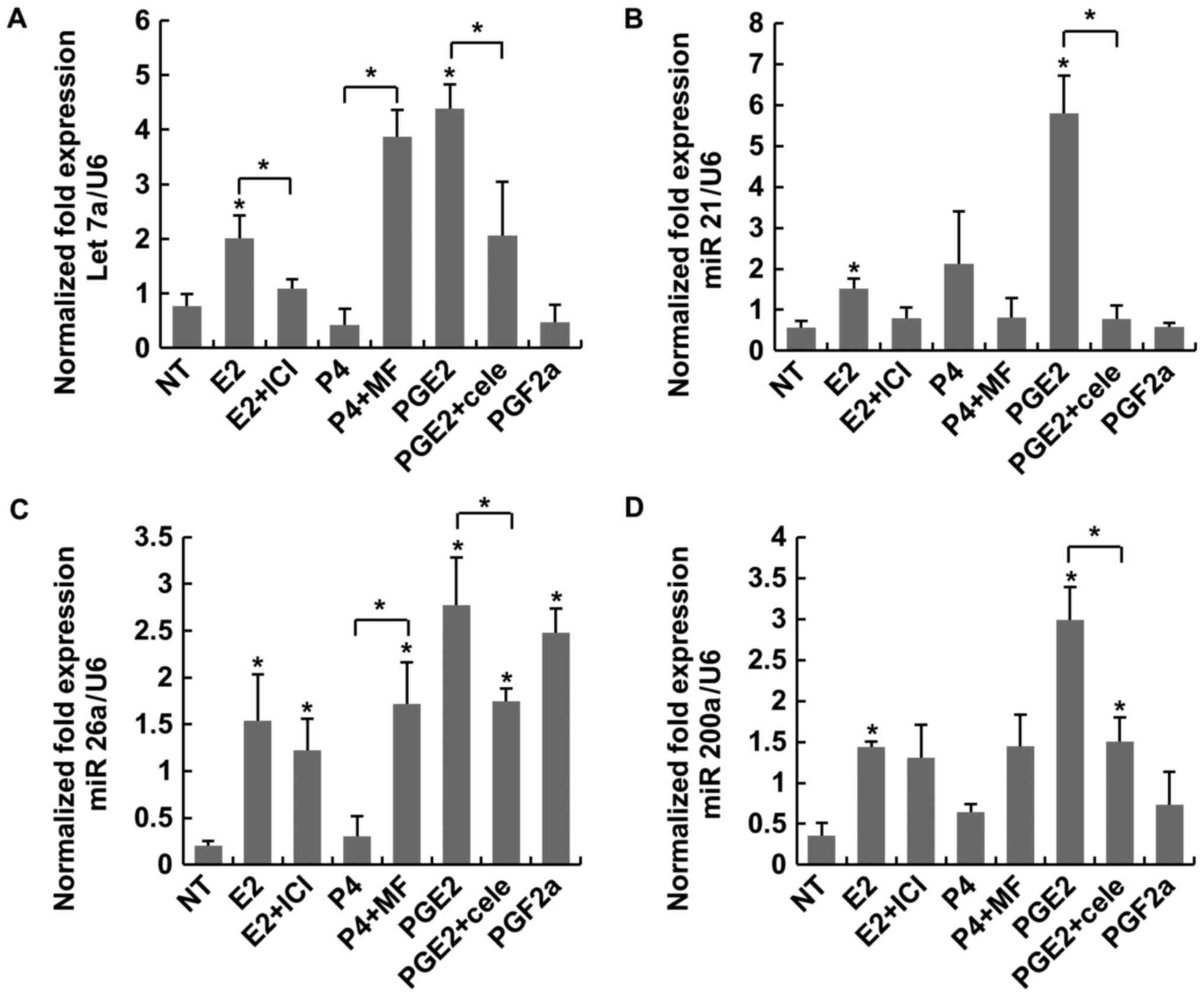
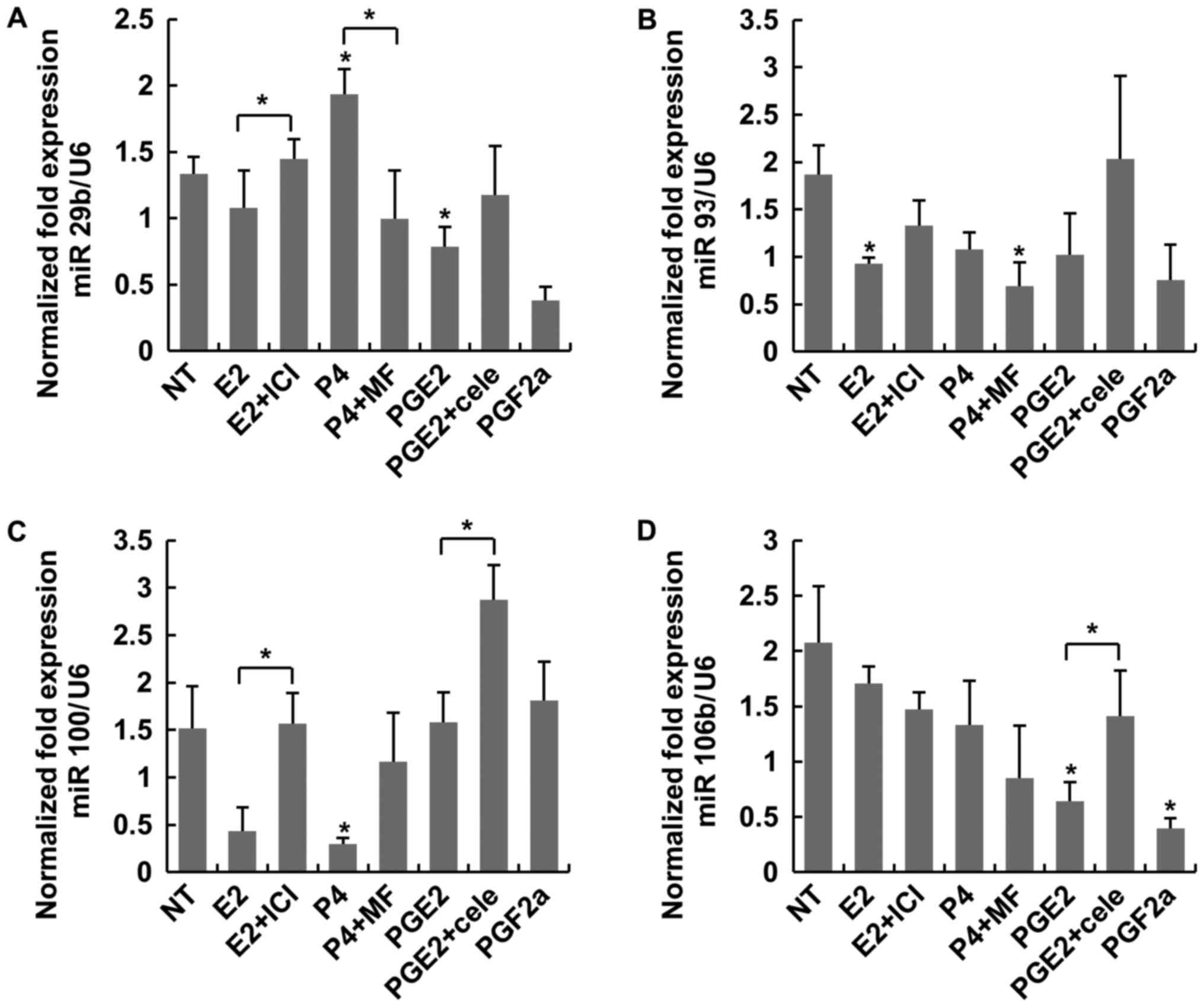
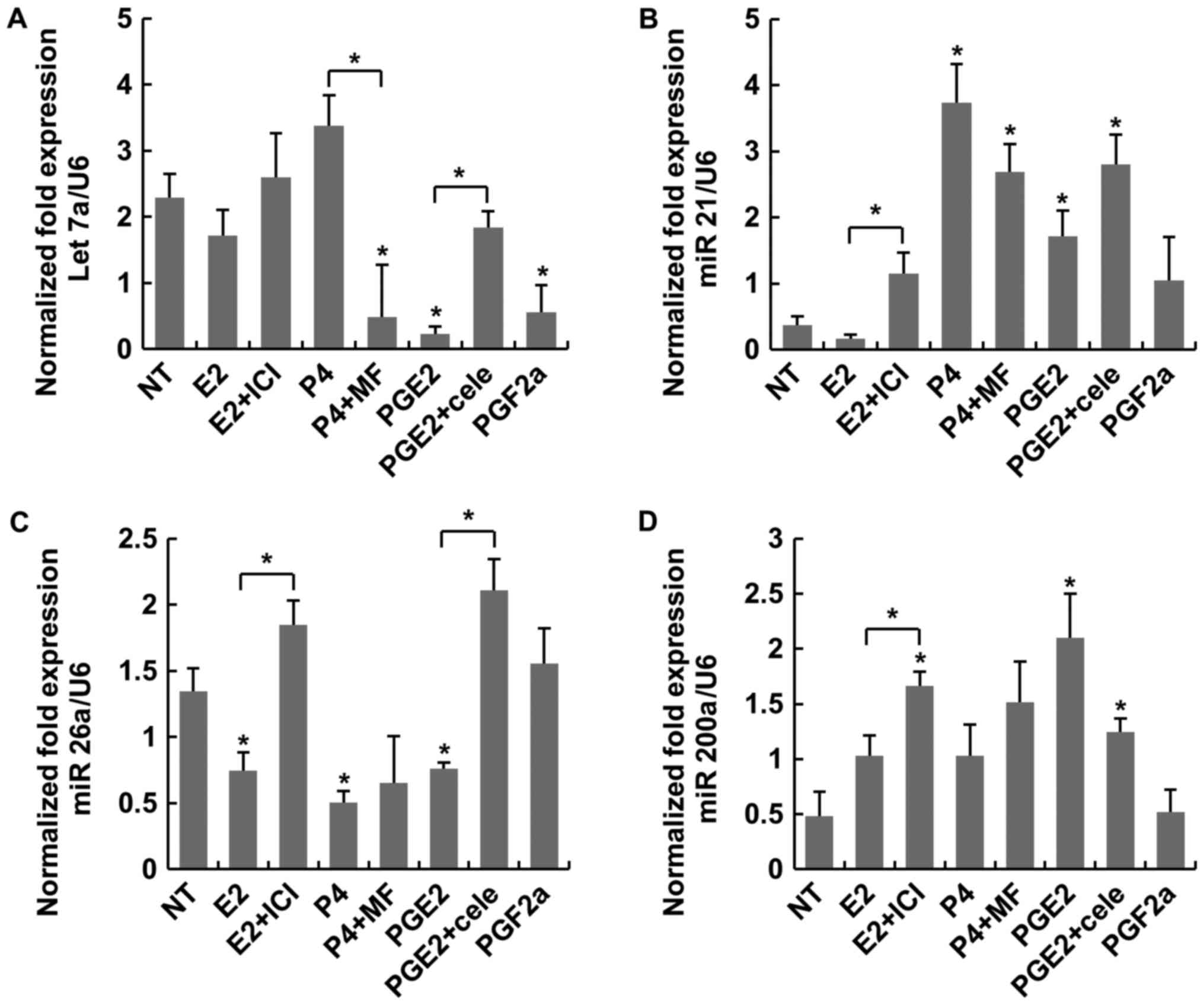
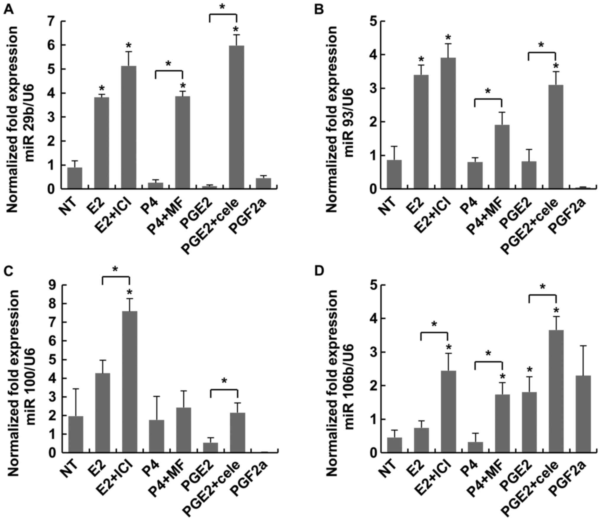
It is well known that prostaglandin (PG) E2 and PGF2α are secreted in copious amounts from the menstruating uterus. The aim of the present study was to determine whether PGs affect the growth of uterine leiomyomas (ULs) to the same extent as estrogen or progesterone (P4). The present study evaluated the expression of eight microRNAs (miRNAs) by reverse transcription‑quantitative polymerase chain reaction (RT‑qPCR) through treatment with estradiol (E2), P4, PGE2, PGF2α and each antagonist or cyclooxygenase‑2 (COX‑2) inhibitor of cultured leiomyoma and myometrial cells (LC and MC, respectively). The eight miRNAs were divided into two groups according to their primary biological action, namely apoptosis‑regulating miRNAs (let‑7a, miR‑21, miR‑26a and miR‑200a) and inflammation‑regulating miRNAs (miR‑29b, miR‑93, miR‑106b and miR‑100b). PGE2 induced significantly higher expression of the 3 anti‑apoptotic miRs, let‑7a, miR‑16a and miR‑200a, in LC when compared with the non‑treated control or E2. PGE2 significantly promoted a greater expression of let‑7a and miR‑26a in LC when compared with P4. Overall, PGE2 exerted the highest anti‑apoptotic and anti‑inflammatory effect in LC, which was comparable with E2. It was not observed among the inflammation‑regulating miRNAs in LC. PGF2α did not exert effects as prominent as those of PGE2. In MC, PGs and sex steroids exerted no similar effects on MC compared with LC. The present study demonstrated that PGE2 levels during menstruation may affect the growth of preexisting ULs without affecting the normal myometrium. Therefore, the control of secretion of PGs from the menstruating uterus or the administration of antagonists may be an alternative therapy for inhibiting the growth of ULs.
View Figures
View References

Related Articles

Journal Cover

August-2018
Volume 18 Issue 2

Print ISSN: 1791-2997
Online ISSN:1791-3004

Sign up for eToc alerts

Recommend to Library

Copy and paste a formatted citation
x
Spandidos Publications style
Kim HG, Jung GY, Park SB, Cho YJ and Han M: Assessment of the effects of prostaglandins on myometrial and leiomyoma cells in vitro through microRNA profiling. Mol Med Rep 18: 2499-2505, 2018
APA
Kim, H.G., Jung, G.Y., Park, S.B., Cho, Y.J., & Han, M. (2018). Assessment of the effects of prostaglandins on myometrial and leiomyoma cells in vitro through microRNA profiling. Molecular Medicine Reports, 18, 2499-2505. https://doi.org/10.3892/mmr.2018.9160
MLA
Kim, H. G., Jung, G. Y., Park, S. B., Cho, Y. J., Han, M."Assessment of the effects of prostaglandins on myometrial and leiomyoma cells in vitro through microRNA profiling". Molecular Medicine Reports 18.2 (2018): 2499-2505.
Chicago
Kim, H. G., Jung, G. Y., Park, S. B., Cho, Y. J., Han, M."Assessment of the effects of prostaglandins on myometrial and leiomyoma cells in vitro through microRNA profiling". Molecular Medicine Reports 18, no. 2 (2018): 2499-2505. https://doi.org/10.3892/mmr.2018.9160