Introduction
Umbilical cord blood transplantation (UCBT) is
increasingly performed as an alternative treatment for
hematological malignancies, including leukemia and lymphomas
(1,2). Hematopoietic stem cells in cord blood
(CB) are immature and limited in number, which may correlate to the
relatively high incidence of engraftment failure and delays in UCBT
(3). Hematopoietic stem cell
activity is routinely estimated by flow cytometric detection of
cluster of differentiation (CD)34+ cells, and by
colony-forming unit (CFU) assays in which the ability of blood
cells to form colonies in a semisolid medium is tested, for
example, formation of granulocyte/macrophage (GM) colonies in the
CFU-GM assay. Predictive markers of CB engraftment are important
for successful UCBT, and reports suggest that increased numbers of
total nucleated cells (TNC), CD34+ cells (3), and CFU-GM or total CFUs (4) in the CB graft are associated with
better engraftment and survival in UCBT.
CB for transplantation is currently provided by the
CB bank (CBB), where CB units (CBUs) are cryopreserved following
harvesting during full term delivery. As cryopreservation causes a
decrease in the absolute number of TNCs and CD34+ cells
(5), a quality assessment
following thawing is performed on selected CBUs (6). Because the cryopreserved CB in the
main bag is shipped for transplantation, the CB in the attached
segment provides the only source for indirect assessment of the CB
quality in the man bag (7).
Aldehyde dehydrogenase (ALDH) is a cytosolic enzyme
is responsible for the oxidation of intracellular aldehydes. In an
ALDH activity detection system (ALDEFLUOR kit) (8), ALDH converts fluorescently-labelled
aminoacetaldehyde, an ALDH substrate which freely diffuses into
cells, into fluorescently-labelled aminoacetate, a negatively
charged product that is retained inside cells. Therefore, cells
with high ALDH activity accumulate increasing amounts of the
fluorescent aminoacetate and are subsequently detected as
ALDH-bright (ALDHbr) cells by flow cytometry (8). ALDH is considered to be a detoxifying
enzyme that protects stem cells from cytotoxic effects, and its
activity is reported to be high in primitive hematopoietic stem
cells (8,9). Therefore, ALDH activity is considered
to be a marker of stem cell activity. In peripheral blood stem cell
(PBSC) transplantation, the number of ALDHbr cells in
mobilized PBSC correlates with engraftment success following
autologous transplantation (10).
In UCBT, ALDHbr cells are correlated with CFU-GM,
suggesting that ALDH activity may be suitable for quality
assessment of CBUs (11).
In the present study, the association between TNCs,
ALDHbr cells, CD34+ cells and CFUs was
examined in 16 CBUs. In addition, the number of ALDHbr
cells in the cryopreserved main bag were compared with those in the
attached segment. The present results suggested that evaluation of
ALDH activity in the cryopreserved CB cells of the attached
segments may be useful for quality assessment prior to UCBT.
Materials and methods
CB collection and processing
Umbilical CB was collected into a 200 ml bag
containing 28 ml citrate phosphate dextrose-adenine anticoagulant
by venipuncture of the umbilical cord following full-term delivery,
in cooperation with the Obstetric Clinic with Hyogo Cord Blood Bank
(Nishinomiya, Japan). Informed consent was obtained from the mother
to donate to the Cord Blood Unit (CBU) following CB transplantation
or for medical research if not suitable for transplantation. CBUs
that were not suitable for transplantation were selected for this
study between November 2012 and December 2014. CB was mixed with 6%
hydroxyethyl starch (Nipro Pharma Corporation, Osaka, Japan) at a
ratio of 5:1 for 5 min, and centrifuged at 50 × g for 5 min at room
temperature. The buffy coat layer was subsequently transferred to a
new bag. The buffy coat was cryopreserved in 5% dimethylsulfoxide,
6% hydroxyethyl starch (Kyokuto Pharmaceutical Industrial Co.,
Ltd., Tokyo, Japan) and 4% human serum albumin (Mitsubishi Tanabe
Pharma Corporation, Osaka, Japan) in a final volume of 25 ml in the
main bag, and ~0.4 ml in the attached tube which was divided into
four attached segments (each volume was ~0.125, 0.055, 0.11 and
0.11 ml) by heat sealing. CBUs were cryopreserved at −80°C without
rate-controlled freezing overnight (12), and were subsequently transferred
and stored in liquid nitrogen. For quality assessment, main bags
and attached segments were thawed in a 37°C water bath, and TNC and
CD34+ cell quantification, CFU assay and ALDH analysis
were performed subsequently. The present study was approved by the
institutional review board of Hyogo College of Medicine and Hyogo
Cord Blood Bank (Nishinomiya, Japan).
Quantification of TNC and CD34+
cells
Quantification of TNCs was determined using an
automated cell counter (XE-5000; Sysmex Corporation, Kobe, Japan).
Quantification of CD34+ hematopoietic stem and
progenitor cells was determined using a BD Stem Cell Enumeration
kit (cat. no. 344563; BD Biosciences, San Jose, CA, USA), which
included a fluorescein isothiocyanate-conjugated anti-CD45
monoclonal antibody (clone 2D1), a phycoerythrin-conjugated
anti-CD34 antibody (clone 8G12) and 7-Amino-actinomycin D (7AAD).
Cells at 2–20×106/ml were incubated with these
antibodies and 7AAD for 20 min at room temperature in the dark,
then erythrocytes were lysed with ammonium chloride solution.
Samples were analyzed using a FACSCanto™ II system and
BD FACSCanto™ Clinical software version 2.4 (BD
Biosciences). CD34+ cells were identified under
sequential gating of the 7AAD− and CD45dim+
cell populations according to the single platform guidelines of the
International Society of Hematotherapy and Graft Engineering
(13).
CFU assay
CFU assays were performed using a commercially
available methylcellulose medium (MethoCult H4034 Optimum; Stemcell
Technologies, Inc., Vancouver, BC, Canada) according to the
manufacturer's protocol. Briefly, 2–4×10⁴ CB cells were
cultured in semisolid methylcellulose medium containing recombinant
human (rh) stem cell factor, rh granulocyte-macrophage colony
stimulating factor (GM-CSF), rh G-CSF and rh interleukin-3 and rh
erythropoietin in 35-mm petri dishes for 14 days at 37°C in a
humidified atmosphere of 5% CO2. CFU-GM,
CFU-granulocyte/erythroid/macrophage/megakaryocyte (GEMM) and
burst-forming units-erythroid (BFU-E) were identified by
observation of GM, GEMM and erythroid colonies, respectively, using
an inverted microscope as described previously (14). The number of each CFU was
calculated as the mean number of colonies in three dishes. Total
CFUs were calculated by summation of mean CFU-GM, BFU-E and
CFU-GEMM values.
ALDH analysis
The ALDEFLUOR kit (Stemcell Technologies, Inc.) was
used for detection of intracellular ALDH enzyme activity by flow
cytometry, according to the manufacturer's protocol. Briefly,
boron-dipyrromethene (BODIPY) fluorescent dye-labeled
aminoacetaldehyde (BAAA) is a substrate for ALDH that diffuses
freely into cells. This is converted by ALDH into BODIPY
fluorescent dye-labeled aminoacetate (BAA), which remains trapped
intracellularly, thereby emitting a green fluorescence that enables
flow cytometric detection of ALDHbr cells (8). As a negative control, the background
fluorescence of the ALDHbr cells was obtained by
inhibition of ALDH activity by diethylaminobenzaldehyde. Analysis
of ALDHbr cells was performed using a FACSCanto II
system.
Statistical analysis
Data are expressed as the mean ± standard deviation
or as the median (range). Paired t-tests were used to assess
differences between the main bag and the attached segment. Linear
regression analysis was performed to assess the correlation between
the two groups. All statistical analyses were performed using
GraphPad Prism version 6.0 for Windows (GraphPad Software, Inc., La
Jolla, CA, USA). P<0.05 was considered to indicate a
statistically significant difference.
Results
Characteristics of thawed cord blood
units
In UCBT, CD34+ cell counts and CFU counts
in the CB graft are known to be good predictors for engraftment and
patient survival. Therefore, in the present study,
ALDHbr cell counts for UBC quality assessment were
evaluated by comparison with these parameters. The number of TNCs,
CD34+, and ALDHbr cells and total CFUs were
analyzed in the CB from the main bags of 16 CBUs, and detailed
results are provided in Table I.
The number of TNCs and ALDHbr cells were
8.06±1.70×10⁸/CBU and 2.61±2.78×10⁵/CBU,
respectively.
 | Table I.Characteristics of thawed cord blood
units in the main bags. |
Table I.
Characteristics of thawed cord blood
units in the main bags.
| Variables | Mean ± SD | Median | Range |
|---|
| TNCs
(×108/unit) | 8.06±1.70 | 7.76 | 5.50–12.25 |
| CD34+
cells (×105/unit) | 13.77±8.91 | 9.33 | 2.06–35.00 |
| ALDHbr
cells (×105/unit) | 2.61±2.78 | 1.16 | 0.29–9.17 |
| CFU-GM
(×105/unit) | 4.85±2.99 | 4.24 | 0.72–12.42 |
| BFU-E
(×105/unit) | 3.18±1.98 | 3.37 | 0.72–7.69 |
| CFU-GEMM
(×105/unit) | 0.04±0.14 | 0.14 | 0–0.54 |
| Total CFUs
(×105/unit) | 8.70±4.56 | 6.82 | 1.44–17.46 |
Association between ALDHbr
cell count and CD34+ cells and total CFU counts
The association between TNCs, ALDHbr
cells, CD34+ cells and CFUs was subsequently examined by
linear regression analysis. The number of TNCs were not
significantly correlated with the number of CD34+ cells
(Pearson's correlation coefficient R=0.1458, P>0.05) or the
number of ALDHbr cells (R=0.07254, P>0.05; data not
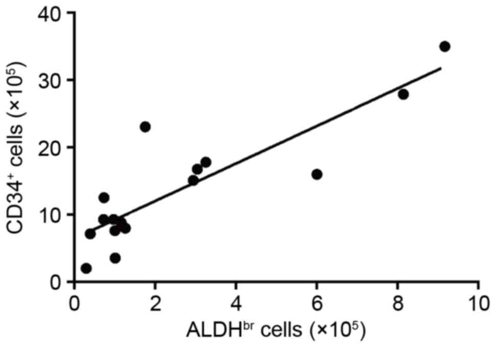
shown). As presented in Fig. 1,
the number of ALDHbr cells were significantly correlated
with CD34+ cell counts (R=0.8686, P<0.0001) in
post-thaw CB in the main bag. Next, three different colony-forming
unit assays were performed for myeloid lineage (CFU-GM), erythroid
lineage (BFU-E) and mixed myeloid and erythroid lineage cells
(CFU-GEMM). The association between ALDHbr cell counts
and CFU-GM, BFU-E or total CFUs (as the sum of the CFU-GM, BFU-E
and CFU-GEMM assays) in thawed CB in the main bag was examined. The
number of ALDHbr cells correlated with the number of
CFU-GM (R=0.6608, P=0.0053; Fig.
2A) and BFU-E (R=0.6964, P=0.0027; Fig. 2B). Notably, the number of
ALDHbr cells exhibited a stronger correlation to the
number of total CFUs (R=0.7502, P=0.0008; Fig. 2C). Additionally, the number of
total CFUs was positively correlated with the number of
CD34+ cells (R=0.7224, P=0.0016; Fig. 2D). The significant correlation of
ALDHbr cell count with CD34+ cell and total
CFU counts supports the hypothesis that measuring ALDHbr
cell numbers may serve as an indicator of CB quality following
cryopreservation.
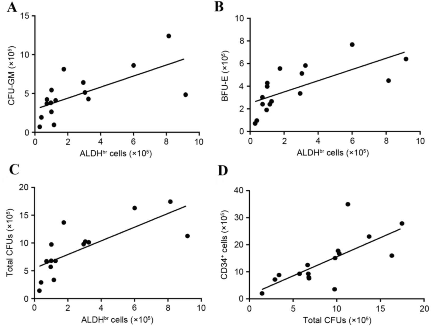 | Figure 2.Correlation among the numbers of
ALDHbr cells, CD34+ cells and CFUs in thawed
cord blood cells in the main bag. Scatter plots of
ALDHbr cell numbers and (A) CFU-GM (Pearson's
correlation coefficient R=0.6608, P=0.0053), (B) BFU-E (R=0.6964,
P=0.0027), and (C) total CFU numbers (R=0.7502, P.0008). (D)
Scatter plot of total CFU and CD34+ cell numbers
(R=0.7224, P=0.0016). Results represent analysis of 16 cord blood
units. ALDHbr, aldehyde dehydrogenase-bright; CFU,
colony forming unit; GM, granulocyte/macrophage; BFU-E,
burst-forming units-erythroid; CD34, cluster of differentiation
34. |
Comparison of ALDHbr cells
in the main bag and the attached segment
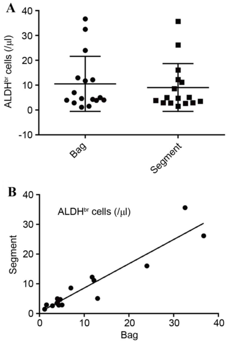
The concentration of ALDHbr cells was
subsequently evaluated in the main bags and attached segments of
the CBUs. As presented Fig. 3A,
the concentration of ALDHbr cells in the main bag and in
the attached segment was 10.05±1.76 and 9.06±1.76 cells/µl of CB,
respectively, which was not significantly different (P=0.1731).
Furthermore, the concentration of ALDHbr cells exhibited
a high linear correlation between the main bags and attached
segments in the 16 CBUs tested in the present study (R=0.9399,
P<0.0001; Fig. 3B). These
results suggested that ALDHbr cell counts in the
cryopreserved attached segments are similar to the main bag, and
therefore may successfully serve as an indicator in CB quality
assessment.
Discussion
Umbilical CB cells are an alternative source of
hematopoietic stem cells for transplantation. TNC, CD34+
cell and CFU counts in a CBU have been previously demonstrated to
positively correlate with good engraftment rate and improved
survival following CBT (3);
therefore, these parameters have been adopted as essential criteria
for assessing UCB quality prior to transplantation. In the present
study, ALDH activity in the CB was examined in parallel with TNC,
CD34+ cell and CFU counts, in order to evaluate it as a
novel marker for UCB quality.
ALDH oxidizes intracellular aldehydes to carboxylic
acid, and this process is involved in cellular detoxification,
proliferation, differentiation and drug resistance (10). ALDHbr cell presence is a
novel marker for human hematopoietic stem cells (8). In the present study, the number of
ALDHbr cells was highly correlated with the number of
CD34+ cells. Storms et al (9) reported that the
ALDHbr/CD34+ cell population contains
primitive hematopoietic stem cells and progenitor cells. The same
study addiitonally reported that ALDH−/CD34+
cells do not contain primitive stem cells but contain a few
progenitor cells, while ALDHbr/CD34− cells
exhibit no hematopoietic activity but contain erythroid cells
(9). Thus, ALHDbr cells
represent primitive hematopoietic stem and committed progenitor
cells. The results of the present study suggested that
ALDHbr cells may serve as a novel indicator of CB
quality; however, whether ALDHbr cells are more reliable
indicators than CD34+ cells requires further
clarification.
Lee et al (11) reported that ALDHbr cells
are correlated with post-thaw CFU-GM counts in CB. The present
study supported this finding and further demonstrated that
ALDHbr cells were correlated with BFU-E and total CFU
counts. In particular, total CFU counts exhibited the strongest
correlation coefficient among CFUs. Previous reports have used
either CFU-GM (11) or total CFU
(4) counts for assessment of
hematopoietic activity in transplantation. The quality and quantity
of mobilized PBSCs he been assessed by CFU-GM counts (15). In unpublished results from our
group, CFU assay of mobilized PBSCs gave rise to >90% of CFU-GM
colonies and only a few BFU-E and CFU-GEMM colonies (data not
shown). This finding may explain why CFU-GM is commonly used for
quality assessment of mobilized PBSCs in PBSC transplantation. By
contrast, CB cells gave rise to approximately 40% of CFU-GM, 50% of
BFU-E and 10% of CFU-GEMM colonies (data not shown). Therefore,
CFU-GM represent <50% total CFUs and the proportion of CFU-GM
and non-CFU-GM (i.e., the sum of BFU-E and CFU-GEMM) differ
considerably among individuals, indicating that CFU-GM may not be a
suitable indicator of CFUs. Accordingly, in the present study, the
number of ALDHbr cells was better correlated with total
CFUs than CFU-GM. Thus, total CFUs may be a better marker than
CFU-GM in the quality assessment of CB.
Attached segments of CBUs are routinely used for
quality assessment of the unit selected for transplantation.
Rodríguez et al (7)
demonstrated that there are no differences in the calculated number
of TNCs, CD34+ cells and CFUs between the main bag and
the attached segment; thus, the attached segment is considered to
be representative of the CBU in the main bag. However, another
report demonstrated that the attached segment exhibited higher
numbers of CD34+ cells and CFU-GM than the main bag
(16). Furthermore, the attached
segment is much smaller in volume compared with the main bag, and
extra caution is required in handling the attached segment to avoid
a rise in temperature that may be harmful to CB cells. Therefore, a
comparison of ALDHbr cell concentration in the main bag
and the attached segment was performed in the present study, to
ascertain the suitability of the attached segment as representative
for the main bag. The results demonstrated that the concentration
of ALDHbr cells in the attached segment was comparable
to that in the main bag.
In conclusion, the present study examined the use of
ALDH activity in the attached segments as a novel indicator of UBC
quality assessment in the main bag prior to transplantation.
Shoulars et al (17) have
recently reported similar results that ALHDbr cells in
the segment are associated with CFUs and may be utilized for
quality assays prior to shipping from a cord blood bank. Larger
clinical studies will be required in the future to examine the
correlation between the number of ALDHbr cells and
patient survival following UCBT.
Acknowledgements
The authors would like to thank Mrs Mayuko Ohno and
Mrs. Tika Tanihara (Hyogo Cord Blood Bank, Nishinomiya, Japan) for
their technical assistance. The authors would also like to thank
Mr. Kazuya Ashida (Hyogo Cord Blood Bank, Nishinomiya, Japan) for
the technical advice in the preparation of this manuscript.
Funding
The present study was supported by the research
grant from the Hyogo College of Medicine (grant received by
YF).
Availability of data and materials
The analyzed datasets generated during the study are
available from the corresponding author on reasonable request.
Authors' contributions
JI and TK performed the experiments using umbilical
cord blood cells. SY analyzed and interpreted the data. SK and TK
contributed for collecting and processing of umbilical cord blood.
YF conceived and design the study and was a major contributor in
writing the manuscript. All authors read and approved the final
manuscript.
Ethics approval and consent to
participate
The study was approved by the institutional review
board of Hyogo College of Medicine and Hyogo Cord Blood Bank
(Nishinomiya, Japan). Informed consent was obtained from the mother
to donate cord blood to Hyogo Cord Blood Bank for CB
transplantation or for medical research if not suitable for
transplantation.
Patient consent for publication
Not applicable.
Competing interests
The authors declare that they have no competing
interests.
References
|
1
|
Gluckman E, Rocha V, Boyer-Chammard A,
Locatelli F, Arcese W, Pasquini R, Ortega J, Souillet G, Ferreira
E, Laporte JP, et al: Outcome of cord-blood transplantation from
related and unrelated donors. Eurocord Transplant Group and the
European Blood and Marrow Transplantation Group. N Engl J Med.
337:373–381. 1997. View Article : Google Scholar : PubMed/NCBI
|
|
2
|
Laughlin MJ, Eapen M, Rubinstein P, Wagner
JE, Zhang MJ, Champlin RE, Stevens C, Barker JN, Gale RP, Lazarus
HM, et al: Outcomes after transplantation of cord blood or bone
marrow from unrelated donors in adults with leukemia. N Engl J Med.
351:2265–2275. 2004. View Article : Google Scholar : PubMed/NCBI
|
|
3
|
Wagner JE, Barker JN, De For TE, Baker KS,
Blazar BR, Eide C, Goldman A, Kersey J, Krivit W, MacMillan ML, et
al: Transplantation of unrelated donor umbilical cord blood in 102
patients with malignant and nonmalignant diseases: Influence of
CD34 cell dose and HLA disparity on treatment-related mortality and
survival. Blood. 100:1611–1618. 2002.PubMed/NCBI
|
|
4
|
Page KM, Zhang L, Mendizabal A, Wease S,
Carter S, Gentry T, Balber AE and Kurtzberg J: Total colony-forming
units are a strong, independent predictor of neutrophil and
platelet engraftment after unrelated umbilical cord blood
transplantation: A single-center analysis of 435 cord blood
transplants. Biol Blood Marrow Transplant. 17:1362–1374. 2011.
View Article : Google Scholar : PubMed/NCBI
|
|
5
|
Broxmeyer HE, Srour EF, Hangoc G, Cooper
S, Anderson SA and Bodine DM: High-efficiency recovery of
functional hematopoietic progenitor and stem cells from human cord
blood cryopreserved for 15 years. Proc Natl Acad Sci USA.
100:645–650. 2003. View Article : Google Scholar : PubMed/NCBI
|
|
6
|
Kudo Y, Minegishi M, Seki O, Takahashi H,
Suzuki A, Narita A, Sato Y, Abe M, Ishioka N, Harigae H and
Tsuchiya S: Quality assessment of umbilical cord blood units at the
time of transplantation. Int J Hematol. 93:645–651. 2011.
View Article : Google Scholar : PubMed/NCBI
|
|
7
|
Rodríguez L, García J and Querol S:
Predictive utility of the attached segment in the quality control
of a cord blood graft. Biol Blood Marrow Transplant. 11:247–251.
2005. View Article : Google Scholar : PubMed/NCBI
|
|
8
|
Storms RW, Trujillo AP, Springer JB, Shah
L, Colvin OM, Ludeman SM and Smith C: Isolation of primitive human
hematopoietic progenitors on the basis of aldehyde dehydrogenase
activity. Proc Natl Acad Sci USA. 96:9118–9123. 1999. View Article : Google Scholar : PubMed/NCBI
|
|
9
|
Storms RW, Green PD, Safford KM,
Niedzwiecki D, Cogle CR, Colvin OM, Chao NJ, Rice HE and Smith CA:
Distinct hematopoietic progenitor compartments are delineated by
the expression of aldehyde dehydrogenase and CD34. Blood.
106:95–102. 2005. View Article : Google Scholar : PubMed/NCBI
|
|
10
|
Fallon P, Gentry T, Balber AE, Boulware D,
Janssen WE, Smilee R, Storms RW and Smith C: Mobilized peripheral
blood SSCloALDHbr cells have the phenotypic and functional
properties of primitive haematopoietic cells and their number
correlates with engraftment following autologous transplantation.
Br J Haematol. 122:99–108. 2003. View Article : Google Scholar : PubMed/NCBI
|
|
11
|
Lee HR, Shin S, Yoon JH, Roh EY, Kim BJ
and Song EY: Aldehyde dehydrogenase-bright cells correlated with
the colony-forming unit-granulocyte-macrophage assay of thawed cord
blood units. Transfusion. 54:1871–1875. 2014. View Article : Google Scholar : PubMed/NCBI
|
|
12
|
Makino S, Harada M, Akashi K, Taniguchi S,
Shibuya T, Inaba S and Niho Y: A simplified method for
cryopreservation of peripheral blood stem cells at −80 degrees C
without rate-controlled freezing. Bone Marrow Transplant.
8:239–244. 1991.PubMed/NCBI
|
|
13
|
Keeney M, Chin-Yee I, Weir K, Popma J,
Nayar R and Sutherland DR: Single platform flow cytometric absolute
CD34+ cell counts based on the ISHAGE guidelines.
International Society of Hematotherapy and Graft Engineering.
Cytometry. 34:61–70. 1998. View Article : Google Scholar : PubMed/NCBI
|
|
14
|
Fujimori Y, Hara H and Nagai K: Effect of
lymphokine-activated killer cell fraction on the development of
human hematopoietic progenitor cells. Cancer Res. 48:534–538.
1988.PubMed/NCBI
|
|
15
|
Fu SQ, Abboud CN, Brennan JK,
Ifthikharuddin JJ, Nichols D and Liesveld JL: Impact of mobilized
blood progenitor cell quality determined by the CFU-GM/CD34+ ratio
on rapid engraftment after blood stem cell transplantation. Blood
Cells Mol Dis. 28:315–321. 2002. View Article : Google Scholar : PubMed/NCBI
|
|
16
|
Lee HR, Shin S, Yoon JH, Roh EY, Song EY,
Han KS and Kim BJ: Attached segment has higher CD34+ cells and
CFU-GM than the main bag after thawing. Cell Transplant.
24:305–310. 2015. View Article : Google Scholar : PubMed/NCBI
|
|
17
|
Shoulars K, Noldner P, Troy JD, Cheatham
L, Parrish A, Page K, Gentry T, Balber AE and Kurtzberg J:
Development and validation of a rapid, aldehyde dehydrogenase
bright-based cord blood potency assay. Blood. 127:2346–2354. 2016.
View Article : Google Scholar : PubMed/NCBI
|