Introduction
Osteonecrosis of the femoral head (ONFH) is a
progressive disease that may cause collapse of the femoral head,
pain and gait disorders. During the continuous disease progression,
collapse of the femoral head eventually leads to secondary
osteoarthritis of the hip joint. The incidence of collapse of the
femoral head and osteoarthritis in untreated patients is 70–80%
(1). The disease usually affects
young adults, and 5–12% of patients require total hip arthroplasty
(2). It has been demonstrated that
engraftment therapy of autologous bone marrow may be used as a
method of treatment (3).
Mesenchymal stem cell (MSC) treatment has also received much
interest.
MSCs have the ability to differentiate into a number
of cell types, including osteoblasts, adipocytes and chondrocytes
(4). Studies involving MSCs are
progressively developing towards clinical application phases. MSCs
are the most commonly utilized cell type in tissue engineering (TE)
fields (5). For example, in order
to treat avascular necrosis of the femoral head, skeletal stem
cells and impaction bone grafting were combined to produce a
mechanically stable living bone composite (6). MSCs have been isolated from various
tissues including bone marrow (BM), adipose tissue, dental pulp,
hair follicle, cornea, umbilical cord and placenta (7). The current gold standard is MSCs that
have been harvested from BM (5).
BMMSCs may be isolated and cultured from animals and humans. A
total of 4 randomized trials demonstrated decreased time to
collapse, improvements in pain score and decreased lesion sizes
with core decompression and BMMSCs treatment, compared with core
decompression alone to cure ONFH (8–11).
The most significant barrier for MSC-based therapies
is the decreased number of MSCs in the tissues of interest. Given
the necrotic nature of ONFH, the lesions associated with
osteonecrosis have poor innate regenerative capacity with few or no
viable MSCs (5).
A number of attempts have been made to solve this
problem. Among these applied methods, the strategy of affinity
peptides of BMMSCs obtained through phage display technology is
popular (12). Phage display is a
selection technique in which a library of peptide or protein
variants is expressed on the outside of a phage virion, while the
genetic material encoding each variant resides on the inside. In
order to recruit the seed cells and elevate the treatment
efficiency, a large number of biological materials have been
modified using these affinity peptides (12–18).
In the present study, an affinity peptide of mouse
bone marrow-derived MSCs was acquired using the phage display
technique. The aim was to conjugate this peptide on the surface of
materials. As peptide sequences have a certain level of
conservation in different species, we hypothesize that the
materials modified by the affinity peptide may be applied to the
therapy of the ONFH in TE and increase the adhesive efficiency of
BMMSCs (12).
Materials and methods
Cell culture
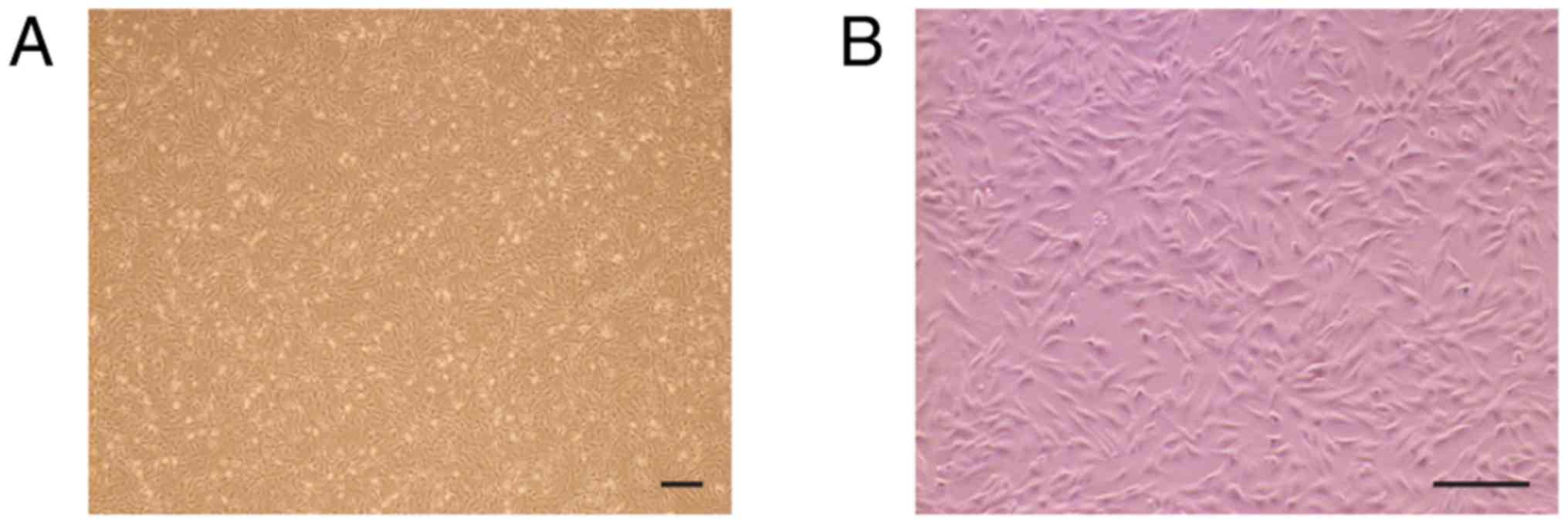
C57BL/6 mouse BMMSCs (cat. no. MUBMX-01001) were
obtained from Cyagen Biosciences, Inc., (Santa Clara, CA, USA). The
cells were cultured in cell culture flasks in a humidified
atmosphere with 5% CO2 at 37°C (Fig. 1). Cells were cultured in
low-glucose Dulbecco's modified Eagle's medium (DMEM; Biological
Industries, Ltd., Kibbutz Beit-Haemek, Israel; cat. no. 01-051-1A)
with L-glutamine containing 10% fetal bovine serum (Gibco; Thermo
Fisher Scientific, Inc., Waltham, MA, USA; cat. no., 10099141) and
antibiotics [100 U/ml penicillin and 0.1 mg/ml streptomycin (P/S);
Gibco; Thermo Fisher Scientific, Inc.; cat. no. 15140122]. The
medium was changed every 2–3 days. Cells were dissociated and
passaged at 80–90% confluence. Mouse BMMSCs at passage 4–5 were
used for subsequent experiments.
Phage display biopanning
Phage display biopanning, an affinity selection
technique that selects peptides through binding to a given target,
was performed according to a previously described protocol, with
certain modifications (14).
Peptide sequences with an affinity for C57BL/6 mouse bone
marrow-derived MSCs were identified by screening the Ph.D.C7C™
Phage Display Library (New England Biolabs, Inc., Ipswich, MA, USA;
cat. no., E8120S) against clonally-derived mouse BMMSCs at passage
4–5. The phage display library consisted of 109
different phages. Cells plated in 60×15 mm petri dishes were rinsed
twice with PBS (Biological Industries, Ltd.; cat. no., 02-024-1A)
and pre-blocked with DMEM containing 0.5% BSA (albumin bovine
fraction-V; NeoFroxx, Einhausen, Germany; cat. no., GRM105-5G)
without supplements at 37°C and 5% CO2 for 1 h. The
Ph.D.C7C™ Phage Display Library (1×10¹¹ PFU) was then
introduced to the cells. Non-binding phages were then discarded,
and the cells were washed 5 times in cold PBS. The phages binding
to the cells were eluted with 1 ml Glycine/HCl (pH 2.2) with 1
mg/ml BSA for 20 min at room temperature while being gently shaken.
The eluted phages were collected and neutralized with 0.15 ml 1 M
Tris-HCl (pH 9.1). A total of 3 rounds of panning were performed
for each sample.
Sequencing
Following 3 rounds of panning, blue plaques were
randomly selected for sequencing. Sequencing of phage DNA was
performed by GENEWIZ, Inc., (Suzhou, China). Firstly, the phage DNA
was enriched using the rolling circle amplification technique. This
technique uses Phi29 DNA polymerase and random hexamer primers to
amplify circular DNA molecules. Subsequently, the amplified phage
DNA products were used for Sanger sequencing. The bioinformatics
analysis of the generated data was carried out using Chromas 2.6.5
software (Technelysium Pty Ltd., South Brisbane, Australia).
Synthesis of peptides
A specific peptide was identified in the C57BL/6
mouse BMMSC-affinity clones and designated as D7. The peptide was a
loop-constrained heptapeptide with a pair of cysteine residues
conjugated to each terminus of 7 amino acids. The two cysteine
residues formed an intramolecular disulfide linkage. A peptide (Aa
sequence CVAVQNDSC) with the same amino acids as D7 in a scrambled
order was used as the negative control and designated as V7. A
peptide comprising 3 amino acids (arginine, glycine and aspartic
acid) was used as the positive control and termed RGD. All the
peptides were synthesized through solid-phase peptide synthesis
using fluorenylmethyloxyccarbonyl chemistry (Scilight-Peptide,
Inc., Beijing, China). An extra 6-aminocaproic acid molecule was
attached at the amino (N) terminal of all peptides to facilitate
fluorescein-5-isothiocyanate (FITC) labeling. The FITC-labeled
peptides were stored in −20°C. A 1 mg/ml concentration was obtained
by dissolving the peptides in PBS prior to use.
Peptide-affinity assay via flow
cytometry
The C57BL/6 mouse BMMSCs were washed twice with PBS,
and dissociated with 0.25% trypsin-EDTA (Gibco; Thermo Fisher
Scientific, Inc.). The cell suspension was then centrifuged at 250
× g for 5 min at room temperature to collect cell sedimentation.
The cells were incubated with 10 µM FITC-labeled peptides (Beijing
Scilight-Peptide Ltd., Co.) for 1 h at 37°C to allow cell binding
and internalization. The mouse BMMSC affinity properties of the
peptides were analyzed quantitatively using flow cytometry (BD LSR
Fortessa; Becton, Dickinson and Company, Franklin Lakes, NJ, USA)
at a wavelength of 488 nm and FlowJo 7.6.1 software (Tree Star,
Inc., Ashland, OR, USA).
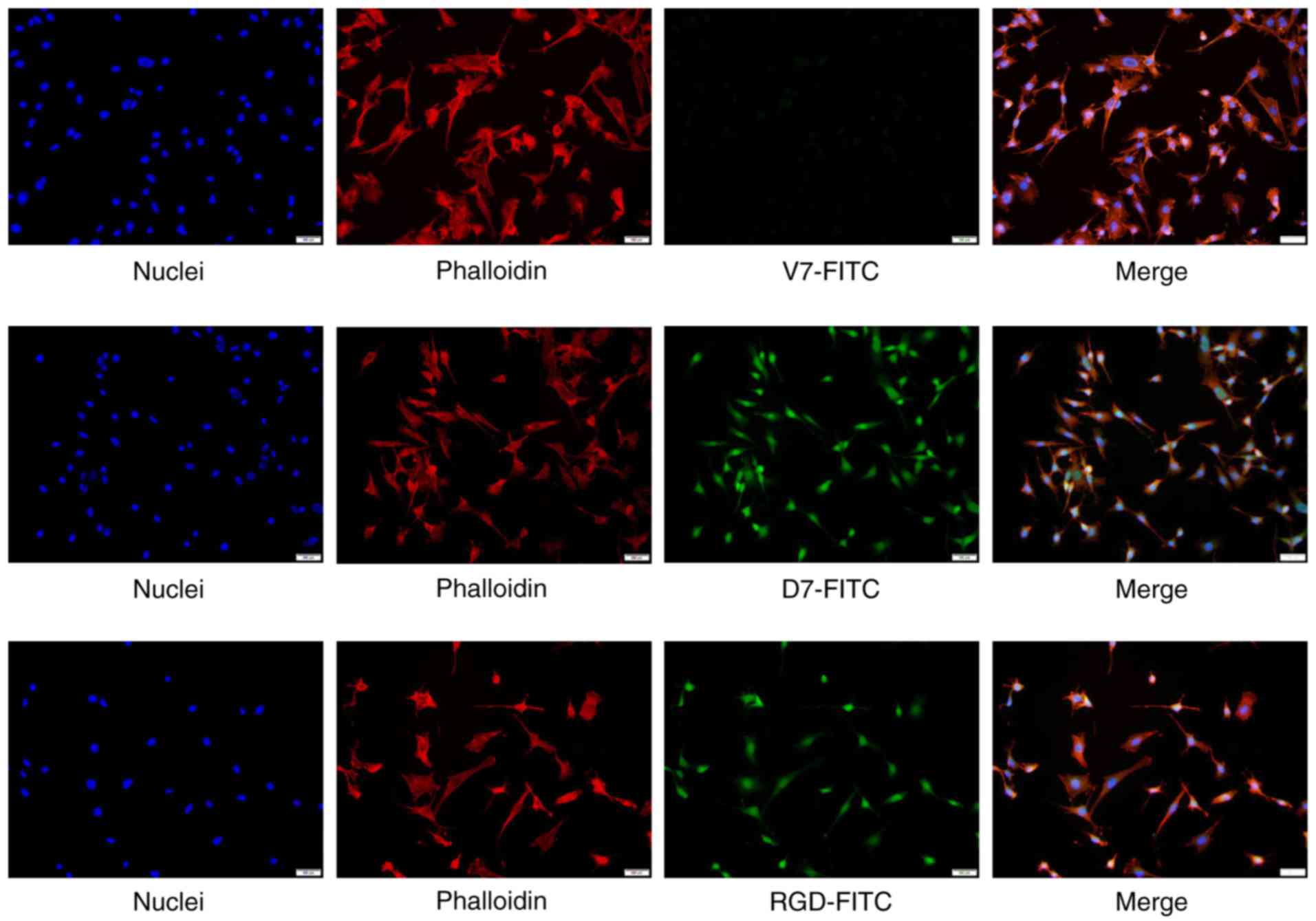
Peptide-affinity assay via
fluorescence cytochemistry
The C57BL/6 mouse BMMSCs were cultured in 24-well
dishes until 70–90% confluence was achieved. Then, the cells were
incubated with 10 µM FITC-labeled peptides for 1 h at 37°C. The
mouse BMMSCs were also incubated with 160 nM rhodamine phalloidin
(Beijing Solarbio Science and Technology, Inc., Beijing, China) for
30 min at room temperature to visualize the cytoskeleton. The
nuclei were then counterstained with 10 µg/ml DAPI (Beijing
Solarbio Science and Technology, Inc.) for 10 min at room
temperature. The cells on the round coverslips were examined under
a fluorescence microscope at magnification, ×200.
Binding efficiency by competition
assay
The affinity peptide was synthesized by a company
(Scilight-Peptide, Ltd., Co.). A total of 7×10⁵ C57BL/6
mouse BMMSCs were cultured on 60×15 mm petri dishes until 70–90%
confluence was reached. Then, 1×10⁹ phages with the D7
sequence were added to the cells. The synthetic peptide was
introduced to cells at a concentration of 100 µM prior to the
addition of the phages during the process of biopanning, as
aforementioned. The level of binding of phages to mouse BMMSCs was
calculated comparing the output titer of phages selected with the
competition of the affinity peptide with the output titer of phages
selected without the addition of the affinity peptide.
Statistical analysis
In the present study, data is expressed as mean ±
standard deviation. A one-way analysis of variance was performed to
evaluate the differences between multiple groups followed by
Dunnett's post-hoc test using IBM SPSS Statistics 24.0 software
(IBM Corp., Armonk, NY, USA). P<0.05 was considered to indicate
a statistically significant difference.
Results
Recovery efficiency following
biopanning increases round by round
Following each round of biopanning, the recovery
efficiencies of the specific phage clones were calculated as the
output titer divided by the input titer of the phages. The input
titer of the phages was 1×10¹¹ PFU for three rounds. As
indicated in Table I, the best
recovery efficiency was attained in the third round, which was
44.4-fold higher compared with that in the first round. The phage
clones remaining after the third round of biopanning were selected
for peptide sequencing.
 | Table I.Recovery efficiency of phage display
biopanning increases round by round. |
Table I.
Recovery efficiency of phage display
biopanning increases round by round.
| Round no. | Input titer, pfu | Output titer,
pfu | Recovery
efficiency | Fold increase |
|---|
| 1 |
1.0×10¹¹ |
5.175×10⁴ |
5.175×10⁻⁷ | 1 |
| 2 |
1.0×10¹¹ |
1.84×10⁶ |
1.84×10⁻⁵ | 35.6 |
| 3 |
1.0×10¹¹ |
2.3×10⁶ |
2.3×10⁻⁵ | 44.4 |
Peptide sequencing
The C57BL/6 mouse BMMSC affinity phage clones were
isolated following three rounds of phage display biopanning.
Subsequent to these three rounds, 10 blue plaques were randomly
selected for sequencing. With the exception of empty phages, there
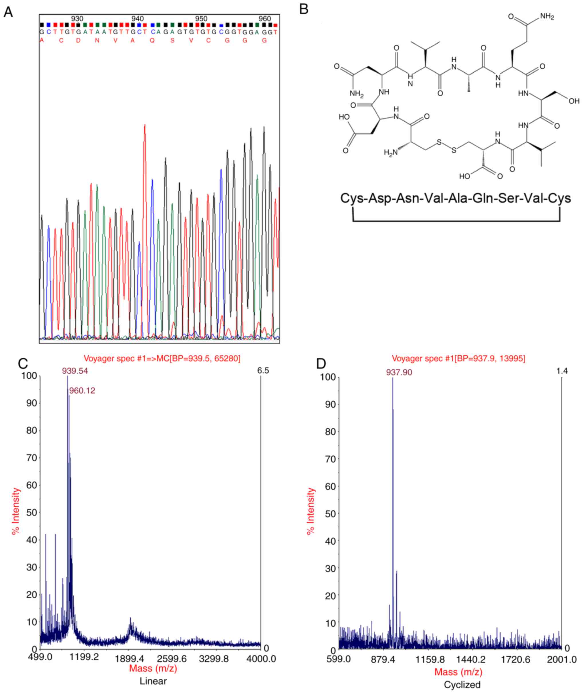
was one type of phage clone that appeared 6 times. Using DNA
sequencing, the sequence, DNVAQSV, was identified via Sanger
sequencing applying Rolling Circle Amplification technique and
Chromas analysis software (Table
II and Fig. 2A). Corresponding
cyclic peptides (Fig. 2B)
indicated a high affinity for the mouse BMMSCs through the
subsequent experiments.
| Table II.Peptide sequences. |
Table II.
Peptide sequences.
| Round no. | Peptide sequence | Expression, %
(n=10) |
|---|
| 1 | Not sequenced | – |
| 2 | Not sequenced | – |
| 3 | DNVAQSV | 60 |
|
| KTSPWAK | 10 |
|
| Empty phage | 30 |
Identified loop-constrained
heptapeptide has a high specific affinity for mouse BMMSCs
The identified peptide (CDNVAQSVC) was designated as
a mouse BMMSCs affinity peptide (D7). The molecular weight of the
linear precursor of D7 peptide was determined to be 939.54 Da by
mass spectrometry (Fig. 2C). The
molecular weight of D7 peptide decreased to 937.90 Da (Fig. 2D). This indicated that the thiol
groups of the two cysteine residues formed an intramolecular
disulfide linkage, and that the cyclic peptide D7 was successfully
synthesized. A peptide (CVAVQNDSC) with the same amino acids as D7
in a scrambled order was used as the negative control and termed
V7. RGD, a peptide composed of 3 amino acids (arginine, glycine and
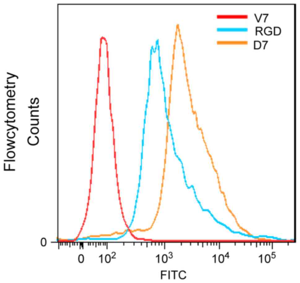
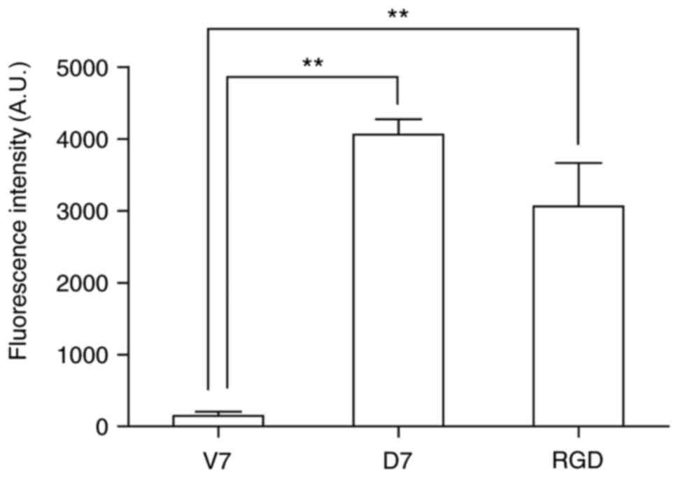
aspartic acid), was used as the positive control. The C57BL/6 mouse
BMMSCs were incubated for 1 h with FITC-labeled D7, V7 or RGD, and
measured via flow cytometry. The average fluorescence intensity was
4,059.0±213.5 for the mouse BMMSCs incubated with FITC-D7,
3,058.5±604.6 for the mouse BMMSCs incubated with FITC-RGD, and
146.5±60.1 for the mouse BMMSCs incubated with FITC-V7. The average
fluorescence intensity for the mouse BMMSCs incubated with FITC-D7
and FITC-RGD was increased than that of FITC-V7, and the difference
was statistically significant (n=3; P<0.01; Figs. 3 and 4). The average fluorescence intensity of
cells incubated with FITC-D7 was 31.2-fold higher compared with
that of the FITC-V7 cells. The cells were also observed under a
fluorescence microscope. Marked fluorescent signals were observed
in the cells incubated with FITC-D7 and FITC-RGD, whereas weak or
no fluorescent signals were observed in the cells incubated with
FITC-V7 (Fig. 5). These results
suggested that D7 exhibited a high affinity for mouse BMMSCs.
No significant difference is
identified in the affinity peptide competition assay
A synthetic peptide containing the CDNVAQSVC
sequence competing with phages for the binding of C57BL/6 mouse
BMMSCs was examined. In the experiment, the phages expressed the
loop-constrained D7 peptide on the outside. Using 100 µM D7
peptide, the following results were obtained. The output titer of
phages selected with the competition of the affinity peptide was
4.14×10⁴, while the output titer of phages selected
without adding the affinity peptide was 4.025×10⁴. It
was identified that there was no significant difference in the
binding of phages to target cells. A total of 3 independent
experiments were performed and the same trend was observed in each
repeat. This was consistent with the phenomenon of the appearance
of non-loaded phages following 3 rounds of panning.
Discussion
In the present study, following 3 rounds of
biopanning, empty phages appeared. The enrichment of insertless
clones is not unusual. The Ph.D. libraries were constructed by
cloning a randomized DNA insert into a phage vector, so all of the
libraries contain a small percentage (0.1–0.5%) of clones with no
insert. Insertless clones begin to predominate panning experiments
if an excessive number of rounds of amplification have been
performed. In the case of the C7C library, the presence of the 2
cysteines in the peptide sufficiently attenuated phage propagation
such that the empty vector had a growth advantage. Concomitantly,
the disulfide linkage in the C7C increased the affinity of peptide
sequences that bind the target in a conformation allowed by the
disulfide constraint. Therefore, the clones containing the insert
that were present following 3 rounds of biopanning were considered
to be optimal for subsequent analysis. In the present study, the
insertless clones were disregarded in favor of the clones
containing the insert.
In phage display biopanning, as unconjugated phages
are gradually eliminated, the recovery efficiencies should
increase. In the present study, following 3 rounds of biopanning,
the recovery efficiencies increased round by round. The recovery
efficiency increased by 44.4-fold following 3 rounds of screening.
These data confirmed that at this point, the phages binding to
C57BL/6 mouse BMMSCs were effectively enriched.
The lack of effective therapy for ONFH is a
difficulty to be overcome in clinical practice, and the clinical
results of traditional methods have not been satisfactory. At
present, TE strategies are being investigated. One of the most
common strategies currently used in TE is the combination of a
biodegradable matrix (scaffold), exogenous or endogenous living
cells and/or biologically active molecules to form a construct that
promotes tissue repair and regeneration. BMMSCs are considered to
be a good cell source for bone regeneration, due to their high
proliferation rates and good osteogenic potential, as demonstrated
by a number of previous studies (6,19,20).
However, there are few or no viable MSCs in necrotic femoral head
sites (5). Additionally, several
previous studies have indicated that only a small percentage of
stem cells remain at the injury site after 2–3 weeks post-injection
(21,22). Therefore, promoting the precise and
efficient homing and entrapment of stem cells into injured tissues
to form ‘stem cell nests’ should be a primary aim in TE therapy.
Abundant BMMSCs are required in order to build ‘stem cell niches’,
to constantly facilitate repair during the entire regeneration
process. However, the problems of homing and entrapment arise when
BMMSCs are adopted as a cell source for bone repair and
regeneration. Therefore, the low efficiency of homing and
entrapment of MSCs becomes a key problem to be overcome in
endogenous stem cell-based TE, and recruitment of endogenous BMMSCs
to the injured sites becomes important.
A number of methods have been examined to solve this
problem. Recruiting BMMSCs using affinity peptides is popular.
Biomaterials modified by affinity peptides for target cells
including BMMSCs have been studied increasingly. Phage display
provides a novel method for searching for affinity peptides highly
specific to BMMSCs. For example, a peptide sequence (DPIYALSWSGMA)
with high affinity for human BMMSCs, was identified using phage
display technology (14). A novel
peptide with the amino acid sequence of EPLQLKM (E7) with high
specific affinity for BMMSCs was also identified via phage display
technology. The E7 peptide was covalently conjugated onto
polycaprolactone electrospun meshes to fabricate functional
scaffolds to cure cartilage defect of rat knee joints (12). E7 peptides were also conjugated to
demineralized bone matrix to create functional biomaterials
combined with chitosan solution to cure the osteochondral defect
(23). RGD is best known for cell
adhesion (24). The peptide
derives from fibronectin in the extracellular matrix. As the RGD
peptide was identified to promote cell adhesion in 1984 (25), a number of materials modified by
RGD have been applied for academic studies or clinical therapies
(26–28). However, this peptide lacks
specificity, as fibronectin receptors exist in all cells. In the
present study, the cyclic peptide D7 identified may also be used to
modify appropriate materials to fabricate functional scaffolds. In
addition, the disulfide linkage in the cyclic peptide will increase
the affinity of peptide sequences that bind the target in a
conformation allowed by the disulfide constraint.
However, the exact mechanism of this affinity
peptide was not investigated in the present study. Therefore,
future studies will aim to identify the receptor of the affinity
peptide on the surface of mouse BMMSCs and explore the potential
mechanism. Core decompression is recommended as the first surgical
treatment option in symptomatic small to medium-sized pre-collapsed
lesions of ONFH (29). A bone
tunnel from the greater trochanter to the femoral head is built,
decreasing intraosseous pressure and pain. In light of the age of
the patient population, this type of treatment represents a
reasonable initial surgical intervention. Core decompression has
been combined with a number of bone-graft substitutes that are
filled into the bone tunnel to treat ONFH. A previous study
described the effect of core decompression combined with calcium
phosphate composite scaffolds containing bone morphogenetic
protein-vascular endothelial growth factor-loaded
poly-lactic-co-glycolic acid microspheres in the treatment of ONFH
(30). Therefore, future studies
will aim to select appropriate materials and modify them using the
affinity cyclic peptide identified in the present study to
fabricate functional biomaterials. Then, core decompression will be
combined with these functional biomaterials to recruit endogenous
BMMSCs and increase the efficiency of TE therapy in ONFH.
In conclusion, through phage display panning
technology, the D7 peptide sequence, a loop-constrained
heptapeptide with a pair of cysteine residues conjugated to each
terminus of 7 amino acids, was identified. The peptide exhibited a
specific affinity for C57BL/6 mouse BMMSCs. Subsequent in
vitro experiments further confirmed the high affinity of the D7
peptide for mouse BMMSCs. These data suggest that the D7 peptide
can be used as a potent motif for recruitment of BMMSCs in
MSC-based TE therapy.
Acknowledgements
Not applicable.
Funding
The present study was supported by the National
Natural Science Foundation of China (grant no., 81271966).
Availability of data and materials
The data sets generated and analyzed during the
current study are available from the corresponding author on
reasonable request.
Authors' contributions
GW, ZM and SS designed the experiments. GW performed
the experiments. GW, NZ, HX, YL and TS analyzed the data. GW wrote
the manuscript. GW revised the manuscript. All authors reviewed the
manuscript.
Ethics approval and consent to
participate
Not applicable.
Patient consent for publication
Not applicable.
Competing interests
The authors declare that they have no competing
interests.
References
|
1
|
Hernigou P, Poignard A and Nogier A: Fate
of very small asymptomatic stage-I osteonecrotic lesions of the
hip. J Bone Joint Surg Am. 86-A:2589–2593. 2004. View Article : Google Scholar : PubMed/NCBI
|
|
2
|
Mont MA, Carbone JJ and Fairbank AC: Core
decompression versus nonoperative management for osteonecrosis of
the hip. Clin Orthop Relat Res. 169–178. 1996. View Article : Google Scholar : PubMed/NCBI
|
|
3
|
Hernigou P, Flouzat-Lachaniette CH,
Delambre J, Poignard A, Allain J, Chevallier N and Rouard H:
Osteonecrosis repair with bone marrow cell therapies: State of the
clinical art. Bone. 70:102–109. 2015. View Article : Google Scholar : PubMed/NCBI
|
|
4
|
Pittenger MF, Mackay AM, Beck SC, Jaiswal
RK, Douglas R, Mosca JD, Moorman MA, Simonetti DW, Craig S and
Marshak DR: Multilineage potential of adult human mesenchymal stem
cells. Science. 284:143–147. 1999. View Article : Google Scholar : PubMed/NCBI
|
|
5
|
Tatara AM: Tissue engineering in
orthopaedics. J Bone Joint Surg Am. 98:1132–1139. 2016. View Article : Google Scholar : PubMed/NCBI
|
|
6
|
Aarvold A, Smith JO, Tayton ER, Jones AM,
Dawson JI, Lanham S, Briscoe A, Dunlop DG and Oreffo RO: A tissue
engineering strategy for the treatment of avascular necrosis of the
femoral head. Surgeon. 11:319–325. 2013. View Article : Google Scholar : PubMed/NCBI
|
|
7
|
Hass R, Kasper C, Böhm S and Jacobs R:
Different populations and sources of human mesenchymal stem cells
(MSC): A comparison of adult and neonatal tissue-derived MSC. Cell
Commun Signal. 9:122011. View Article : Google Scholar : PubMed/NCBI
|
|
8
|
Zhao D, Cui D, Wang B, Tian F, Guo L, Yang
L, Liu B and Yu X: Treatment of early stage osteonecrosis of the
femoral head with autologous implantation of bone marrow-derived
and cultured mesenchymal stem cells. Bone. 50:325–330. 2012.
View Article : Google Scholar : PubMed/NCBI
|
|
9
|
Sen RK, Tripathy SK, Aggarwal S, Marwaha
N, Sharma RR and Khandelwal N: Early results of core decompression
and autologous bone marrow mononuclear cells instillation in
femoral head osteonecrosis: A randomized control study. J
Arthroplasty. 27:679–686. 2012. View Article : Google Scholar : PubMed/NCBI
|
|
10
|
Gangji V, De Maertelaer V and Hauzeur JP:
Autologous bone marrow cell implantation in the treatment of
non-traumatic osteonecrosis of the femoral head: Five year
follow-up of a prospective controlled study. Bone. 49:1005–1009.
2011. View Article : Google Scholar : PubMed/NCBI
|
|
11
|
Lim YW, Kim YS, Lee JW and Kwon SY: Stem
cell implantation for osteonecrosis of the femoral head. Exp Mol
Med. 45:e612013. View Article : Google Scholar : PubMed/NCBI
|
|
12
|
Shao Z, Zhang X, Pi Y, Wang X, Jia Z, Zhu
J, Dai L, Chen W, Yin L, Chen H, et al: Polycaprolactone
electrospun mesh conjugated with an MSC affinity peptide for MSC
homing in vivo. Biomaterials. 33:3375–3387. 2012. View Article : Google Scholar : PubMed/NCBI
|
|
13
|
Meng Q, Man Z, Dai L, Huang H, Zhang X, Hu
X, Shao Z, Zhu J, Zhang J, Fu X, et al: A composite scaffold of MSC
affinity peptide-modified demineralized bone matrix particles and
chitosan hydrogel for cartilage regeneration. Sci Rep. 5:178022015.
View Article : Google Scholar : PubMed/NCBI
|
|
14
|
Ramaraju H, Miller SJ and Kohn DH:
Dual-functioning peptides discovered by phage display increase the
magnitude and specificity of BMSC attachment to mineralized
biomaterials. Biomaterials. 134:1–12. 2017. View Article : Google Scholar : PubMed/NCBI
|
|
15
|
Li Q, Xing D, Ma L and Gao C: Synthesis of
E7 peptide-modified biodegradable polyester with the improving
affinity to mesenchymal stem cells. Mater Sci Eng C Mater Biol
Appl. 73:562–568. 2017. View Article : Google Scholar : PubMed/NCBI
|
|
16
|
Man Z, Yin L, Shao Z, Zhang X, Hu X, Zhu
J, Dai L, Huang H, Yuan L, Zhou C, et al: The effects of
co-delivery of BMSC-affinity peptide and rhTGF-β1 from coaxial
electrospun scaffolds on chondrogenic differentiation.
Biomaterials. 35:5250–5260. 2014. View Article : Google Scholar : PubMed/NCBI
|
|
17
|
Shao Z, Zhang X, Pi Y, Yin L, Li L, Chen
H, Zhou C and Ao Y: Surface modification on polycaprolactone
electrospun mesh and human decalcified bone scaffold with
synovium-derived mesenchymal stem cells-affinity peptide for tissue
engineering. J Biomed Mater Res A. 103:318–329. 2015. View Article : Google Scholar : PubMed/NCBI
|
|
18
|
Pi Y, Zhang X, Shi J, Zhu J, Chen W, Zhang
C, Gao W, Zhou C and Ao Y: Targeted delivery of non-viral vectors
to cartilage in vivo using a chondrocyte-homing peptide identified
by phage display. Biomaterials. 32:6324–6332. 2011. View Article : Google Scholar : PubMed/NCBI
|
|
19
|
Udehiya RK, Amarpal, Aithal HP,
Kinjavdekar P, Pawde AM, Singh R and Taru Sharma G: Comparison of
autogenic and allogenic bone marrow derived mesenchymal stem cells
for repair of segmental bone defects in rabbits. Res Vet Sci.
94:743–752. 2013. View Article : Google Scholar : PubMed/NCBI
|
|
20
|
Chen M, Le DQ, Kjems J, Bünger C and
Lysdahl H: Improvement of distribution and osteogenic
differentiation of human mesenchymal stem cells by hyaluronic acid
and β-tricalcium phosphate-coated polymeric scaffold in vitro.
Biores Open Access. 4:363–373. 2015. View Article : Google Scholar : PubMed/NCBI
|
|
21
|
Discher DE, Mooney DJ and Zandstra PW:
Growth factors, matrices, and forces combine and control stem
cells. Science. 324:1673–1677. 2009. View Article : Google Scholar : PubMed/NCBI
|
|
22
|
Karp JM and Leng Teo GS: Mesenchymal stem
cell homing: The devil is in the details. Cell Stem Cell.
4:206–216. 2009. View Article : Google Scholar : PubMed/NCBI
|
|
23
|
Huang HJ, Zhang X, Hu X, Shao Z, Zhu J,
Dai L, Man Z, Yuan L, Chen H, Zhou C and Ao Y: A functional
biphasic biomaterial homing mesenchymal stem cells for in vivo
cartilage regeneration. Biomaterials. 35:9608–9619. 2014.
View Article : Google Scholar : PubMed/NCBI
|
|
24
|
Ruoslahti E and Pierschbacher MD:
Arg-Gly-Asp: A versatile cell recognition signal. Cell. 44:517–518.
1986. View Article : Google Scholar : PubMed/NCBI
|
|
25
|
Pierschbacher MD and Ruoslahti E: Cell
attachment activity of fibronectin can be duplicated by small
synthetic fragments of the molecule. Nature. 309:30–33. 1984.
View Article : Google Scholar : PubMed/NCBI
|
|
26
|
Hersel U, Dahmen C and Kessler H: RGD
modified polymers: Biomaterials for stimulated cell adhesion and
beyond. Biomaterials. 24:4385–4415. 2003. View Article : Google Scholar : PubMed/NCBI
|
|
27
|
Zhang H and Hollister S: Comparison of
bone marrow stromal cell behaviors on poly(caprolactone) with or
without surface modification: Studies on cell adhesion, survival
and proliferation. J Biomater Sci Polym Ed. 20:1975–1993. 2009.
View Article : Google Scholar : PubMed/NCBI
|
|
28
|
Zhang H, Lin CY and Hollister SJ: The
interaction between bone marrow stromal cells and RGD-modified
three-dimensional porous polycaprolactone scaffolds. Biomaterials.
30:4063–4069. 2009. View Article : Google Scholar : PubMed/NCBI
|
|
29
|
Mont MA, Cherian JJ, Sierra RJ, Jones LC
and Lieberman JR: Nontraumatic osteonecrosis of the femoral head:
Where do we stand today? A ten-year update. J Bone Joint Surg Am.
97:1604–1627. 2015. View Article : Google Scholar : PubMed/NCBI
|
|
30
|
Zhang HX, Zhang XP, Xiao GY, Hou Y, Cheng
L, Si M, Wang SS, Li YH and Nie L: In vitro and in vivo evaluation
of calcium phosphate composite scaffolds containing BMP-VEGF loaded
PLGA microspheres for the treatment of avascular necrosis of the
femoral head. Mater Sci Eng C Mater Biol Appl. 60:298–307. 2016.
View Article : Google Scholar : PubMed/NCBI
|