Introduction
Cancer remains a problem worldwide and the majority
of patients suffer from the severe side effects of anticancer drugs
(1). There is an urgent
requirement to improve the specificity of cancer drugs to tumors.
At present, aptamers, which are typically small single-stranded
DNA/RNA oligonucleotides or small peptides generated by artificial
systemic evolution of ligands by exponential enrichment (2), have been demonstrated to bind to a
wide variety of targets, including small molecules, proteins, cells
and tissues, with high affinity and selectivity (3–6).
Owing to their various advantages, aptamers have been used in
biomarker identification, disease diagnosis, therapies, and most
promising, as targeting elements for developing novel drug delivery
systems (7,8). Cabazitaxel (Cab) is an antineoplastic
agent with a broad antitumor spectrum; however, its use in the
clinical setting has been hindered by certain adverse effects.
(9) Novel targeting
biotin-conjugated TLS1c Cab liposomes (BioTL-Cab/lipo) have been
designed to investigate whether they may decrease the cytotoxicity
of Cab in mice while retaining its antitumor properties. Increasing
efforts have been made in the field of aptamer-based
tumor-targeting; however, few studies have demonstrated the effects
and characteristics of aptamers in Cab-associated liposomes.
Shangguan et al (10) observed that TLS1c, an aptamer with
a short DNA sequence, specifically targeted MEAR hepatoma cells.
The synthetic nucleic acid nature of TLS1c enables easy and simple
conjugation with biotin, enhancing its stability in biological
fluids (11). The biotinylated
TLS1c (BioTL) was selected as a targeting recognition element for
MEAR hepatoma in the present study. Liposomes, as a traditional
passive targeting system (12),
have been widely investigated in drug administration. Liposomes may
decrease the cytotoxicity of drugs; however, to the best of our
knowledge, Cab liposomes (Cab/lipo) have not been previously
investigated.
In the present study, DNA aptamer TLS1c-modified
Cab/lipo was used to investigate tumor-targeting efficiency, where
the direct comparison between Cab/lipo and BioTL-Cab/lipo was
reported for the first time, to the best of our knowledge. The
cytotoxicity of the prepared liposomes was investigated in Caco-2,
HepG2 and MEAR cell lines, and it was identified that
BioTL-Cab/lipo exhibited higher cytotoxicity in MEAR cells compared
with Caco-2 and HepG2 cells. Consistent results were obtained with
confocal laser scanning microscopy (CLSM) and flow cytometry
analysis; highly specific targeting of the prepared BioTL-Cab/lipo
to MEAR cells was demonstrated. Furthermore, the biodistribution
and in vivo antitumor efficacy were monitored in MEAR
tumor-bearing animal models. The accumulation of BioTL-Cab/lipo in
tumor tissue was demonstrated with real-time images. Collectively,
the results of the present study suggested that TLS1c-modified
Cab/lipo demonstrated a specific and potent antitumor effect in
vivo.
Materials and methods
Materials
Soya phosphatidyl choline (LIPOID S 100; SPC) was
purchased from Lipoid GmbH (Ludwigshafen, Germany). Ethyl
polyenoate (EP) was obtained from Shandong Yuwang Pharmaceutical
Co., Ltd. (Yucheng, China). Cab was supplied by Guangzhou Zhiyuan
Pharmacy Co., Ltd. (Guangzhou, China). Sodium cholesterol sulfate
(SCS) was purchased from Shanghai Haoran Biotechnology Co., Ltd.
(Shanghai, China). BioTL was purchased from Genewiz, Inc. (Beijing,
China). Tris (hydroxymethyl) methyl aminomethane (Tris) was
obtained from Jian Yang Biotechnology Co., Ltd. (Guangzhou, China).
The nucleotide sequence of TLS1c is
5′-ACAGGAGTGATGGTTGTTATCTGGCCTCAGAGGTTCTCGGGTGTGGTCACTCCTG-3′.
TLS1c was purchased from Genewiz, Inc.; biotin was conjugated with
TLS1c at the 3′ end. Shanghai Macklin Biochemical Co., Ltd.
(Shanghai, China) provided 6-carboxy fluorescein (6-FAM). Harvey
Biotech Co., Ltd. (Beijing, China) provided
1,1′-dioctadecyl-3,3,3′,3′-tetramethylindocarbocyanine perchlorate
(DiI). HepG2, macrophages and MEAR cells were provided by Shanghai
Fuxiang Biotechnology Co., Ltd. (Shanghai, China). Caco-2 cells
were obtained from the American Type Culture Collection (Manassas,
VA, USA). The TDL-40B Ultrafiltration centrifuge was supplied by
Shanghai Anting Science Technology Instrument Factory (Shanghai,
China). All other chemical and buffer components were of analytical
grade.
Preparation of liposomes
Liposomes were prepared by reverse-phase evaporation
of vesicles (13). The organic
phase (OP) comprised 8 mg Cab, 120 mg SPC, 12 mg SCS and 16 mg EP
(a surfactant) dissolved in 3 ml dichloromethane. The aqueous phase
(AP) constituted 20 ml Tris-HCl (pH 7.5). The OP was deposited into
the AP with stirring (600 rpm) for 45 min at 25°C. The emulsion was
transferred to a container and BioTL in Tris-HCl was added. Probe
sonication (UH-500A ultrasonic processor; Tianjin Automatic Science
Instruments Co., Ltd., Tianjin, China) was conducted for 15 min in
an ice-water bath. Dichloromethane was evaporated using a Rotary
Evaporator (RE-2000; Shanghai Yarong Bio, Inc., Shanghai, China) at
45°C, for 15 min and 100 r/min. The BioTL-Cab/lipo was freeze-dried
using a Tofflon LY0-0.5 freeze dryer (Shanghai Tofflon Technology
Co., Ltd., Shanghai, China) with 5% (w/v) lactose solution as the
freeze-drying protective additive. Non-BioTL-containing liposomes
(Cab/lipo) were prepared using the same composition and method as
BioTL-Cab/lipo without the addition of BioTL solution. To examine
the behavior of liposomes in vivo and in vitro, Cab
was replaced with 6-FAM (the content was 2 mg 6-FAM/lipo or
BioTL-6-FAM/lipo) by the same procedure.
Physicochemical characterization of
liposomes
The particle size polydispersity index, and the ζ
potential were determined using a Zetasizer Nano ZS analyzer
(Malvern Instruments, Ltd., Malvern, UK). The content of the loaded
Cab was measured by high performance liquid chromatography (HPLC)
analysis (LC-10A, Shimadzu Corporation, Kyoto, Japan) equipped with
an ODS C18 chromatographic column (4.6×250 mm; 5 µm; Thermo Fisher
Scientific, Inc., Waltham, MA, USA). The mobile phase was composed
of methanol, acetonitrile and water (35:40:25; v/v). A constant
flow rate of 1 ml/min and detection wavelength of 240 nm were
applied for analysis at room temperature. The content was
calculated using a linear calibration curve of standard Cab
solutions in MeOH.
The encapsulation efficiency (EE) of Cab or 6-FAM
was calculated as a percentage of the dissociated Cab from the
suspension using 3,500 rpm for 5 min in order for the total Cab
content of the liposomes to be degraded by methanol in the buffer;
the nominal molecular weight limit of the ultrafiltration membrane
was 3 kDa. The following formula was used to calculate the EE% of
liposomes:
EE%=(Total content - Dissociate d
content)Total content×100
The effects of the holding capacity of BioTL on the
EE% of liposomes was validated using an Ultra-Micro ultraviolet
(UV) spectrophotometer (Thermo Fisher Scientific, Inc.). Due to the
nucleic acid characterization of UV absorption, the content of
BioTL was determined by absorption at 260 nm.
Additionally, the optimal ratio of aptamers to SPC
(µg/mg) was determined using a flow cytometer (BD
FACSCanto™ II; BD Biosciences, Franklin Lakes, NJ, USA).
MEAR cells were cultured at 37°C in 5% CO2 and 95%
humidity in Dulbecco's modified Eagle's medium (Gibco; Thermo
Fisher Scientific, Inc.) supplemented with 10% fetal bovine serum
(Gibco; Thermo Fisher Scientific, Inc.). MEAR cells were counted
and seeded on 6-well plates (1×10⁶ cells/well), and
cultured overnight at 37°C in the incubator. Subsequently, cells
were treated with different BioTL-6-FAM/lipo to SPC rates (0.2, 1
and 2 µg/mg) for 1, 3 and 5 h at 37°C to determine the best ratio.
The concentration of BioTL was 1.2, 6, 12 µg/ml, respectively, and
the concentration of SPC used as the control was 6 mg/ml.
The medium was removed at the aforementioned time
points; cells were washed with PBS and subsequently harvested using
0.05% trypsin at 37°C for 1 min. The fluorescence intensity of
6-FAM in cells was determined with excitation and emission
wavelengths of 452 and 521 nm respectively using the flow
cytometer.
Cytotoxicity assays
HepG2, macrophages and MEAR cells were cultured at
37°C in 5% CO2 and 95% humidity in Dulbecco's modified
Eagle's medium (Gibco; Thermo Fisher Scientific, Inc.) supplemented
with 10% fetal bovine serum (Gibco; Thermo Fisher Scientific,
Inc.), Caco-2 cells were cultured under the same conditions with an
additional 1% non-essential amino acids. The cytotoxic effect of
the Cab solution was initially assessed using MEAR cells
(5×10³ cells/well), which were seeded in 96-well plates
(Corning Incorporated, Corning, NY, USA), and cultured overnight.
Cells were incubated with fresh medium containing different
concentrations of Cab (0.0625, 0.125, 0.25, 0.5, 1 and 2 µg/ml).
After 24, 48 or 72 h incubation, MTT (5 mg/ml; 20 µl) was added to
each well for a further incubation of 4 h at 37°C. Subsequently,
the medium was removed and 100 µl dimethyl sulfoxide (DMSO) was
added to dissolve the formazan crystals with 10 min agitation. The
optical density was determined at 490 nm using a spectrophotometer
(Multiskan GO, Thermo Fisher Scientific, Inc.). The cytotoxicity of
the Cab/lipo and BioTL-Cab/lipo was evaluated using Caco-2, HepG2
and MEAR cells. BioTL-Cab/lipo (0.5 µg/ml), Cab/lipo (0.5 µg/ml)
and Cab dissolved in Tris-HCl (0.5 µg/ml) were incubated with cells
24 h later at 37°C. The optical density was determined as described
above.
Cellular uptake studies
Caco-2, HepG2 and MEAR cells were seeded in a
12-well culture plate (2×10⁵ cells/well). After 24 h,
cells were incubated with fresh medium containing 6-FAM/lipo or
BioTL-6-FAM/lipo at 37°C for 3 h. The final concentration of 6-FAM
was 6 µg/ml. After 3 h incubation, cells were washed with cold PBS
three times. Cells were stained with DAPI (Biosharp, Nanjing,
China) at room temperature for 20 min. The cells were washed three
times with PBS under agitation. Cells were fixed on slides with
cold acetone (−20°C) at room temperature and visualized via CLSM
(Carl Zeiss AG, Oberkochen, Germany) at ×400 magnification, images
were acquired of ≥5 fields per view.
Caco-2, HepG2 and MEAR cells (1×10⁶
cells/well) were seeded in the 6-well plates and cultured
overnight. Cells were incubated with fresh medium containing
6-FAM/lipo, BioTL-6-FAM/lipo or Tris-HCl at 37°C for 3 h. The cells
treated with Tris-HCl served as the control group. Flow cytometry
was conducted as described above.
Biodistribution in mice
Male BALB/c nude mice (n=6; 5 weeks-old, ~20 g) were
housed in a temperature-controlled room (22–25°C), with 50–60%
relative humidity under a 12 h light-dark cycle with free access to
food and water. All animal treatments were performed in accordance
with the Regulations of the Administration of Affairs Concerning
Experimental Animals (http://en.pkulaw.cn/display.aspx?cgid=3f3dc921e8e837aebdfb&lib=law)
and were approved by the Animal Research Ethics Committee at
Guangzhou University of Chinese Medicine (Guangzhou, China).
To observe the biodistribution of liposomes in
vivo, the liposomes were first stained with DiI at 37°C for 30
min. MEAR cells suspended in PBS (2×10⁷ cells/ml; 0.2
ml) was subcutaneously injected into the right lower limb of BALB/c
nude mice. The tumor length and width of all mice were monitored
every other day following the subcutaneous injection of MEAR cells.
The tumor volumes were calculated using the following equation,
subsequent to measuring the width and length of each tumor with a
calliper:
Tumor volume(mm3)=width2×length2
Until the tumor volumes reached ~50 mm3,
200 µl BioTL-Cab/lipo/DiI or Cab/lipo/DiI (n=3 mice per group) was
injected intravenously via the tail vein. At different time points
(1, 2, 4, 6 and 24 h) post-injection, mice were anesthetized and
scanned using an IVIS® Lumina XRMS Series III
(PerkinElmer Inc., Waltham, MA, USA) with excitation and emission
wavelengths of 549 and 565 nm, respectively.
Antitumor efficacy in mice
Male BALB/c nude mice (n=36; 5 weeks-old, ~20 g)
were obtained from the Experimental Animal Center at Guangzhou
University of Chinese Medicine (Production license no.
44005800003020). The mice were housed in a temperature-controlled
room (22–25°C), with 50–60% relative humidity under a 12 h
light-dark cycle with free access to food and water. When the MEAR
cell tumor volumes reached ~50 mm3, chemotherapy was
performed on the xenograft mice. Mice were weighed and randomly
divided in 6 groups (n=6 mice per group): i) Negative control group
(NS; 200 µl normal saline solution); ii) drug solution group [200
µl Cab solution (1 mg/ml), equal to 10 mg/kg of Cab to mice]; iii)
high BioTL-Cab/lipo dose group [HBG; 300 µl BioTL-Cab/lipo solution
(1 mg/ml), equal to 15 mg/kg of Cab to mice]; iv) middle
BioTL-Cab/lipo dose group [MBG; 200 µl BioTL-Cab/lipo solution (1
mg/ml), equal to 10 mg/kg of Cab to mice]; v) low BioTL-Cab/lipo
dose group [LBG; 100 µl BioTL-Cab/lipo solution (1 mg/ml), equal to
5 mg/kg of Cab to mice]; and vi) Cab/lipo group [200 µl Cab/lipo
solution (1 mg/ml), equal to 10 mg/kg of Cab to mice]. The mice
received injections every three days via the tail vein for three
times in total. The mice were sacrificed 48 h after the final
injection, and the tumors were removed and weighed.
Statistical analysis
The results were presented as the mean ± standard
deviation of three independent experiments. Statistical analysis
was performed using SPSS software, version 20 (IBM Corp., Armonk,
NY, USA); one-way analysis of variance with a Dunnett's test or a
Student's t-test were performed. P<0.05 was considered to
indicate a statistically significant difference.
Results
Physicochemical characterization of
liposomes
The in vivo characteristics of liposomes is
affected by numerous factors, including particle size and surface
charge (14). Table I demonstrates the size, ζ potential
and EE% of Cab/lipo, BioTL-Cab/lipo, 6-FAM/lipo and
BioTL-6-FAM/lipo. The particle size of the liposomes was 80–100 nm
with an acceptable polydispersity index of 0.246–0.273. The
particle size of the liposomes markedly increased in the presence
of BioTL in Cab/lipo and 6-FAM/lipo. BioTL increased the EE of Cab
by liposomes, but reduced that of 6-FAM by liposomes in the process
of emulsification. BioTL is a single-stranded DNA (ssDNA) with a
surfactant effect that may increase or reduce the EE of drugs by
liposomes. Cab/lipo and BioTL-Cab/lipo exhibited an increased EE%
compared with 6-FAM/lipo or BioTL-6-FAM/lipo. The solubility of
6-FAM is lower than that of Cab in dichloromethane due to the
differing chemical properties of Cab and 6-FAM (15,16).
Generally, all of the liposome samples had a ζ potential <-45
mV, demonstrating that the particles were negatively charged, and
thus, electrostatic interaction may prevent them from
aggregating.
 | Table I.Size and EE of liposomes (n=3). |
Table I.
Size and EE of liposomes (n=3).
| Treatment | Size (nm) | PDI | ζ (mV) | EE (%) |
|---|
| Cab/lipo | 82.73±3.82 | 0.261 | −50.2±4.34 |
93.19±2.29a |
| BioTL-Cab/lipo | 90.10±2.71 | 0.246 | −58.6±2.40 |
97.26±3.18a |
| 6-FAM/lipo | 90.98±1.21 | 0.245 | −45.4±1.50 |
89.63±3.12b |
|
BioTL-6-FAM/lipo | 96.98±1.24 | 0.273 | −55.8±2.04 |
83.63±3.05b |
In the present study, based on the nucleic acid
properties of the aptamer TLS1c (single-stranded DNA), an
Ultra-Micro spectrophotometer was used for the quantification of
free or loosely attached BioTL. A ratio of 1 µg/mg BioTL to SPC had
been applied to the liposomes and the percentage of free BioTL was
1.09±0.91%, suggesting that almost all aptamers were captured by
liposomes (data not shown).
The EE% and the size of liposomes with different
ratios of BioTL to SPC are presented in Table II. The highest EE% and absolute ζ
potential value of liposomes were observed with the BioTL to SPC
ratio of 1 µg/mg. The ratio of 2 µg/mg resulted in the lowest EE%,
which may be due to a large excess of aptamers, preventing
encapsulation of the drug.
| Table II.Effect of BioTL/SPC ratio on size and
EE% of liposomes (n=3). |
Table II.
Effect of BioTL/SPC ratio on size and
EE% of liposomes (n=3).
| BioTL ratio to SPC
(µg/mg) | Size (nm) | PDI | ζ (mV) | EE (%) |
|---|
| 0.2 | 85.87±1.39 | 0.288 | −54.3±2.65 | 93.17±3.91 |
| 1 | 90.10±2.71 | 0.246 | −58.6±2.40 | 97.26±3.18 |
| 2 | 92.76±1.22 | 0.273 | −57.4±1.42 | 91.64±3.27 |
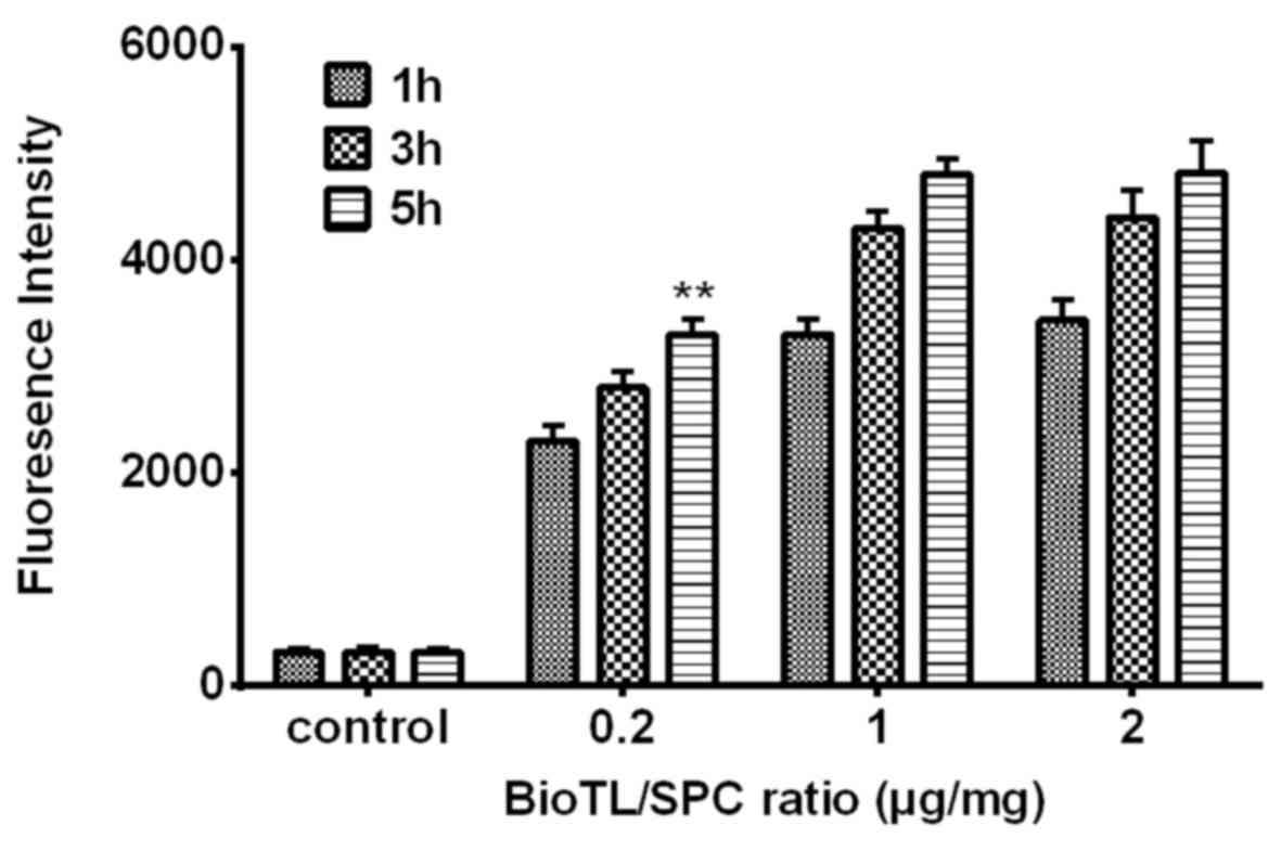
The results from flow cytometry analysis (Fig. 1), combined with the data presented
in Table II, suggested that 1
µg/mg BioTL was the optimal ratio added to the liposomes regarding
the binding rate. As presented Fig.
1, a time-dependent increase in the fluorescence intensity of
MEAR cells was observed within 5 h incubation. At 5 h, the
liposomes modified with 0.2 µg/mg aptamer exhibited lower binding
ability to cells, with a mean fluorescence intensity of 3,066.
Application of 1 µg/mg aptamer revealed increased cell binding
ability, with a mean fluorescence intensity of 4,761, which was
significantly higher compared with 0.2 µg/mg aptamer at 5 h
(P<0.01 vs. the ratios of 1 µg/mg BioTL/SPC at 5 h); however,
with 2 µg/mg aptamer, the mean fluorescence intensity reached
4,805, which was markedly higher compared with 1 µg/mg. These data
suggested that as the ratio exceeded 1 µg/mg, the binding of
liposomes to MEAR cells may be less affected by aptamer
concentration. Considering the EE (%) results in Table II, 1 µg/mg BioTL was determined as
the optimal ratio to modify liposomes.
Cytotoxicity assays
The cytotoxicity of Cab was determined prior to
other cell experiments. As presented in Fig. 2A, the cytotoxicity of Cab on
viability demonstrated a significant dose-dependent effect in MEAR
cells. Cell viability also decreased in all conditions with
increasing incubation time. The inhibitory rate significantly
increased in response to various concentrations (0.0625–2 µg/ml) at
an incubation time of 72 h compared to the control group. The half
maximal inhibitory concentrations of Cab to MEAR were determined to
be 0.65, 0.48 and 0.21 µg/ml for 24, 48 and 72 h of treatment,
respectively.
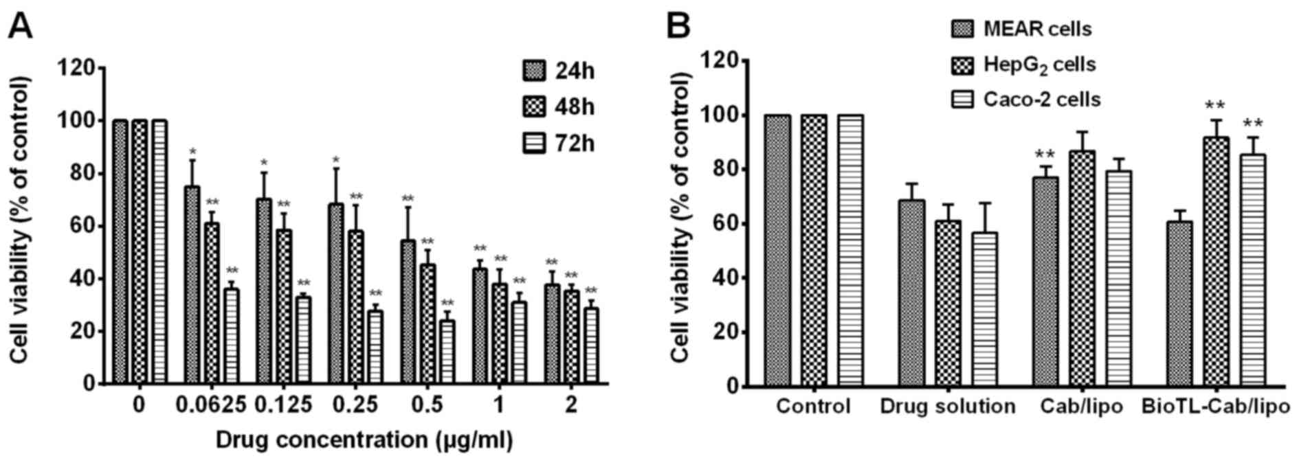 | Figure 2.Effect of Cab solution, Cab/lipo and
BioTL-Cab/lipo on the inhibition of MEAR, HepG2 and Caco-2 cell
viability. (A) Viability of MEAR cells treated with different
concentrations of Cab solution after 24, 48 and 72 h. *P<0.05,
**P<0.01 vs. Control. (B) Viability of MEAR, HepG2 and Caco-2
cells treated with Cab solution, Cab/lipo and BioTL-Cab/lipo after
24 h. All values are presented as the mean ± standard deviation.
**P<0.01 vs. BioTL-Cab/lipo-treated MEAR cells, Cab/lipo or drug
solution. MEAR, BNL 1ME A.7R.1 cells; Cab, cabazitaxel; BioTL,
biotinylated TLS1c; lipo, liposomes. |
As presented in Fig.
2B, cell viability notably increased in response to 0.5 µg/ml
Cab/lipo compared with the drug solution in all the three cell
types, which suggested that liposomes may decrease the cytotoxicity
of the drug directly. Furthermore, it was identified that the
viability of Caco-2 or HepG2 cells incubated with BioTL-Cab/lipo
was significantly increased compared with the drug solution and
Cab/lipo; however, the highest levels of cytotoxicity were observed
in MEAR cells when treated with BioTL-Cab/lipo compared with
Cab/lipo. Based on the results of the cell cytotoxicity assays, it
was speculated that the modification of BioTL to Cab/lipo may
improve specificity for MEAR cells.
Cellular uptake studies
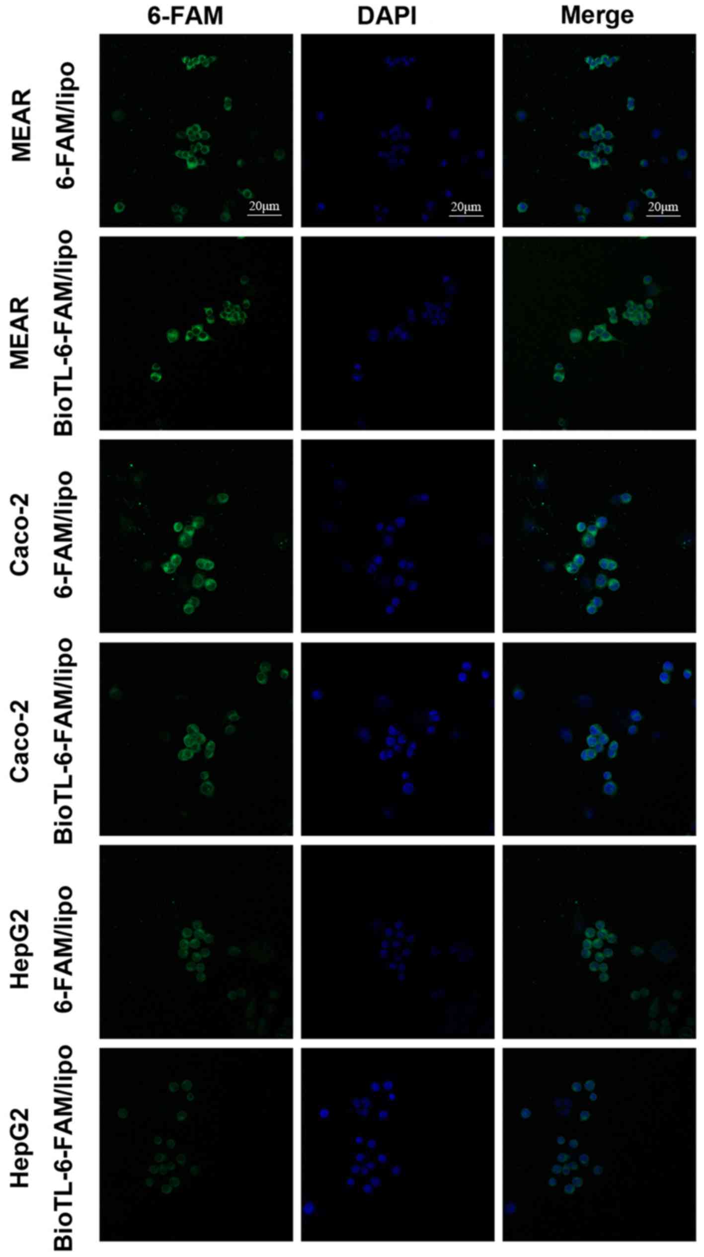
In the preliminary experiments, the cellular uptake
of liposomes in MEAR, Caco-2 and HepG2 cells was determined by CLSM
(Fig. 3). The fluorescence
intensity observed in 6-FAM/lipo-treated MEAR cells was notably
lower compared with BioTL-6-FAM/lipo; the fluorescence intensity of
6-FAM/lipo-treated Caco-2 cells was a notably higher compared with
BioTL-6-FAM/lipo, which demonstrated that 6-FAM/lipo was engulfed
by Caco-2 cells at a higher degree compared with BioTL-6-FAM/lipo.
A similar phenomenon for 6-FAM/lipo-treated HepG2 cells was
observed as the fluorescence intensity was markedly higher compared
with BioTL-6-FAM/lipo-treated HepG2 cells, was detected. The images
supported the observations of the present study that
BioTL-6-FAM/lipo exhibits higher specificity for MEAR cells; the
green fluorescence signals of BioTL-6-FAM/lipo-treated Caco-2 cells
or HepG2 cells were weaker compared with BioTL-6-FAM/lipo-treated
MEAR cells. These results demonstrated the targeting effect of
BioTL-6-FAM/lipo to MEAR cells in vitro. The accumulation of
green fluorescence signals was mainly detected in the cytoplasmic
area of all the cells.
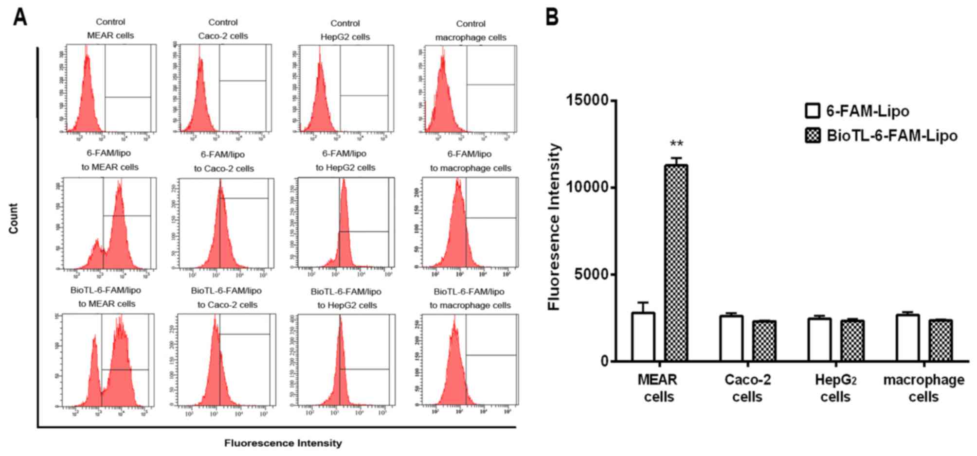
Flow cytometry analysis was performed to
quantitatively measure the cellular uptake of liposomes. MEAR cells
were incubated overnight, and 6-FAM/lipo or BioTL-6-FAM/lipo was
applied for an additional 3 h. The quantitative flow cytometry
analysis demonstrated that the cellular uptake of
6-FAM/lipo-treated MEAR cells was notably decreased compared with
MEAR cells treated with BioTL-6-FAM/lipo (Fig. 4A), possibly due to the targeting
specificity of BioTL to MEAR cells. Additionally, a significant
increase in fluorescence intensity was observed in
BioTL-6-FAM/lipo-treated MEAR cells compared with
6-FAM/lipo-treated MEAR cells (Fig.
4B). This demonstrated the high specificity of BioTL-6-FAM/lipo
to MEAR cells compared with Caco-2, HepG2 cells or macrophages.
These results suggested the selectivity of BioTL-liposomes for MEAR
cells compared with the Caco-2 and HepG2 cells, which is in
accordance with the findings of the cytotoxicity assay (Fig. 2B). Based on the aforementioned
findings, it may be concluded that the aptamer (BioTL)-modified
liposomes exhibited high specificity to the target cells. Although
the cellular internalization process of the TLS1c aptamer has not
been clearly elucidated, these results demonstrated that TLS1c was
able to induce the efficient delivery of liposomes to MEAR cells.
The results of the CLSM and flow cytometry assay demonstrated the
specific targeting of BioTL-Cab/lipo to MEAR cells.
In vivo biodistribution of
BioTL-Cab/lipo
High affinity of liposomes to tumor cells is a
critical factor contributing to high antitumor efficacy in
vivo. The use of fluorescence imaging to monitor local tumor
accumulation and functional activity of magnetic nanoparticles
covalently linked to small interfering RNAs was previously
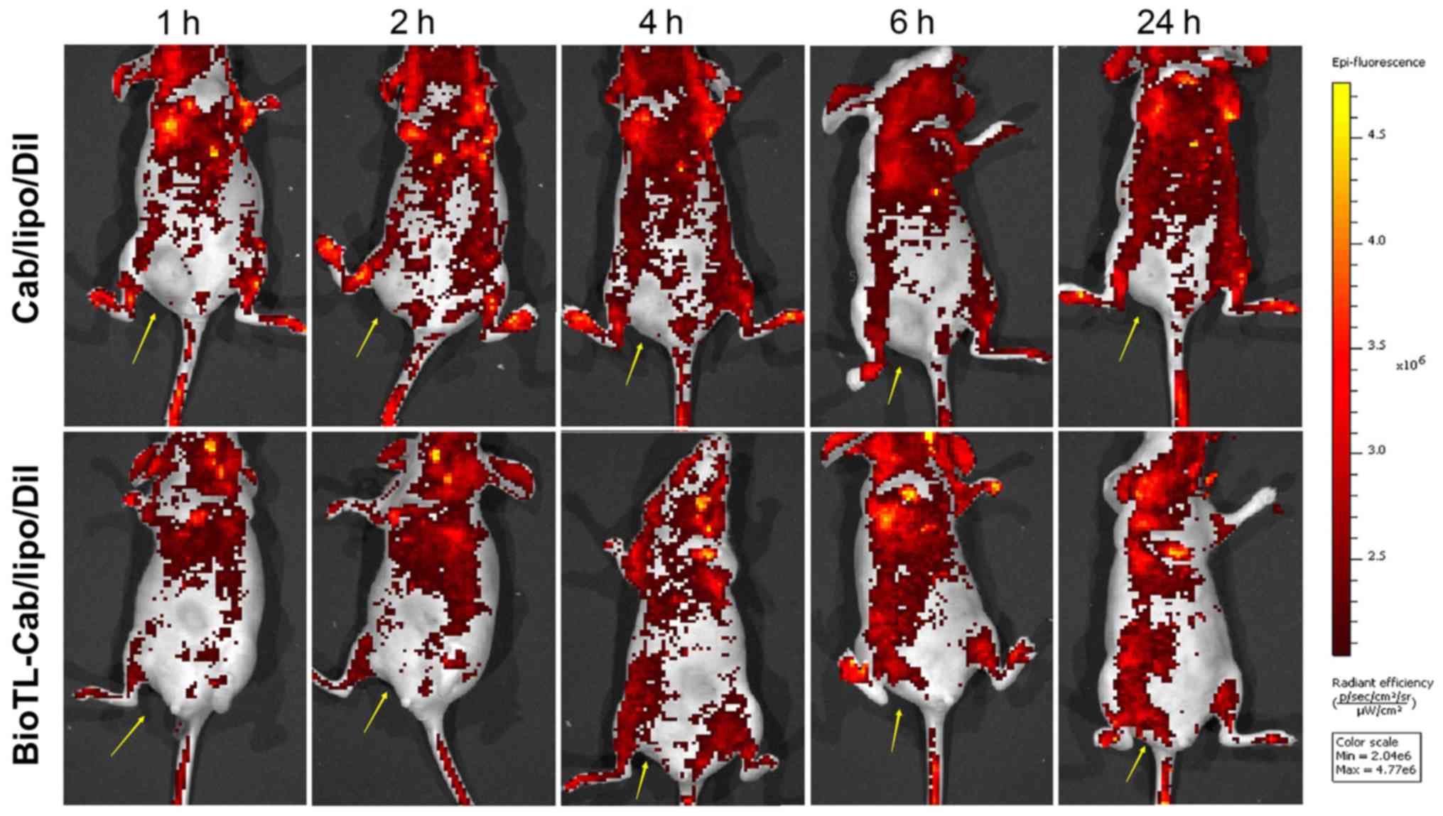
described (17). The real-time
biodistribution of liposomes in tumor-bearing nude mice was
evaluated with in vivo images in the present study. The
tumor-bearing mice were used for the observation of the
biodistribution of BioTL liposomes stained with DiI. In total, 200
µl BioTL-Cab/lipo/DiI or Cab/lipo/DiI were intravenously injected
into the mice. As presented in Fig.
5, strong signals were observed in the blood-rich tissues,
including the brain, heart and lungs. As Cab/lipo/DiI was
intravenously injected in the nude mice, the fluorescence signal at
the tumor location was almost undetectable. Conversely, the
fluorescence signals at the tumor locations in the
BioTL-Cab/lipo/DiI-treated mice gradually increased with time (≤24
h). Cab/lipo/DiI primarily circulated in the bloodstream, but did
not enter the tumor tissue (Fig.
5). On the contrary, the liposomal system of BioTL-Cab/lipo/DiI
accumulated in tumor tissue. Furthermore, time-dependent
fluorescence signals were identified for BioTL-Cab/lipo/DiI,
suggesting that the liposomes modified with BioTL gradually
accumulated in tumor tissue as a function of time. These results
suggested that the BioTL-modified liposomes may deliver antitumor
drugs directly to the tumor with long-term retention, which is
critical for enhancing any antitumor effect.
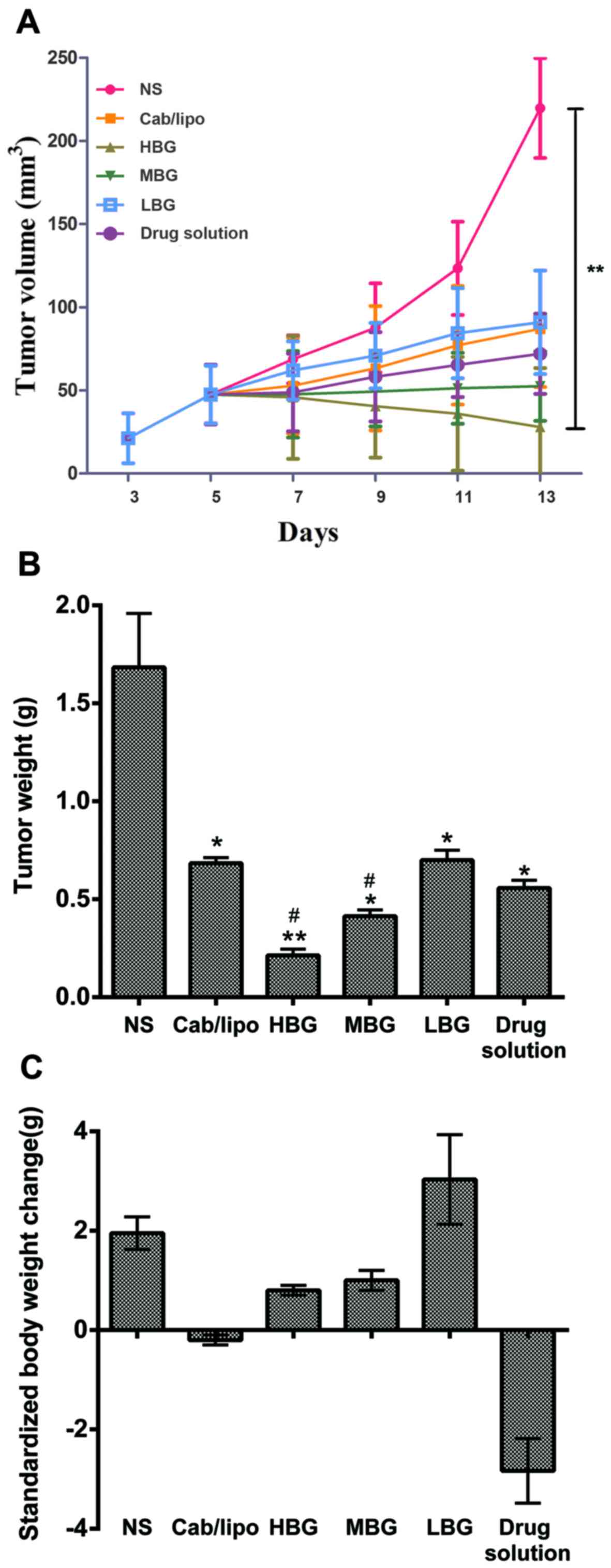
In vivo antitumor efficacy
To investigate the in vivo therapeutic
efficacy of BioTL-Cab/lipo, an antitumor study was performed on
subcutaneously inoculated MEAR tumor-bearing mouse models. The
tumor volume in MEAR tumor-bearing mice as a function of time
(days) during the study was presented in Fig. 6A. The NS group demonstrated a rapid
increase in tumor volume over the whole experimental period.
Conversely, the HBG exhibited the most notable tumor suppression,
in which the tumor volume decreased from 50 mm3 on day 5
to ~28 mm3 on day 13, which demonstrated that the tumor
volume was significantly decreased compared to the NS group.
For tumor weight analysis, mice were sacrificed 48 h
after the last administration. The tumor tissue was removed and
weighed (Fig. 6B), and alterations
in the body weight were associated with the time of treatment
(Table III). Alterations in
standardized body weight of mice administered the drug solution and
Cab/lipo were <0 g, suggesting that the side effects of Cab were
harmful to mice; however, the changes in standardized body weight
of the three groups of mice administered BioTL-Cab/lipo were >0
g, indicating that the toxicity of Cab decreased following
modification with the same amount of aptamer.
| Table III.Alterations in body weight following
treatment (n=3). |
Table III.
Alterations in body weight following
treatment (n=3).
|
| Body weight
(g) |
|---|
|
|
|
|---|
| Point of
analysis | NS | Cab/lipo | HBG | MGB | LBG | Drug solution |
|---|
|
Pre-administration | 20.13±1.18 | 19.72±3.35 | 18.32±3.14 | 16.57±2.24 | 17.21±1.14 | 20.30±3.27 |
|
Post-administration |
23.7±1.70 | 20.17±3.08 | 19.26±1.96 | 17.88±1.68 | 20.97±1.67 | 18.00±1.17 |
| Difference | 3.57 | 0.45 | 0.94 | 1.31 | 3.76 | −2.30 |
The results of the present study suggested that
BioTL-Cab/lipo exerted considerably enhanced inhibitory effects on
tumor growth compared with Cab/lipo or the drug solution in the
MEAR cell-induced tumor xenografts. According to these results,
antitumor drugs, particularly with similar physical/chemical
properties to Cab, may also be successfully delivered by the
developed BioTL/lipo system with similar targeting effects.
Therefore, the results of the present study may provide insight
into the development of BioTL/lipo-based approaches for a variety
of anticancer treatments.
Discussion
The present study demonstrated that aptamer
(TLS1c)-modified Cab liposomes decreased Cab cytotoxicity in mice.
Compared with proteins or peptides, oligonucleotide aptamers are
easier to chemically prepare and modify, and scale up in production
(18–21). Furthermore, aptamers are generally
nonimmunogenic (22,23) and may be rapidly removed from the
blood with minimal system toxicity (24). These advantages suggest aptamers as
alternative targeting elements for preparing ‘immuno-liposomes’.
Therefore, the present study aimed to develop a targeting system
for drug delivery with aptamers. As of the advantages of liposomes,
including high efficiency to encapsulate hydrophilic drugs,
protection of the encapsulated drugs from external conditions,
sustained-release of drugs, ease of surface modification, and most
importantly, the ability of decreasing drug toxicity, they have
been employed as drug delivery vectors in previous
aptamer-targeting studies (25).
Li et al (26) successfully
developed a method of selective delivery using liposomes for the
treatment of malignant melanomas with AS1411, a DNA aptamer with
strong binding affinity for nucleolin, which demonstrated the
potential of aptamer-liposome conjugates in the application of
targeting treatment; however, few studies using aptamer-conjugated
Cab liposomes for cancer cell targeting have been conducted. The
aim of the present study was to develop a novel liposomal delivery
system based on the specificity of the aptamer TLS1c for MEAR cells
to decrease the toxicity of Cab in vitro and in
vivo.
When BioTL is modified on the surface of liposomes,
it may form a film, which improves the specificity of liposomes for
target cells (27). Although, when
the ratio was >1 µg/mg aptamer to SPC, the abundance of aptamers
markedly increased; however, a significant increase in fluorescence
intensity was not observed. This may be explained by the limitation
of targets on cells. As the ratio of aptamer to SPC was 1 µg/mg,
aptamers may combine well with the target. Conversely, upon
application of 2 µg/mg aptamer to SPC, no notable alterations in
targeting ability were reported. Therefore, the modification ratio
of BioTL to SPC was selected as 1 µg/mg.
Investigation into the effects of BioTL-Cab/lipo on
cell viability revealed increased cell viability of the three cell
lines compared with the drug solution. Additionally, these results
suggested that Cab/lipo may not possess the specificity to target
cells. Conversely, the highest cytotoxic effects of BioTL-Cab/lipo
were observed in MEAR cells. These observations suggested that the
targeting ability of Cab/lipo improved following conjugation with
BioTL. The results of CLSM and flow cytometry analyses demonstrated
that BioTL-Cab/lipo specifically targeted MEAR cells. Liposomes are
endocytosed by cells and expel their contents into the cytoplasm
(28,29). In the present study, the
fluorescence signal of 6-FAM was also detected in the cytoplasm.
The accumulation of liposomes with or without aptamers was observed
in the cytoplasm; however, the mechanism underlying the
internalization of BioTL-Cab/lipo into cells remains unclear.
Further investigation into the mechanism of BioTL-Cab/lipo
internalization by MEAR cells may be conducted in the future.
The therapeutic efficacy of drug-loaded
nanoparticles is affected by the accumulation of drugs in tumor
cells in vivo or in vitro; accumulation depends on
the cellular uptake and distribution characteristics of the
nanoparticles (30). In the
present study, analysis of the biodistribution and antitumor
efficacy of BioTL-Cab/lipo in vivo demonstrated the
targeting of MEAR cells. The specific targeting of BioTL-Cab/lipo
to the tumor location was also observed; however, accumulation in
the brain, heart or lungs was reported, which may be due to the
intrinsic high blood perfusion rate of the circulatory system.
Furthermore, the tumor volume significantly decreased in response
to a high dose of BioTL-Cab/lipo. The findings of the present study
supported the initial hypothesis that the cytotoxicity of Cab
decreased with aptamer-modified liposomes.
Accumulating evidence has suggested that BioTL, an
aptamer with a short DNA sequence, specifically targeted MEAR
hepatoma cells (10,12); however, the effects of
BioTL-Cab/lipo on MEAR, Caco-2, macrophages and HepG2 cells remain
unknown. Therefore, the present study aimed to investigate whether
BioTL-Cab/lipo could specifically target MEAR cells, and not
Caco-2, macrophages and HepG2 cells. To the best of our knowledge,
the present study is the first to demonstrate the development of
liposomes co-modified with Cab and the aptamer TLS1c to target MEAR
cells. The results suggested that TLS1c-conjugated liposomes
successfully targeted MEAR cells. In addition, the complexes of
BioTL-Cab/lipo demonstrated significant inhibition of the viability
of MEAR cells in vitro and in vivo. It was proposed
that the aptamer-modified liposome delivery system may be an
effective and safe strategy for the delivery of Cab to MEAR
cells.
In conclusion, a novel liposome delivery system
based on the specificity of aptamer TLS1c for MEAR cells was
developed to deliver Cab to MEAR cells. BioTL-Cab/lipo was designed
as a targeting drug delivery system to decrease the toxicity of
Cab. As a result, BioTL-Cab/lipo was associated with increased
cellular uptake and reduced cytotoxicity in MEAR cells compared
with non-modified liposomes (Cab/lipo). Furthermore, BioTL-Cab/lipo
notably accumulated at the tumor site. Significant decrease in
tumor volume and weight were observed when BioTL-Cab/lipo was
intravenously administered; systemic side effects, such as animal
weight loss in the model inoculated with MEAR cells, were decreased
compared with the drug solution. These results suggested that
BioTL-Cab/lipo may be a promising system for targeting tumor cells
and decreasing drug toxicity.
Acknowledgements
Not applicable.
Funding
The present study was supported by the Guangzhou
Science and Technology Project (grant no. 201604020166; China).
Availability of data and materials
The datasets used during the present study are
available from the corresponding author upon reasonable
request.
Authors' contributions
YC and ZO designed and conceived this study. YC and
ZO performed the experiments. QL, YZ and XZ performed the animal
experiments and cell culture. JY, ZJZ and MH conducted statistical
analysis of the results. YC and ZO wrote the paper. SG reviewed and
edited the manuscript, acquired funds and contributed to the study
design. All authors read and approved the manuscript, and agree to
be accountable for all aspects of the research in ensuring that the
accuracy or integrity of any part of the work are appropriately
investigated and resolved.
Ethics approval and consent to
participate
The present study was approved by the Animal
Research Ethics Committee at Guangzhou University of Chinese
Medicine (Guangzhou, China). All animal treatments were performed
in accordance with the Regulations of the Administration of Affairs
Concerning Experimental Animals.
Patient consent for publication
Not applicable.
Competing interests
The authors declare that they have no competing
interests.
References
|
1
|
Force T and Kerkela R: Cardiotoxicity of
the new cancer therapeutics-mechanisms of, and approaches to, the
problem. Drug Discov Today. 13:778–784. 2008. View Article : Google Scholar : PubMed/NCBI
|
|
2
|
Tuerk C and Gold L: Systematic evolution
of ligands by exponential enrichment: RNA ligands to bacteriophage
t4 DNA polymerase. Science. 249:505–510. 1990. View Article : Google Scholar : PubMed/NCBI
|
|
3
|
Shangguan D, Li Y, Tang Z, Cao ZC, Chen
HW, Mallikaratchy P, Sefah K, Yang CJ and Tan W: Aptamers evolved
from live cells as effective molecular probes for cancer study.
Proc Natl Acad Sci USA. 103:11838–11843. 2006. View Article : Google Scholar : PubMed/NCBI
|
|
4
|
Taghdisi SM, Lavaee P, Ramezani M and
Abnous K: Reversible Targeting and controlled release delivery of
daunorubicin to cancer cells by aptamer-wrapped carbon nanotubes.
Eur J Pharm Biopharm. 77:200–206. 2011. View Article : Google Scholar : PubMed/NCBI
|
|
5
|
Shigdar S, Macdonald J, O'Connor M, Wang
T, Xiang D, Al Shamaileh H, Qiao L, Wei M, Zhou SF, Zhu Y, et al:
Aptamers as theranostic agents: Modifications, serum stability and
functionalisation. Sensors (Basel). 13:13624–13637. 2013.
View Article : Google Scholar : PubMed/NCBI
|
|
6
|
Fang X and Tan W: Aptamers generated from
cell-SELEX for molecular medicine: A chemical biology approach. Acc
Chem Res. 43:48–57. 2010. View Article : Google Scholar : PubMed/NCBI
|
|
7
|
Alibolandi M, Ramezani M, Abnous K and
Hadizadeh F: AS1411 aptamer-decorated biodegradable polyethylene
glycol-poly(lactic-co-glycolic acid) nanopolymersomes for the
targeted delivery of gemcitabine to non-small cell lung cancer in
vitro. J Pharm Sci. 105:1741–1750. 2016. View Article : Google Scholar : PubMed/NCBI
|
|
8
|
Keefe AD, Pai S and Ellington A: Aptamers
as therapeutics. Nat Rev Drug Discov. 9:537–550. 2010. View Article : Google Scholar : PubMed/NCBI
|
|
9
|
Sperlich C and Saad F: Optimal management
of patients receiving cabazitaxel-based chemotherapy. Can Urol
Assoc J. 7 (1-2 Suppl 1):S18–S24. 2013. View Article : Google Scholar : PubMed/NCBI
|
|
10
|
Shangguan D, Meng L, Cao ZC, Xiao Z, Fang
X, Li Y, Cardona D, Witek RP, Liu C and Tan W: Identification of
liver cancer-specific aptamers using whole live cells. Anal Chem.
80:721–728. 2008. View Article : Google Scholar : PubMed/NCBI
|
|
11
|
Ishii M, Koyama A, Iseki H, Narumi H,
Yokoyama N and Kojima N: Anti-allergic potential of
oligomannose-coated liposome-entrapped Cry j I as immunotherapy for
Japanese cesar pollinosis in mice. Int Immunopharmacol.
10:1041–1046. 2010. View Article : Google Scholar : PubMed/NCBI
|
|
12
|
Banghan AD and Horne RW: Negative staining
of phospholipids and their structural modification by-surface
active agents as observed in electron microscope. J Mol Biol.
8:660–668. 1964. View Article : Google Scholar : PubMed/NCBI
|
|
13
|
Szoka F Jr and Papahadjopoulos D:
Procedure for preparation of liposomes with large internal aqueous
space and high capture by reverse-phase evaporation. Proc Natl Acad
Sci USA. 75:4194–4198. 1978. View Article : Google Scholar : PubMed/NCBI
|
|
14
|
He C, Hu Y, Yin L, Tang C and Yin C:
Effects of particle size and surface charge on cellular uptake and
biodistribution of polymeric nanoparticles. Biomaterials.
31:3657–3666. 2010. View Article : Google Scholar : PubMed/NCBI
|
|
15
|
Sethna SM and Shah NM: The chemistry of
coumarins. Chem Rev. 36:1–62. 1945. View Article : Google Scholar
|
|
16
|
Malhotra M, Dhingra R, Sharma T, Deep A,
Narasimhan B, Phogat P and Sharma PC: Cabazitaxel: A novel drug for
hormone-refractory prostate cancer. Mini Rev Med Chem. 13:915–920.
2013. View Article : Google Scholar : PubMed/NCBI
|
|
17
|
Medarova Z, Pham W, Farrar C, Petkova V
and Moore A: In vivo imaging of siRNA delivery and silencing in
tumors. Nat Med. 13:372–377. 2007. View
Article : Google Scholar : PubMed/NCBI
|
|
18
|
Chen Z, Tai Z, Gu F, Hu C, Zhu Q and Gao
S: Aptamer-mediated delivery of docetaxel to prostate cancer
through polymeric nanoparticles for enhancement of antitumor
efficacy. Eur J Pharm Biopharm. 107:130–141. 2016. View Article : Google Scholar : PubMed/NCBI
|
|
19
|
Brown DM and Todd AR: 13. Nucleotides.
Part X. Some observations on the structure and chemical behaviour
of the nucleic acids. J Chem Soc. 52–58. 1952. View Article : Google Scholar
|
|
20
|
Beaucage SL and Caruthers MH:
Deoxynucleoside phosphoramidites-A new class of key intermediates
for deoxypolynucleotide synthesis. Tetrahedron Lett. 22:1859–1862.
1981. View Article : Google Scholar
|
|
21
|
Reese CB: Oligo-and poly-nucleotides: 50
years of chemical synthesis. Org Biomol Chem. 3:3851–3868. 2005.
View Article : Google Scholar : PubMed/NCBI
|
|
22
|
Peer D, Karp JM, Hong S, Farokhzad OC,
Margalit R and Langer R: Nanocarriers as an emerging platform for
cancer therap. Nat Nanotechnol. 2:751–760. 2007. View Article : Google Scholar : PubMed/NCBI
|
|
23
|
Shrivastava G, Hyodo M, Yoshimura SH,
Akita H and Harashima H: Identification of a
nucleoporin358-specific RNA aptamer for use as a nucleus-targeting
liposomal delivery system. Nucleic Acid Ther. 26:286–298. 2016.
View Article : Google Scholar : PubMed/NCBI
|
|
24
|
Scaggiantie B, Dapas B, Farra R, Grassi M,
Pozzato G, Giansante C, Fiotti N, Tamai E, Tonon F and Grassi G:
Aptamers as targeting delivery devices or anti-cancer drugs for
fighting tumors. Curr Drug Metab. 14:565–582. 2013. View Article : Google Scholar : PubMed/NCBI
|
|
25
|
Aravind A, Jeyamohan P, Nair R,
Veeranarayanan S, Nagaoka Y, Yoshida Y, Maekawa T and Kumar DS:
AS1411 aptamer tagged PLGA-lecithin-PEG nanoparticles for tumor
cell targeting and drug delivery. Biotechnol Bioeng. 109:2920–2931.
2012. View Article : Google Scholar : PubMed/NCBI
|
|
26
|
Li L, Hou J, Liu X, Guo Y, Wu Y, Zhang L
and Yang Z: Nucleolin-targeting liposomes guided by aptamer AS1411
for the delivery of siRNA for the treatment of malignant melanoma.
Biomaterials. 35:3840–3850. 2014. View Article : Google Scholar : PubMed/NCBI
|
|
27
|
Chu C, Xu P, Zhao H, Chen Q, Chen D, Hu H,
Zhao X and Qiao M: Effect of surface ligand density on cytotoxicity
and pharmacokinetic profile of docetaxel loaded liposomes. Asian J
Pharm Sci. 11:655–661. 2016. View Article : Google Scholar
|
|
28
|
Batzei S and Korn ED: Interaction of
phospholipid vesicles with cells. Endocytosis and fusion as
alternate mechanisms for the uptake of lipid-soluble and
water-soluble molecules. J Cell Biol. 66:621–634. 1975. View Article : Google Scholar : PubMed/NCBI
|
|
29
|
Szymanowskia F, Hugoa AA, Alvesc P,
Simõesc PN, Gómez-Zavagliaa A and Péreza PF: Endocytosis and
intracellular traffic of cholesterol-PDMAEMA liposome complexes in
human epithelial-like cells. Colloids Surf B Biointerfaces.
156:38–43. 2017. View Article : Google Scholar : PubMed/NCBI
|
|
30
|
Le UM and Cui Z: Biodistribution and
tumor-accumulation of gadolinium (Gd) encapsulated in
long-circulating liposomes in tumor-bearing mice for potential
neutron capture therapy. Int J Pharm. 320:96–103. 2006. View Article : Google Scholar : PubMed/NCBI
|