Introduction
Chronic hypoxia is normal occurrence during
embryonic development (1). In
addition, it is the basic pathophysiological characteristic of
individuals with cyanotic congenital heart disease (CHD) or those
inhabitants who lived on the plateau for a long period of time
(2,3). Circulation overload, chronic hypoxic
stress and other complications on infants with cyanotic CHD results
in mortality as high as 36.4% (4).
Notably, it has been clinically observed that a number of infants
with mild to moderate cyanosis have been able to survive for a
prolonged period, including to adulthood. They tend to be well
adapted to systematic hypoxia and typically demonstrate good
cardiac function during the perioperative period of cardiac surgery
(5). The observation suggested
that the heart may respond to chronic hypoxia, with some protective
adaptation. In addition, previous studies have demonstrated that
the myocardium may develop a set of endogenous protective
mechanisms to attenuate cardiomyocyte apoptosis and to promote
potential proliferation during chronic hypoxia (6–8).
However, this ability to replenish the lost cardiomyocytes during
infarct or in cardiac overload disorders is extremely limited in
adults (9). The molecular
mechanisms underlying the hypoxic myocardial injury and adaptation
remain unclear, which limits clinical treatment of myocardial
hypoxia, ischemia and infarction. Therefore, exploring and
understanding the molecular mechanisms may aid in providing
protection against chronic hypoxic-ischemic heart disease,
including coronary heart disease and cyanotic CHD.
Microarray and high-throughput sequencing have been
widely used in the diagnosis and treatment of diseases. Among them,
next-generation sequencing (NGS) technology is frequently applied
to assess cardiovascular diseases. Notably, NGS can be used to
identify potential genetic mutations in hereditary heart diseases,
analyze transcriptome changes, screen biological targets of disease
and identify molecular pathways involved in cardiovascular
development and disease (10). The
vast amount of data produced by the high-throughput platforms rely
on analysis and interpretation using bioinformatics, which is a
cutting-edge interdisciplinary approach that links biology,
computer science and information technology. Accordingly, the
combination of NGS technologies and bioinformatics has made data
processing and data mining a reality, and has been extensively
applied in the analysis of biological sequences, genome,
transcriptome, proteome, metabolome and systems biology (11). Grunert et al (12) performed a comparative study between
myocardial specimens of healthy organ donors and patients with
tetralogy of Fallot (TOF) congenital cardiac malformations by
DNA-targeted resequencing and mRNA-sequencing technology. The study
examined the genetic origin and variation of TOF, but did not
elucidate the biological significance of these differential
expressed genes (DEGs) under chronically hypoxic conditions.
TOF is the most common cyanotic CHD, and chronic
hypoxia is an important pathophysiological feature of this disease,
in addition to genetic variations and hemodynamic abnormalities
(13). Thus, based on the raw NGS
data (GSE36761) uploaded by Grunert et al (12) to the Gene Expression Omnibus (GEO)
database, the present study identified DEGs in TOF myocardium
compared with healthy specimens. Furthermore, Gene Ontology (GO)
and Kyoto Encyclopedia of Genes and Genomes (KEGG) pathway
enrichment analyses on these DEGs were performed. The present
findings may provide indications to molecular mechanisms underlying
the injury and adaptation of chronically hypoxic
cardiomyocytes.
Materials and methods
NGS data
Raw NGS data (GSE36761) were downloaded from GEO
(http://www.ncbi.nlm.nih.gov/geo;
downloaded on April 15, 2017); the data are in the Sequence Read
Archive (SRA) format. These data, which included 8 healthy
myocardia and 22 TOFs, were subjected to single-end read
sequencing, which was performed on the GPL9052 platform [Illumina
Genome Analyzer (Homo sapiens)]. Only 7 original data sets
of the healthy specimens were obtained from the GEO database.
Data processing and identification of
DEGs
The SRA files were converted into fastq using
fastq-dump in the sratoolkit version 2.8.2 (http://www.ncbi.nlm.nih.gov/sra). The data were
subjected to quality control using fastqc (http://www.bioinformatics.babraham.ac.uk/projects/fastqc)
in the Ubuntu version 16.04 LTS system (http://www.ubuntu.com). The adapter and null base
sequences were removed with fastx_clipper and the low-quality reads
were filtered using a fastq_quality_filter in the FASTX toolkit
version 0.0.14 (http://hannonlab.cshl.edu/fastx_toolkit), followed by
further quality control checks. In addition, the differential
analysis of the preprocessed data was conducted using the pipeline
of hierarchical indexing for spliced alignment of transcripts
(HISAT2)-high-throughput sequencing (HTSeq)-differential expression
sequencing (DESeq2) (Fig. 1)
(14). First, raw reads were
aligned to the University of California Santa Cruz (UCSC;
http://genome.ucsc.edu) human reference genome
(hg38) using the splicing aligner HISAT2 v2.0.5 (http://ccb.jhu.edu/software/hisat2/index.shtml), which
generated Sequence Alignment Map (SAM) formatted data. HISAT2 is a
highly efficient system for aligning reads from RNA sequencing
experiments. It is the fastest system currently available, with
less memory usage and equal or better accuracy than other methods.
The command ‘grep’ was used to select the uniquely aligned reads,
and the SAM data was subsequently converted to Binary Alignment Map
(BAM) format using samtools version 1.4 (http://www.htslib.org). The aligned reads were counted
using HTSeq version 0.7.2 (http://htseq.readthedocs.io). The DESeq2 package in R
version 3.5.0 (http://www.r-project.org) was used to convert the
above count data to the data frame of gene expression in a Windows
7 system. Pearson's correlation coefficient was calculated, and the
command ‘hclust’ was applied in the hierarchical clustering
analysis of the unweighted pair group method with arithmetic mean
(UPGMA). DESeq2 was subsequently used for differential expression
analysis (P<0.05; fold changes >2), following the removal of
the discrete specimens. The heat map and volcano plot of the DEGs
were plotted using pheatmap and ggplot2, respectively.
 | Figure 1.Bioinformatics pipeline for
next-generation sequencing. BAM, Binary Alignment Map data format;
DEGs, differentially expressed genes; DESeq2, differential
expression sequencing; GO, Gene Ontology; hg38, human reference
genome; HISAT2, hierarchical indexing for spliced alignment of
transcripts; HTSeq, high-throughput sequencing; KEGG, Kyoto
Encyclopedia of Genes and Genomes; PPI, protein-protein
interaction; SAM, Sequence Alignment Map data format; SAR, Sequence
Read Archive; UCSC, University of California Santa Cruz. |
GO and KEGG pathway enrichment
analyses of DEGs
GO is a bioinformatics program that develops a
common language to annotate and classify the biological functions
of genes and their products of all species (15). It is divided into three categories:
Molecular function, cellular component and biological process (BP).
The present study focused on the biological process of these DEGs,
thus only the BP terms were analyzed. KEGG is a database that
systematically analyzes gene functions and associates genes with
different biological signaling pathways for annotation (16). Gene symbols were converted into
Entrez IDs by the ‘org.Hs.eg.db’ package in R, and GO_BP and KEGG
pathway enrichment analyses of these DEGs were performed using
‘clusterProfiler’ package in R using the following parameters:
Adjusted P-values and q-values, both <0.05, and the minimum
number of genes enriched for each signal pathway was >10
(17).
Protein-protein interaction (PPI) and
network analyses of DEGs
Search Tool for the Retrieval of Interacting
Genes/Proteins (STRING) is a database and online analysis software
for studying known and predicted PPIs (18). STRING (version 10.0; accessed on
May 15, 2018) contains 9.6 million pieces of protein information
from 2,031 species, and information on 1.38 billion PPIs. To
evaluate the interactions between DEGs, gene symbols of DEGs were
inputted to STRING, the confidence was set at 0.7 and the source of
PPI was set as default, including text mining, experiments,
databases, co-expression, neighborhood, gene fusion and
co-occurrence. The obtained PPI data were entered into the
Cytoscape version 3.5.0 (http://cytoscape.org) network analyzer, and the
degrees of association between a node (gene) and neighboring nodes
(genes) were calculated with the NetworkAnalyzer Tool.
Results
Identification of DEGs
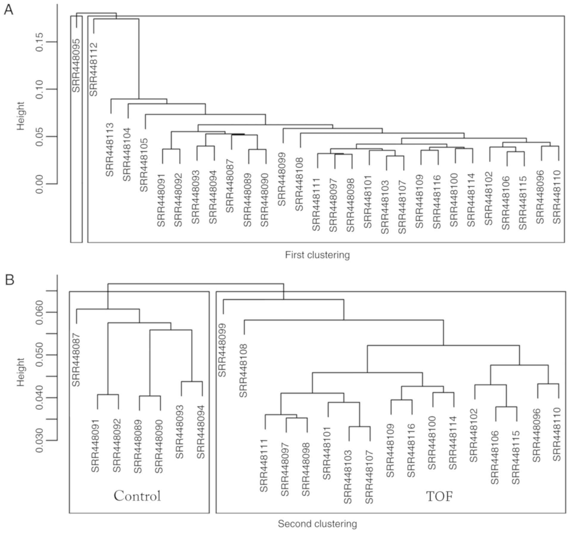
A total of 29 myocardial specimens were analyzed,
including 7 healthy myocardia (Control) and 22 TOFs (Fig. 2A). Following data cleaning, all
specimens qualified, and through hierarchical clustering analysis,
five discrete specimens from the TOF group were excluded, including
SRR448095, SRR448104, SRR448105, SRR448112 and SRR448113. Following
the two cluster analyses, 7 specimens were included in the Control
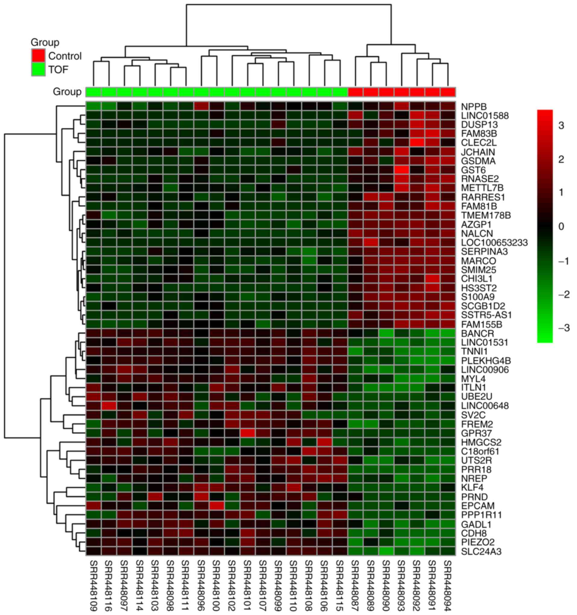
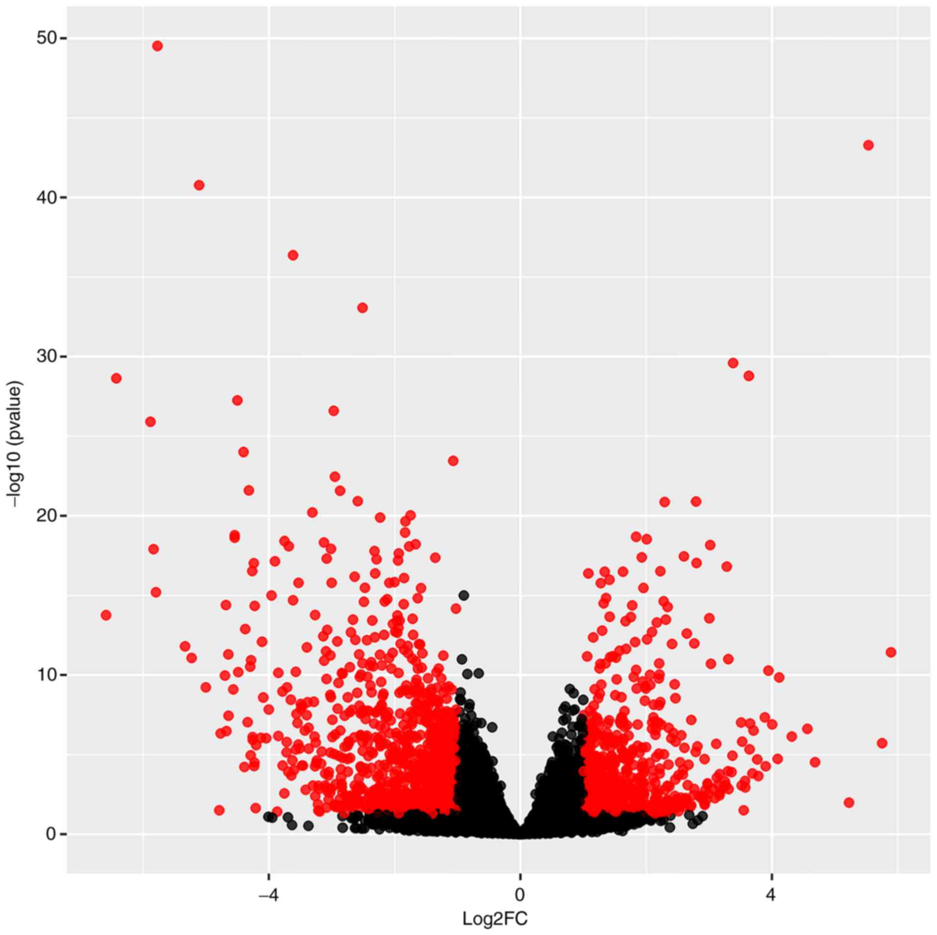
group and 17 specimens were included in TOF group (Fig. 2B). Differential analysis using
DESeq2 revealed that a total of 1,260 genes, including 494
upregulated and 766 downregulated genes, exhibited differential
expression by over 2-fold. The top 25 upregulated and downregulated
genes were indicated on the heat map according to the fold changes
(Fig. 3), and all DEGs were
plotted on a volcano plot (Fig.
4).
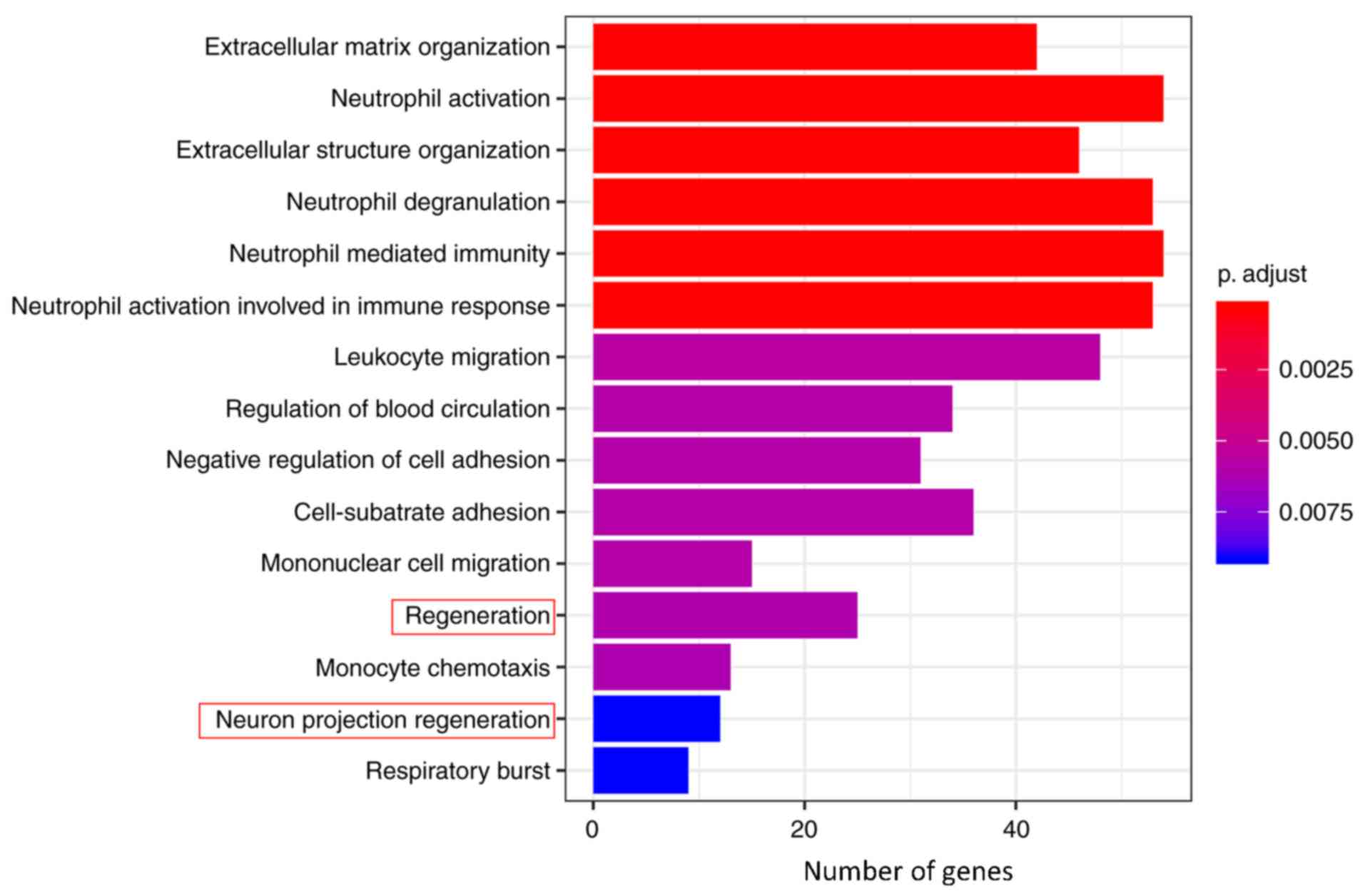
GO and KEGG pathway enrichment
analyses
GO_BP and KEGG pathway enrichment analyses were
performed on the 1,260 genes using ‘clusterProfiler’ to understand
the primary enrichment of these DEGs in BPs and signaling pathways.
A total of 926 DEGs were enriched in 83 GO_BP terms, including
extracellular matrix (ECM) organization, neutrophil activation,
extracellular structure organization, neutrophil degranulation,
neutrophil mediated immunity, neutrophil activation involved in the
immune response, leukocyte migration, regulation of blood
circulation, negative regulation of cell adhesion, cell-substrate
adhesion, mononuclear cell migration, regeneration, monocyte
chemotaxis, neuron projection regeneration and respiratory burst
(Table I). Furthermore, a total of
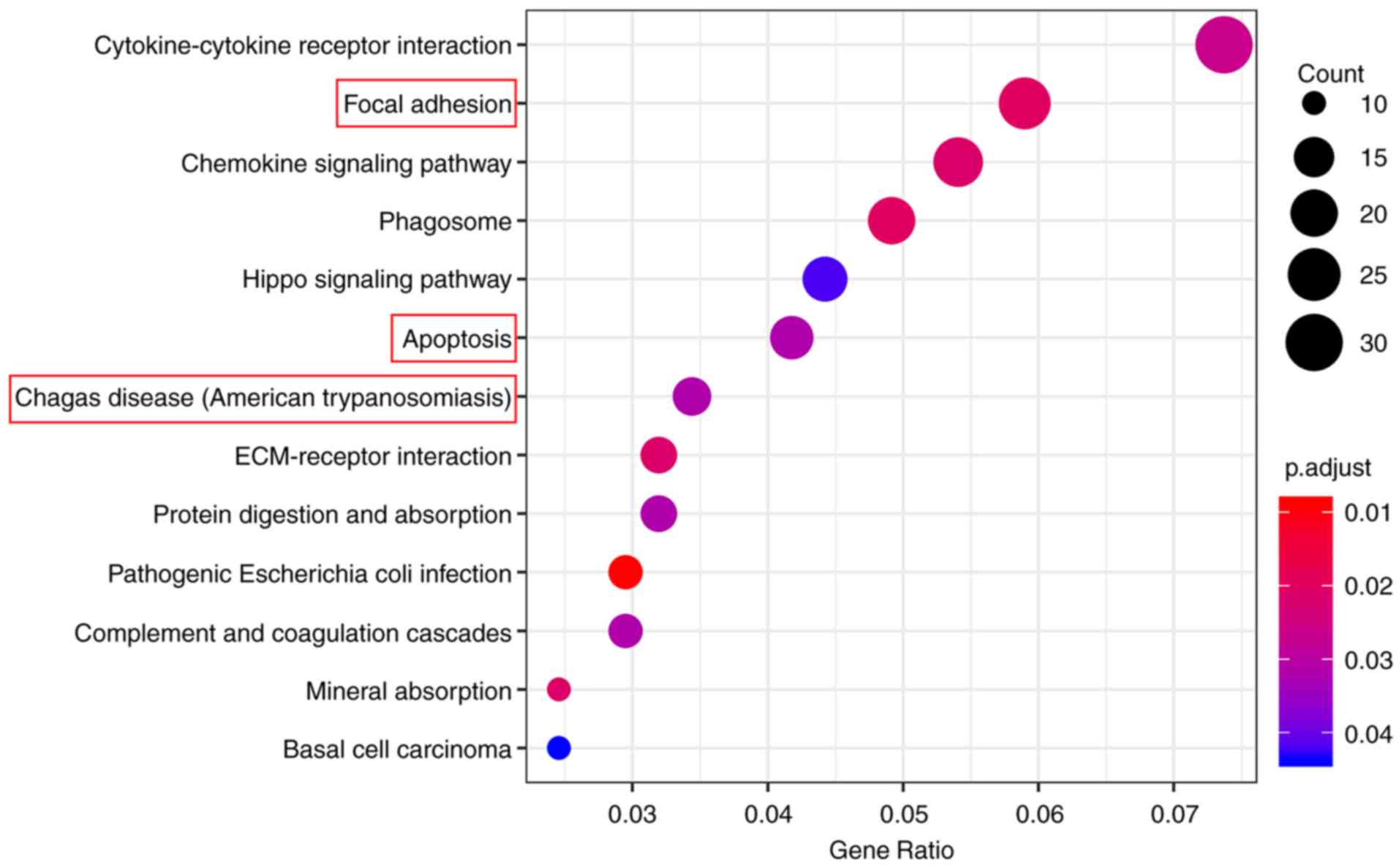
406 DEGs were enriched in 13 KEGG pathways, including
cytokine-cytokine receptor interaction, focal adhesion, chemokine
signaling pathway, phagosome, Hippo signaling pathway, apoptosis,
Chagas disease (American trypanosomiasis), ECM-receptor
interaction, protein digestion and absorption, pathogenic
Escherichia coli infection, complement and coagulation
cascades, mineral absorption and basal cell carcinoma (Table II). The top 15 GO_BP terms and all
13 KEGG pathways were graphically demonstrated according to the P-
and q-values (Figs. 5 and 6, respectively).
 | Table I.GO BP analysis of differentially
expressed genes in hypoxic myocardium. |
Table I.
GO BP analysis of differentially
expressed genes in hypoxic myocardium.
| GO_BP ID | Description | Gene ratio | P-value |
|---|
| GO:0030198 | Extracellular
matrix organization | 42/926 |
2.40×10−07 |
| GO:0042119 | Neutrophil
activation | 54/926 |
3.35×10−07 |
| GO:0043062 | extracellular
structure organization | 46/926 |
3.36×10−07 |
| GO:0043312 | Neutrophil
degranulation | 53/926 |
3.37×10−07 |
| GO:0002446 | Neutrophil mediated
immunity | 54/926 |
3.80×10−07 |
| GO:0002283 | Neutrophil
activation involved in immune response | 53/926 |
4.09×10−07 |
| GO:0050900 | Leukocyte
migration | 48/926 |
7.90×10−06 |
| GO:1903522 | Regulation of blood
circulation | 34/926 |
9.80×10−06 |
| GO:0007162 | Negative regulation
of cell adhesion | 31/926 |
1.16×10−05 |
| GO:0031589 | Cell-substrate
adhesion | 36/926 |
1.27×10−05 |
| GO:0071674 | Mononuclear cell
migration | 15/926 |
1.29×10−05 |
| GO:0031099 | Regeneration | 25/926 |
1.45×10−05 |
| GO:0002548 | Monocyte
chemotaxis | 13/926 |
1.58×10−05 |
| GO:0031102 | Neuron projection
regeneration | 12/926 |
2.66×10−05 |
| GO:0045730 | Respiratory
burst | 9/926 |
2.72×10−05 |
| Table II.KEGG pathway analysis of
differentially expressed genes in hypoxic myocardium. |
Table II.
KEGG pathway analysis of
differentially expressed genes in hypoxic myocardium.
| KEGG ID | Description | Gene ratio | P-value | Genes |
|---|
| hsa05130 | Pathogenic
Escherichia coli infection | 12/406 |
3.09×10⁻⁵ | WAS, ACTB, TUBA4A,
CTNNB1, TLR5, ARPC1B, TUBA3C, TUBA1B, TUBB4B, TUBA1C, TUBA3D and
TUBA3E |
| hsa04510 | Focal adhesion | 24/406 |
2.00×10⁻⁴ | COL1A1, COL4A3,
BCL2, VTN, ITGA11, ACTB, COL6A6, BIRC3, CAV2, PIK3R1, CTNNB1,
COL9A2, TNC, ITGA3, JUN, MAP2K1, RAC2, VASP, ITGA10, LAMC3, SHC3,
MYL7, TNN and COL9A1 |
| hsa04145 | Phagosome | 20/406 |
2.11×10⁻⁴ | C3, CYBA, MPO,
CYBB, ACTB, TUBA4A, ATP6V1B1, C1R, MRC1, MSR1, SFTPD, CTSS, TUBA3C,
TLR6, TUBA1B, TUBB4B, MARCO, TUBA1C, TUBA3D and TUBA3E |
| hsa04978 | Mineral
absorption | 10/406 |
3.60×10⁻⁴ | FTL, ATP1B3,
SLC31A1, HMOX1, MT1A, MT1X, MT2A, SLC26A9, ATP1A4 and MT1M |
| hsa04062 | Chemokine signaling
pathway | 22/406 |
4.46×10⁻⁴ | JAK3, WAS, CCR2,
PIK3R1, CCR1, PF4, PPBP, MAP2K1, RAC2, CCL2, CCL11, CCL17, CCL21,
CXCL14, DOCK2, CCL13, CCL8, CCL26, CCL19, SHC3, NFKBIA and
GNB4 |
| hsa04512 | ECM-receptor
interaction | 13/406 |
4.52×10⁻⁴ | COL1A1, COL4A3,
VTN, ITGA11, COL6A6, COL9A2, TNC, ITGA3, ITGA10, LAMC3, SV2C, TNN
and COL9A1 |
| hsa04060 | Cytokine-cytokine
receptor interaction | 30/406 |
6.49×10⁻⁴ | ACVRL1, AMH, IL10,
GDF6, TNFRSF1B, CCR2, IL15RA, CCR1, BMP6, BMP8B, NGFR, TNFRSF11B,
PF4, PPBP, CCL2, CCL11, CCL17, CCL21, TNFSF9, IL18RAP, CXCL14,
CCL13, CCL8, CCL26, CCL19, BMP10, TNFRSF12A, IL17RB, BMP5 and
CSF1 |
| hsa04610 | Complement and
coagulation cascades | 12/406 |
1.10×10⁻³ | C3, C6, F13A1, F5,
SERPIND1, C1QB, SERPINE1, BDKRB2, SERPINA5, VTN, C1R and F3 |
| hsa04974 | Protein digestion
and absorption | 13/406 |
1.12×10⁻³ | COL1A1, COL3A1,
COL4A3, COL6A6, ATP1B3, COL9A2, CPA3, XPNPEP2, KCNE3, SLC7A8,
COL14A1, COL9A1 and ATP1A4 |
| hsa04210 | Apoptosis | 17/406 |
1.12×10⁻³ | BCL2, ACTB, BIRC3,
PIK3R1, TUBA4A, CTSH, CTSC, JUN, MAP2K1, CTSS, TUBA3C, TUBA1B,
GADD45G, NFKBIA, TUBA1C, TUBA3D and TUBA3E |
| hsa05142 | Chagas disease
(American trypanosomiasis) | 14/406 |
1.22×10⁻³ | C3, C1QB, IL10,
SERPINE1, BDKRB2, ACE, PIK3R1, GNA15, JUN, CCL2, TLR6, PPP2R2C,
NFKBIA and GNAO1 |
| hsa04390 | Hippo signaling
pathway | 18/406 |
1.76×10⁻³ | AMH, SERPINE1,
GDF6, ACTB, DLG2, DVL1, CTNNB1, FZD2, BMP6, BMP8B, SNAI2, WNT9A,
FZD1, FZD7, WNT6, PPP2R2C, BMP5 and NKD1 |
| hsa05217 | Basal cell
carcinoma | 10/406 |
2.00×10⁻³ | DVL1, CTNNB1, FZD2,
WNT9A, FZD1, FZD7, SMO, WNT6, GADD45G and HHIP |
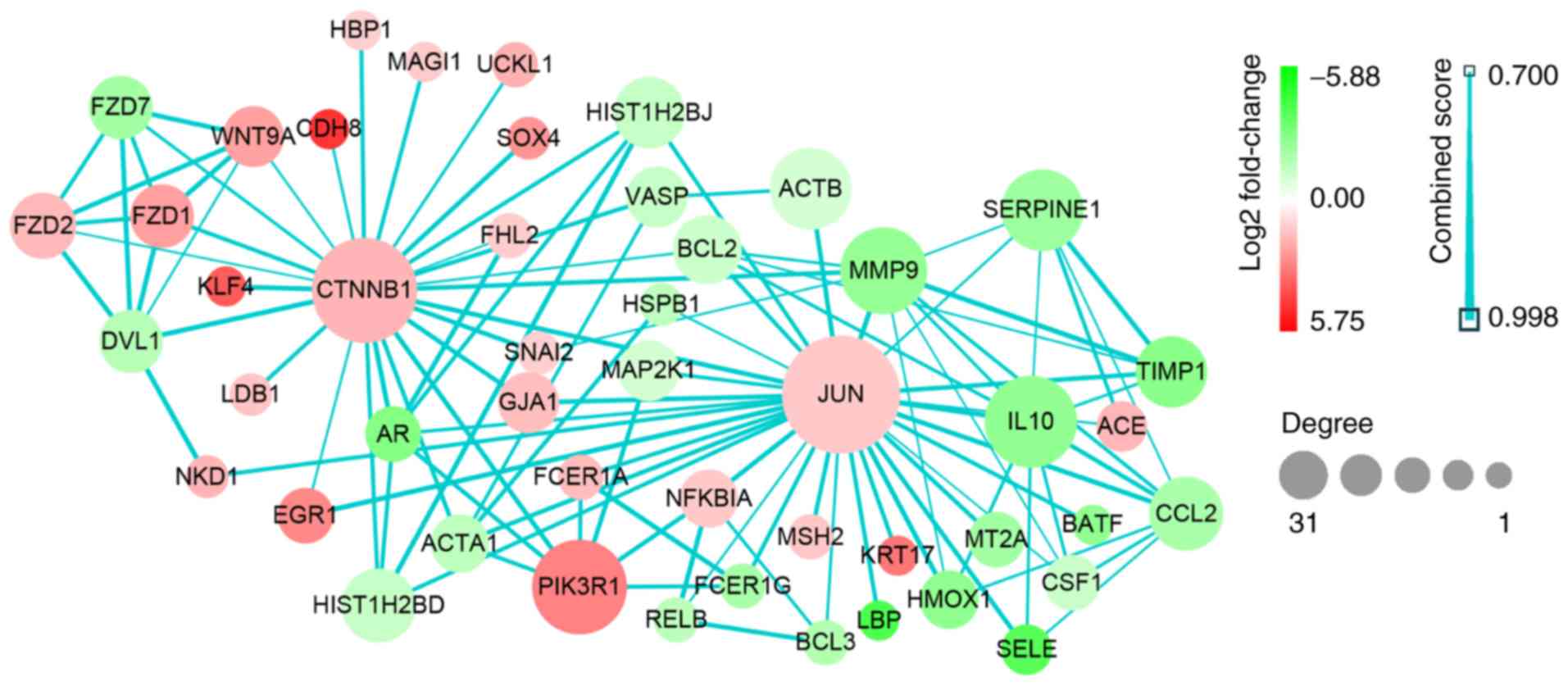
Network analysis from the PPI
data
Based on the data from PPI analysis, six hub genes
with correlated degree scores >25 were identified: G protein
subunit β 4 (GNB4), C-C motif chemokine receptor (CCR)2, CCR1,
platelet factor 4 (PF4), catenin β1 (CTNNB1) and Jun proto-oncogene
(JUN), although the fold changes of these genes did not reach the
top 25 genes in the heat map (Fig.
3). Notably, CTNNB1 and JUN were upregulated (Table III; Fig. 7). Following GO and KEGG analyses,
JUN was indicated to be primarily enriched in the GO_BP terms of
regeneration and neuronal projection regeneration, and in the
signaling pathways of focal adhesion, apoptosis and Chagas disease
(American trypanosomiasis; Figs. 5
and 6).
| Table III.Hub genes identified by Cytoscape
network analysis. |
Table III.
Hub genes identified by Cytoscape
network analysis.
| Hub gene | Degree | Regulation |
|---|
| JUN | 31 | Up |
| CTNNB1 | 26 | Up |
| GNB4 | 32 | Down |
| CCR2 | 27 | Down |
| CCR1 | 25 | Down |
| PF4 | 25 | Down |
Discussion
Chronic hypoxia is the basic pathophysiological
state of cyanotic CHD; the heart forms endogenous mechanisms to
protect cardiomyocytes against injuries caused by hypoxia (6–8). To
identify the effects of chronic hypoxia on the heart, the present
study analyzed DEGs in chronically hypoxic myocardium. NGS data was
extracted from the GSE36761 data set, and 1,260 DEGs between
healthy and chronically hypoxic myocardium were identified using
bioinformatics. The DEGs that co-existed or were co-expressed under
chronically hypoxic conditions were likely to be involved in
similar biological processes (19). Therefore, for better understanding
of the interactions between these DEGs, enrichment analysis was
performed, which suggested that they were predominantly enriched in
GO_BPs such as inflammatory response, cell regeneration and
apoptosis. Further analysis of the PPI network also identified six
hub genes underlying the myocardial adaptation to chronic
hypoxia.
GO analysis indicated that hypoxia in the myocardium
was associated with inflammatory responses activated by neutrophils
and monocytes, which involved changes in ECM organization, cell
substrate adhesion, monocyte migration and chemotaxis. Consistent
with previous studies (20–24),
this indicated that chronic hypoxia was associated with
inflammation. In accordance with the findings of hypoxia-activated
cardiomyocyte mitosis and regeneration (25), GO analysis revealed that the DEGs
in hypoxic myocardium were also enriched in cell regeneration.
KEGG pathway analysis indicated that the DEGs
identified in the present study were predominantly enriched in
cytokine-cytokine receptor interactions, chemokine signaling
pathways, ECM-receptor interaction and signaling pathways, such as
focal adhesion and apoptosis. Focal adhesions are multi-protein
structures that contain integrin that can mechanically connect the
cytoplasmic actin skeleton of different cells to the ECM (26,27).
Thus, ECM signals can be transmitted by focal adhesion kinase (FAK)
into the cell, thereby mediating cell adhesion, migration,
survival, cell cycle regulation, proliferation and apoptosis
(28). Notably, hypoxia induces
the activation of the FAK signaling pathway in cardiomyocytes,
promotes the adhesive interaction between cardiomyocytes and ECMs
(29) and may result in cell
proliferation and survival. However, the DEGs in the present study
were also enriched in the apoptosis pathway, which also supports
the findings that hypoxia may induce cardiomyocyte apoptosis
(30). Therefore, it is notable
that these DEGs were associated with regeneration, focal adhesion
and apoptosis, which was indicative of the homeostasis between loss
and renewal of cardiomyocytes exposed to hypoxia.
To further understand the intrinsic significance of
the 1,260 DEGs, that is, the interactions between them, a PPI
network was constructed. The data identified six hub genes with
correlated degree scores of >25 through the network correlation
analysis, including GNB4, CCR2, CCR1, PF4, CTNNB1 and JUN. Notably,
JUN and CTNNB1 were identified to be upregulated. GNB4 is the β-4
subunit of guanine nucleotide-binding protein (G protein), an
important regulator of the α subunit of G protein and some signal
transduction receptors and effectors (31). It is typically co-expressed with
the γ subunit to form dimers that regulate important effector
systems, including the N-type Ca2+ channel, the G
protein-gated inwardly-rectifying K+ channel and
phospholipase (32,33). Downregulation of GNB4 in
chronically hypoxic myocardium is associated with ion
channel-associated heart rate and contractility (34), reduction of G-protein-coupled
receptors such as chemokine receptors (20), and apoptosis (35). CCR2 and CCR1 are predominantly
expressed on the surface of inflammatory cells, including
lymphocytes, monocytes and macrophages; they are types of G
protein-coupled chemokine receptors that mediate the infiltration
of neutrophils following ischemia (36). The downregulation of CCR2 and CCR1
in response to hypoxia was in accord with a previous study
(20). The reduction of chemokine
receptor expression seemed to imply that less inflammatory cells
(such as mononuclear macrophages and neutrophils) migrated into the
hypoxic myocardium, thereby alleviating inflammation, improving
adverse cardiac remodeling and reducing apoptosis (37,38).
This may possibly be considered a protective adaptation of the
myocardium to chronic hypoxia (39,40).
PF4 is released from α-granules of activated platelets; not only
are the activated platelets associated with cell apoptosis and
survival (41), but also PF4
itself has been linked to apoptosis. Notably, PF4 inhibits tumor
growth and tumor angiogenesis (42) and induces tumor cell apoptosis
(43). Furthermore, as the small
CXC chemokine and agonist of CCR1, PF4 promotes chemotaxis in
inflammatory cells, including neutrophils and monocytes (44,45).
Downregulation of PF4 under chronic hypoxia may be connected with
apoptosis and the reduction of chemokine receptors in myocardium.
Cytoplasmic β-catenin, encoded by the CTNNB1 gene, is a key protein
in the Wnt signaling pathway; this is a conserved pathway during
animal evolution and development, yet the specific genes it
regulates depend on the cell type and background (46). In adult hearts, the Wnt signaling
pathway is quiescent under normal conditions but is activated
following myocardial injury and participates in cardiac remodeling
(47). Once exposed to hypoxia,
hypoxia inducible factor-1α (HIF-1α) competitively inhibits the
formation of β-catenin/T-cell factor-4 complex and the downstream
transcriptional activities in colorectal tumor cells, resulting in
G1 arrest in the cell cycle. However, β-catenin also
enhances HIF-1α-mediated transcription and helps the tumor cells
with survival and adaptation to hypoxia (48). A previous study has reported that
the Wnt/β-catenin signaling pathway may promote proliferation and
protect cardiomyocytes from apoptosis, though little direct
evidence exists to date (49).
Therefore, the present study hypothesized that activation of the
Wnt/β-catenin pathway may have an inhibitory effect on apoptosis
under chronic hypoxia.
In the present study, the role of JUN in hypoxic
myocardium became the primary focus. The transcription factor
c-Jun, which is encoded by the oncogene JUN and belongs to the
basic-region leucine zippers (bZIP) family, constitutes the
activator protein-1 (AP-1) in the form of homodimers or
heterodimers with other bZIPs, thereby regulating transcription and
cellular activities, including proliferation, apoptosis,
tumorigenesis and tissue morphogenesis (50). Through the GO and KEGG pathway
enrichment analyses, it was identified that c-Jun was enriched in
the biological function of ‘regeneration’ and the signaling
pathways of ‘apoptosis’ and ‘focal adhesion’. It seemed paradoxical
that c-Jun functioned in promoting survival as well as apoptosis in
hypoxic myocardium. As an important endpoint of mitogen-activated
protein kinase (MAPK) cascade activation, the DNA binding and
transcriptional activity of c-Jun are enhanced following activation
by JNK phosphorylation at Ser63/73. Following this, it forms AP-1,
transcribes the downstream proteins (51) and mediates the expression or
activation of pro-apoptotic genes, including Fas ligand,
insulin-like growth factor binding protein 4, tumor necrosis factor
(TNF)-α, B cell lymphoma-2 homologous antagonist/killer (52), p53 upregulated modulator of
apoptosis (53) and Bim (54). Conversely, c-Jun has also been
demonstrated to have anti-apoptotic properties. Endogenous c-Jun
could upregulate survivin, counteract apoptosis, maintain mammary
epithelial cell survival (55) and
prevent TNF-α-induced hepatocyte apoptosis by antagonizing the
activity of the pro-apoptotic gene p53 (56). Additionally, it works with signal
transducer and activator of transcription 3 to inhibit Fas
transcription, thereby reducing the sensitivity of tumor cells to
apoptosis (57). Another study
identified that c-Jun transcriptionally inhibits the tumor
suppressor phosphatase and tensin homolog, which in turn activates
the Akt signaling pathway and promotes fibroblast survival
(58). In addition to its
functions in apoptosis, c-Jun also promotes cell cycle progression
through direct transcriptional activation of cyclin D1 (59) and repression of p53 (60). Previous studies have demonstrated
that the MAPK subfamily JNK and its target c-Jun are activated by
phosphorylation, which induces cardiomyocyte apoptosis under
hypoxia stress (6,7). However, the downstream mechanisms
underlying apoptosis caused by c-Jun remains to be elucidated.
Therefore, whether c-Jun takes part in anti-apoptotic effects
during hypoxic myocardial apoptosis is worthy of further
investigation. One possible explanation for the opposing nature of
pro- and anti-apoptosis (or pro-survival) is that c-Jun may
activate anti-apoptotic programs under hypoxia to inhibit the
excessive cardiomyocyte apoptosis caused by c-Jun itself and other
factors. In addition, c-Jun may even promote cell cycle progression
and proliferation to replenish the lost cardiomyocytes subjected to
hypoxia. In this way, a new cellular homeostasis under hypoxia
could be maintained. Therefore, the role of c-Jun in promoting
survival and apoptosis may be the adaptation of myocardial tissue
to chronic hypoxia, that is, some mechanism of self-protection and
self-repair.
In conclusion, data mining was used in the present
study to reveal the biological significance of DEGs in hypoxic
myocardium, and genes associated with the life or death of
myocardium were screened. The results provided a series of
potential targets for studying mechanisms underlying the cellular
adaptation to chronic hypoxia. However, the specific molecular
mechanisms require further biological validation in in vitro
or in in vivo experiments.
Acknowledgements
Not applicable.
Funding
The present study was supported by The National
Science Foundation of China (grant nos. 81700277 and 81270228).
Availability of data and materials
The original data and program codes are available
from the corresponding author on reasonable request or from the
research data repositories Figshare (https://figshare.com/s/d7d700afae1d4194c391).
Authors' contributions
YX conceived and supervised the study; FW performed
the primary bioinformatics analysis and was a major contributor in
writing the manuscript; FG mainly contributed in the analysis of
the biological significance of hub genes and picture editing; and
SH was involved in interpretation of parts of the hub genes and
English language editing. All authors read and approved the final
manuscript.
Ethics approval and consent to
participate
Not applicable.
Patient consent for publication
Not applicable.
Competing interests
The authors declare that they have no competing
interests.
References
|
1
|
Compernolle V, Brusselmans K, Franco D,
Moorman A, Dewerchin M, Collen D and Carmeliet P: Cardia bifida,
defective heart development and abnormal neural crest migration in
embryos lacking hypoxia-inducible factor-1alpha. Cardiovasc Res.
60:569–579. 2003. View Article : Google Scholar : PubMed/NCBI
|
|
2
|
Burchell HB, Taylor BE, et al: Circulatory
adjustments to the hypoxemia of congenital heart disease of the
cyanotic type. Circulation. 1:404–414. 1950. View Article : Google Scholar : PubMed/NCBI
|
|
3
|
Fan X, Ma L, Zhang Z, Li Y, Hao M, Zhao Z,
Zhao Y, Liu F, Liu L, Luo X, et al: Associations of high-altitude
polycythemia with polymorphisms in PIK3CD and COL4A3 in Tibetan
populations. Hum Genomics. 12:372018. View Article : Google Scholar : PubMed/NCBI
|
|
4
|
Humayun KN and Atiq M: Clinical profile
and outcome of cyanotic congenital heart disease in neonates. J
Coll Physicians Surg Pak. 18:290–293. 2008.PubMed/NCBI
|
|
5
|
Rafiee P, Shi Y, Kong X, Pritchard KA Jr,
Tweddell JS, Litwin SB, Mussatto K, Jaquiss RD, Su J and Baker JE:
Activation of protein kinases in chronically hypoxic infant human
and rabbit hearts: Role in cardioprotection. Circulation.
106:239–245. 2002. View Article : Google Scholar : PubMed/NCBI
|
|
6
|
He S, Liu P, Jian Z, Li J, Zhu Y, Feng Z
and Xiao Y: miR-138 protects cardiomyocytes from hypoxia-induced
apoptosis via MLK3/JNK/c-jun pathway. Biochem Biophys Res Commun.
441:763–769. 2013. View Article : Google Scholar : PubMed/NCBI
|
|
7
|
He S, Liu S, Wu X, Xin M, Ding S, Xin D,
Ouyang H and Zhang J: Protective role of downregulated MLK3 in
myocardial adaptation to chronic hypoxia. J Physiol Biochem.
73:371–380. 2016. View Article : Google Scholar : PubMed/NCBI
|
|
8
|
Liu B, Zhang HG, Zhu Y, Jiang YH, Luo GP,
Tang FQ, Jian Z and Xiao YB: Cardiac resident macrophages are
involved in hypoxiainduced postnatal cardiomyocyte proliferation.
Mol Med Rep. 15:3541–3548. 2017. View Article : Google Scholar : PubMed/NCBI
|
|
9
|
Laflamme MA and Murry CE: Heart
regeneration. Nature. 473:326–335. 2011. View Article : Google Scholar : PubMed/NCBI
|
|
10
|
Churko JM, Mantalas GL, Snyder MP and Wu
JC: Overview of high throughput sequencing technologies to
elucidate molecular pathways in cardiovascular diseases. Circ Res.
112:1613–1623. 2013. View Article : Google Scholar : PubMed/NCBI
|
|
11
|
Manzoni C, Kia DA, Vandrovcova J, Hardy J,
Wood NW, Lewis PA and Ferrari R: Genome, transcriptome and
proteome: The rise of omics data and their integration in
biomedical sciences. Brief Bioinform. 19:286–302. 2018. View Article : Google Scholar : PubMed/NCBI
|
|
12
|
Grunert M, Dorn C, Schueler M, Dunkel I,
Schlesinger J, Mebus S, Alexi-Meskishvili V, Perrot A, Wassilew K,
Timmermann B, et al: Rare and private variations in neural crest,
apoptosis and sarcomere genes define the polygenic background of
isolated Tetralogy of Fallot. Hum Mol Genet. 23:3115–3128. 2014.
View Article : Google Scholar : PubMed/NCBI
|
|
13
|
Ghorbel MT, Cherif M, Jenkins E, Mokhtari
A, Kenny D, Angelini GD and Caputo M: Transcriptomic analysis of
patients with tetralogy of Fallot reveals the effect of chronic
hypoxia on myocardial gene expression. J Thorac Cardiovasc Surg.
140:337–345.e26. 2010. View Article : Google Scholar : PubMed/NCBI
|
|
14
|
Ahmed M, Nguyen HQ, Hwang JS, Zada S, Lai
TH, Kang SS and Kim DR: Systematic characterization of
autophagy-related genes during the adipocyte differentiation using
public-access data. Oncotarget. 9:15526–15541. 2018. View Article : Google Scholar : PubMed/NCBI
|
|
15
|
Ashburner M, Ball CA, Blake JA, Botstein
D, Butler H, Cherry JM, Davis AP, Dolinski K, Dwight SS, Eppig JT,
et al: Gene ontology: Tool for the unification of biology. The Gene
Ontology Consortium. Nat Genet. 25:25–29. 2000. View Article : Google Scholar : PubMed/NCBI
|
|
16
|
Kanehisa M and Goto S: KEGG: Kyoto
encyclopedia of genes and genomes. Nucleic Acids Res. 28:27–30.
2000. View Article : Google Scholar : PubMed/NCBI
|
|
17
|
Yu G, Wang LG, Han Y and He QY:
clusterProfiler: An R package for comparing biological themes among
gene clusters. OMICS. 16:284–287. 2012. View Article : Google Scholar : PubMed/NCBI
|
|
18
|
Szklarczyk D, Franceschini A, Wyder S,
Forslund K, Heller D, Huerta-Cepas J, Simonovic M, Roth A, Santos
A, Tsafou KP, et al: STRING v10: Protein-protein interaction
networks, integrated over the tree of life. Nucleic Acids Res.
43:Database Issue. D447–D452. 2015. View Article : Google Scholar : PubMed/NCBI
|
|
19
|
Roy S, Bhattacharyya DK and Kalita JK:
Reconstruction of gene co-expression network from microarray data
using local expression patterns. BMC Bioinformatics. 15 (Suppl
7):S102014. View Article : Google Scholar : PubMed/NCBI
|
|
20
|
Bosco MC, Puppo M, Santangelo C, Anfosso
L, Pfeffer U, Fardin P, Battaglia F and Varesio L: Hypoxia modifies
the transcriptome of primary human monocytes: Modulation of novel
immune-related genes and identification of CC-chemokine ligand 20
as a new hypoxia-inducible gene. J Immunol. 177:1941–1955. 2006.
View Article : Google Scholar : PubMed/NCBI
|
|
21
|
Murdoch C, Muthana M and Lewis CE: Hypoxia
regulates macrophage functions in inflammation. J Immunol.
175:6257–6263. 2005. View Article : Google Scholar : PubMed/NCBI
|
|
22
|
Hoenderdos K, Lodge KM, Hirst RA, Chen C,
Palazzo SG, Emerenciana A, Summers C, Angyal A, Porter L, Juss JK,
et al: Hypoxia upregulates neutrophil degranulation and potential
for tissue injury. Thorax. 71:1030–1038. 2016. View Article : Google Scholar : PubMed/NCBI
|
|
23
|
Bartels K, Grenz A and Eltzschig HK:
Hypoxia and inflammation are two sides of the same coin. Proc Natl
Acad Sci USA. 110:18351–18352. 2013. View Article : Google Scholar : PubMed/NCBI
|
|
24
|
Taylor CT and Colgan SP: Regulation of
immunity and inflammation by hypoxia in immunological niches. Nat
Rev Immunol. 17:774–785. 2017. View Article : Google Scholar : PubMed/NCBI
|
|
25
|
Nakada Y, Canseco DC, Thet S, Abdisalaam
S, Asaithamby A, Santos CX, Shah AM, Zhang H, Faber JE, Kinter MT,
et al: Hypoxia induces heart regeneration in adult mice. Nature.
541:222–227. 2017. View Article : Google Scholar : PubMed/NCBI
|
|
26
|
Chen CS, Alonso JL, Ostuni E, Whitesides
GM and Ingber DE: Cell shape provides global control of focal
adhesion assembly. Biochem Biophys Res Commun. 307:355–361. 2003.
View Article : Google Scholar : PubMed/NCBI
|
|
27
|
Zaidel-Bar R, Cohen M, Addadi L and Geiger
B: Hierarchical assembly of cell-matrix adhesion complexes. Biochem
Soc Trans. 32:416–420. 2004. View Article : Google Scholar : PubMed/NCBI
|
|
28
|
Vadali K, Cai X and Schaller MD: Focal
adhesion kinase: An essential kinase in the regulation of
cardiovascular functions. IUBMB Life. 59:709–716. 2007. View Article : Google Scholar : PubMed/NCBI
|
|
29
|
Seko Y, Takahashi N, Sabe H, Tobe K,
Kadowaki T and Nagai R: Hypoxia induces activation and subcellular
translocation of focal adhesion kinase (p125(FAK)) in cultured rat
cardiac myocytes. Biochem Biophys Res Commun. 262:290–296. 1999.
View Article : Google Scholar : PubMed/NCBI
|
|
30
|
Jung F, Weiland U, Johns RA, Ihling C and
Dimmeler S: Chronic hypoxia induces apoptosis in cardiac myocytes:
A possible role for Bcl-2-like proteins. Biochem Biophys Res
Commun. 286:419–425. 2001. View Article : Google Scholar : PubMed/NCBI
|
|
31
|
Smrcka AV: G protein β subunits: Central
mediators of G protein-coupled receptor signaling. Cell Mol Life
Sci. 65:2191–2214. 2008. View Article : Google Scholar : PubMed/NCBI
|
|
32
|
Ruiz-Velasco V, Ikeda SR and Puhl HL:
Cloning, tissue distribution, and functional expression of the
human G protein beta 4-subunit. Physiol Genomics. 8:41–50. 2002.
View Article : Google Scholar : PubMed/NCBI
|
|
33
|
Rosskopf D, Nikula C, Manthey I, Joisten
M, Frey U, Kohnen S and Siffert W: The human G protein beta4
subunit: Gene structure, expression, Ggamma and effector
interaction. FEBS Lett. 544:27–32. 2003. View Article : Google Scholar : PubMed/NCBI
|
|
34
|
Clapham DE and Neer EJ: G protein beta
gamma subunits. Annu Rev Pharmacol Toxicol. 37:167–203. 1997.
View Article : Google Scholar : PubMed/NCBI
|
|
35
|
Adams JW and Brown JH: G-proteins in
growth and apoptosis: Lessons from the heart. Oncogene.
20:1626–1634. 2001. View Article : Google Scholar : PubMed/NCBI
|
|
36
|
Reichel CA, Khandoga A, Anders HJ,
Schlondorff D, Luckow B and Krombach F: Chemokine receptors Ccr1,
Ccr2, and Ccr5 mediate neutrophil migration to postischemic tissue.
J Leukoc Biol. 79:114–122. 2006. View Article : Google Scholar : PubMed/NCBI
|
|
37
|
Liehn EA, Merx MW, Postea O, Becher S,
Djalali-Talab Y, Shagdarsuren E, Kelm M, Zernecke A and Weber C:
Ccr1 deficiency reduces inflammatory remodelling and preserves left
ventricular function after myocardial infarction. J Cell Mol Med.
12:496–506. 2008. View Article : Google Scholar : PubMed/NCBI
|
|
38
|
Majmudar MD, Keliher EJ, Heidt T,
Leuschner F, Truelove J, Sena BF, Gorbatov R, Iwamoto Y, Dutta P,
Wojtkiewicz G, et al: Monocyte-directed RNAi targeting CCR2
improves infarct healing in atherosclerosis-prone mice.
Circulation. 127:2038–2046. 2013. View Article : Google Scholar : PubMed/NCBI
|
|
39
|
Hayasaki T, Kaikita K, Okuma T, Yamamoto
E, Kuziel WA, Ogawa H and Takeya M: CC chemokine receptor-2
deficiency attenuates oxidative stress and infarct size caused by
myocardial ischemia-reperfusion in mice. Circ J. 70:342–351. 2006.
View Article : Google Scholar : PubMed/NCBI
|
|
40
|
Zhou L, Azfer A, Niu J, Graham S,
Choudhury M, Adamski FM, Younce C, Binkley PF and Kolattukudy PE:
Monocyte chemoattractant protein-1 induces a novel transcription
factor that causes cardiac myocyte apoptosis and ventricular
dysfunction. Circ Res. 98:1177–1185. 2006. View Article : Google Scholar : PubMed/NCBI
|
|
41
|
Gawaz M and Vogel S: Platelets in tissue
repair: Control of apoptosis and interactions with regenerative
cells. Blood. 122:2550–2554. 2013. View Article : Google Scholar : PubMed/NCBI
|
|
42
|
Tanaka T, Manome Y, Wen P, Kufe DW and
Fine HA: Viral vector-mediated transduction of a modified platelet
factor 4 cDNA inhibits angiogenesis and tumor growth. Nat Med.
3:437–442. 1997. View Article : Google Scholar : PubMed/NCBI
|
|
43
|
Liang P, Cheng SH, Cheng CK, Lau KM, Lin
SY, Chow EY, Chan NP, Ip RK, Wong RS and Ng MH: Platelet factor 4
induces cell apoptosis by inhibition of STAT3 via up-regulation of
SOCS3 expression in multiple myeloma. Haematologica. 98:288–295.
2013. View Article : Google Scholar : PubMed/NCBI
|
|
44
|
Deuel TF, Senior RM, Chang D, Griffin GL,
Heinrikson RL and Kaiser ET: Platelet factor 4 is chemotactic for
neutrophils and monocytes. Proc Natl Acad Sci USA. 78:4584–4587.
1981. View Article : Google Scholar : PubMed/NCBI
|
|
45
|
Fox JM, Kausar F, Day A, Osborne M,
Hussain K, Mueller A, Lin J, Tsuchiya T, Kanegasaki S and Pease JE:
CXCL4/Platelet Factor 4 is an agonist of CCR1 and drives human
monocyte migration. Sci Rep. 8:94662018. View Article : Google Scholar : PubMed/NCBI
|
|
46
|
Lam AP and Gottardi CJ: β-catenin
signaling: A novel mediator of fibrosis and potential therapeutic
target. Curr Opin Rheumatol. 23:562–567. 2011. View Article : Google Scholar : PubMed/NCBI
|
|
47
|
Bergmann MW: WNT signaling in adult
cardiac hypertrophy and remodeling: Lessons learned from cardiac
development. Circ Res. 107:1198–1208. 2010. View Article : Google Scholar : PubMed/NCBI
|
|
48
|
Kaidi A, Williams AC and Paraskeva C:
Interaction between beta-catenin and HIF-1 promotes cellular
adaptation to hypoxia. Nat Cell Biol. 9:210–217. 2007. View Article : Google Scholar : PubMed/NCBI
|
|
49
|
Ozhan G and Weidinger G: Wnt/β-catenin
signaling in heart regeneration. Cell Regen (Lond).
4:32015.PubMed/NCBI
|
|
50
|
Meng Q and Xia Y: c-Jun, at the crossroad
of the signaling network. Protein Cell. 2:889–898. 2011. View Article : Google Scholar : PubMed/NCBI
|
|
51
|
Davis RJ: Signal transduction by the JNK
group of MAP kinases. Cell. 103:239–252. 2000. View Article : Google Scholar : PubMed/NCBI
|
|
52
|
Fan M and Chambers TC: Role of
mitogen-activated protein kinases in the response of tumor cells to
chemotherapy. Drug Resist Updat. 4:253–267. 2001. View Article : Google Scholar : PubMed/NCBI
|
|
53
|
Zhao Z, Wang J, Tang J, Liu X, Zhong Q,
Wang F, Hu W, Yuan Z, Nie C and Wei Y: JNK- and Akt-mediated Puma
expression in the apoptosis of cisplatin-resistant ovarian cancer
cells. Biochem J. 444:291–301. 2012. View Article : Google Scholar : PubMed/NCBI
|
|
54
|
Tomicic MT, Meise R, Aasland D, Berte N,
Kitzinger R, Kramer OH, Kaina B and Christmann M: Apoptosis induced
by temozolomide and nimustine in glioblastoma cells is supported by
JNK/c-Jun-mediated induction of the BH3-only protein BIM.
Oncotarget. 6:33755–33768. 2015. View Article : Google Scholar : PubMed/NCBI
|
|
55
|
Katiyar S, Casimiro MC, Dettin L, Ju X,
Wagner EF, Tanaka H and Pestell RG: C-jun inhibits mammary
apoptosis in vivo. Mol Biol Cell. 21:4264–4274. 2010. View Article : Google Scholar : PubMed/NCBI
|
|
56
|
Eferl R, Ricci R, Kenner L, Zenz R, David
JP, Rath M and Wagner EF: Liver tumor development. c-Jun
antagonizes the proapoptotic activity of p53. Cell. 112:181–192.
2003. View Article : Google Scholar : PubMed/NCBI
|
|
57
|
Ivanov VN, Bhoumik A, Krasilnikov M, Raz
R, Owen-Schaub LB, Levy D, Horvath CM and Ronai Z: Cooperation
between STAT3 and c-jun suppresses Fas transcription. Mol Cell.
7:517–528. 2001. View Article : Google Scholar : PubMed/NCBI
|
|
58
|
Hettinger K, Vikhanskaya F, Poh MK, Lee
MK, de Belle I, Zhang JT, Reddy SA and Sabapathy K: c-Jun promotes
cellular survival by suppression of PTEN. Cell Death Differ.
14:218–229. 2007. View Article : Google Scholar : PubMed/NCBI
|
|
59
|
Wisdom R, Johnson RS and Moore C: c-Jun
regulates cell cycle progression and apoptosis by distinct
mechanisms. EMBO J. 18:188–197. 1999. View Article : Google Scholar : PubMed/NCBI
|
|
60
|
Schreiber M, Kolbus A, Piu F, Szabowski A,
Möhle-Steinlein U, Tian J, Karin M, Angel P and Wagner EF: Control
of cell cycle progression by c-Jun is p53 dependent. Genes Dev.
13:607–619. 1999. View Article : Google Scholar : PubMed/NCBI
|