Introduction
Osteosarcoma is the most commonly diagnosed primary
malignant bone tumor, and can lead to tumor-related mortality among
children and adolescents (1).
Osteosarcoma is the most common form of bone and soft tissue
malignant tumor, and is characterized by high tumor cell
proliferation, high mortality and rapid metastasis (2). Unfortunately, the prognosis of
patients with metastatic osteosarcoma remains poor (3). In addition, only a slight improvement
in the survival rate of osteosarcoma patients has been achieved
even following the development of novel therapeutic technologies,
such as radiotherapy, adjuvant chemotherapy and surgery (4,5). To
date, there are few available biomarkers to manage the tumor burden
and also the treatment response for osteosarcoma. Thus, prognostic
assessment and early diagnosis of osteosarcoma are of great
importance. Therefore, the prognostic evaluation and early
diagnosis of osteosarcoma are important for the initiation of early
treatment and thus patient survival can be improved
accordingly.
Chromobox protein homolog 3 (CBX3) belongs to the
heterochomatin protein 1 (HP1) family, which is involved in
transcriptional activation or repression, cell differentiation and
growth and epigenetic modifications (6,7).
Additionally, it has been reported that CBX3 is upregulated in many
cancer types, which is of great significance for the development of
cancer and the regulation of growth-related genes (8,9).
Thus, it is expected that CBX3 plays an extremely significant role
in the progression of tumors. However, the functional role of CBX3
in osteosarcoma progression, tumorigenesis and prognosis requires
further elucidation.
The present study is the first to investigate the
functional role and prognostic value of CBX3 in osteosarcoma. This
study demonstrated that there was a high expression of CBX3 in
sarcoma and osteosarcoma tissues. In addition, the clinical
significance of CBX3 expression in human osteosarcoma was also
explored. Meanwhile, the impact of CBX3 on cell cycle distribution,
apoptosis and proliferation of osteosarcoma cells was also
investigated.
Materials and methods
Bioinformatic analysis through
Oncomine databases
Oncomine database (https://www.oncomine.org) was used for the assessment
of the differences in CBX3 mRNA expression between matched normal
tissues and sarcoma tissues, based on the following filtering
conditions: P-value <0.0001, gene rank (top 10%), fold change
>2, data type (mRNA), analysis type (cancer vs. normal analysis)
and gene (CBX3).
Patients and tissue samples
A total of 98 osteosarcoma patients (56 males and 42
females; mean age, 18 years) who underwent initial surgery at the
First Affiliated Hospital of Xinxiang Medical University (Xinxiang,
China) between January 2008 and December 2017 were enrolled in this
study. All patients were diagnosed with osteosarcoma at the
Pathology Department of the First Affiliated Hospital of Xinxiang
Medical University. Written informed consent for the participation
in this study was obtained from all the patients. The Medical
Ethics Committee of the First Affiliated Hospital of Xinxiang
Medical University approved the use of the samples. All specimens
were immediately frozen and stored in liquid nitrogen (−196°C).
Every patient was treatment-naïve until the resection.
Cell lines
The human osteosarcoma cell line MG63 was purchased
from the Shanghai Cell Bank (Shanghai, China). Dulbecco's modified
Eagle's medium (DMEM; Gibco; Thermo Fisher Scientific, Inc.,
Waltham, MA, USA) and 10% fetal bovine serum (FBS; Invitrogen;
Thermo Fisher Scientific, Inc.) were used to culture all the cells
at 37°C in 5% CO2.
Reverse transcription-quantitative
polymerase chain reaction (RT-qPCR) assay
TRIzol was used to extract all the RNA from the
osteosarcoma cells and tissues according to the manufacturer's
protocol (Shanghai Invitrogen Biotechnology Co., Ltd., Shanghai,
China). The qPCR was used to detect the expression levels of CBX3
mRNA by One-Step RT-PCR kit (Takara Bio, Inc., Otsu, Japan) based
on the manufacturer's protocol. The thermocycling conditions were
as follows: Initial denaturation at 95°C for 30 sec, followed by 40
amplification cycles of 95°C for 5 sec and 60°C for 34 sec. The
CBX3 primers were purchased from Shanghai Genechem Co., Ltd.
(Shanghai, China). The internal reference was glyceraldehyde
3-phosphate dehydrogenase (GAPDH).
The primers used in the present study were as
follows: CBX3 forward, 5′-TGGCCTCCAACAAAACTACA-3′ and reverse,
5′-TCCCATTCACTACACGTCGA-3′; and GAPDH forward,
5′-GGAGTCAACGGATTTGGTCGTAT-3′ and reverse,
5′-AGCCTTCTCCATGGTGGTGAAGAC-3′. The 2−ΔΔCq method
(10) was used to quantify the
CBX3 mRNA expression levels. Then, the GAPDH mRNA expression level
was used to normalize it.
Transfection of osteosarcoma MG63
cells
The negative control siRNA (NC siRNA) and CBX3 small
interfering RNA (siRNA) were provided by Shanghai GeneChem
Biotechnology Co., Ltd. (Shanghai, China). The sequences of the
siRNAs used in the present study were: Si-CBX3,
5′-GCGTTTCTTAACTCTCAGAAA-3′ and negative control (NC),
5′-TTCTCCGAACGTGTCACGT-3′. The cells were cultured to 60%
confluence, and were then plated in a 6-well plate at the density
of 5×10⁵/well. The cells were then transfected with 50
nM NC siRNA or CBX3 siRNA. Transfection was performed using
Lipofectamine 2000 (Invitrogen; Thermo Fisher Scientific, Inc.)
according to the manufacturer's protocol. qPCR and western blotting
were used to validate the transfection efficiency of CBX3 siRNA 48
h after transfection.
Western blot analysis
The cells and tissues were harvested according to
the manufacturer's instructions. A BCA protein assay kit (Pierce;
Thermo Fisher Scientific, Inc.) was used to determine the protein
concentration. Afterwards, equal amounts of total protein (50
µg/lane) were separated by 12.5% SDS-PAGE, and transferred onto
polyvinylidene difluoride membranes. Membranes were blocked with 5%
skim milk powder in TBS-Tween (0.1%) for 2 h at room temperature
and then incubated with a 1:800 dilution of rabbit anti-CBX3 (cat.
no. ab10480) antibody or a 1:2,500 dilution of rabbit anti-GAPDH
(cat. no. ab9485) antibody (Abcam, Cambridge, MA, USA).
Additionally, the secondary antibody was horseradish
peroxidase-conjugated goat anti-rabbit IgG (1:10,000; cat. no.
ab6721; Abcam). The positive bands were detected using the Enhanced
Chemiluminescence plus western blotting detection system (GE
Healthcare, Chicago, IL, USA). Band intensities were quantified by
densitometry using ImageJ Software version 1.6 (National Institutes
of Health, Bethesda, MD, USA). GAPDH was applied as the internal
reference for the normalization of the expression levels of
CBX3.
Cell proliferation assay and flow
cytometry
After being transfected with NC siRNA or CBX3 siRNA,
the cells at the exponential phase were cultured in 96-wells at a
density of 4×10³ cells/well. The Cell-Counting Kit-8
(CCK-8; Beijing Transgen Biotech Co., Ltd., Beijing, China) was
used to determine the cell proliferation ability according to the
manufacturer's instructions.
In addition, the MG63 cells were transfected with NC
siRNA or CBX3 siRNA and incubated at 37°C for 48 h. Afterwards, the
transfected osteosarcoma cells were harvested and washed twice with
phosphate-buffered saline (PBS). Then, 75% ice-cold ethanol was
used for the fixation. Afterwards, there was management of the
cells via the Cell Cycle Staining kit (Multi Sciences Biotech Co.,
Ltd., Hangzhou, China). The MG63 cells were harvested and incubated
at room temperature for 15 min with FITC-conjugated Annexin V and
propidium iodide (PI) to determine the impact of CBX3 siRNA on cell
apoptosis according to the manufacturer's instructions (Annexin
V-FITC Apoptosis Detection Kit; Nanjing KeyGen Biotech Co., Ltd.,
Nanjing, China). Flow cytometry was performed using a FACScan flow
cytometer and the data were analyzed using the BD CellQuest
software (version 5.1; BD Biosciences, San Jose, CA, USA).
Statistical analysis
The SPSS 22.0 software (IBM Corp., Armonk, NY, USA)
was applied for the processing of the data and also for the
statistical analysis. The data values are expressed as mean ±
standard deviation (SD). The Chi-square test was used to analyze
the enumeration data. The Student's t-test was used for the
analysis of the measurement data between two groups. The one-way
analysis of variance (ANOVA) was used for group comparisons.
Kaplan-Meier analysis was used to evaluate the survival. The
log-rank test was used to compare the differences in the survival
rates. In addition, the multivariate and univariate analysis
applied the Cox regression analysis. Statistical significance was
set at a P-value of <0.05.
Results
CBX3 is overexpressed in human sarcoma
tissues
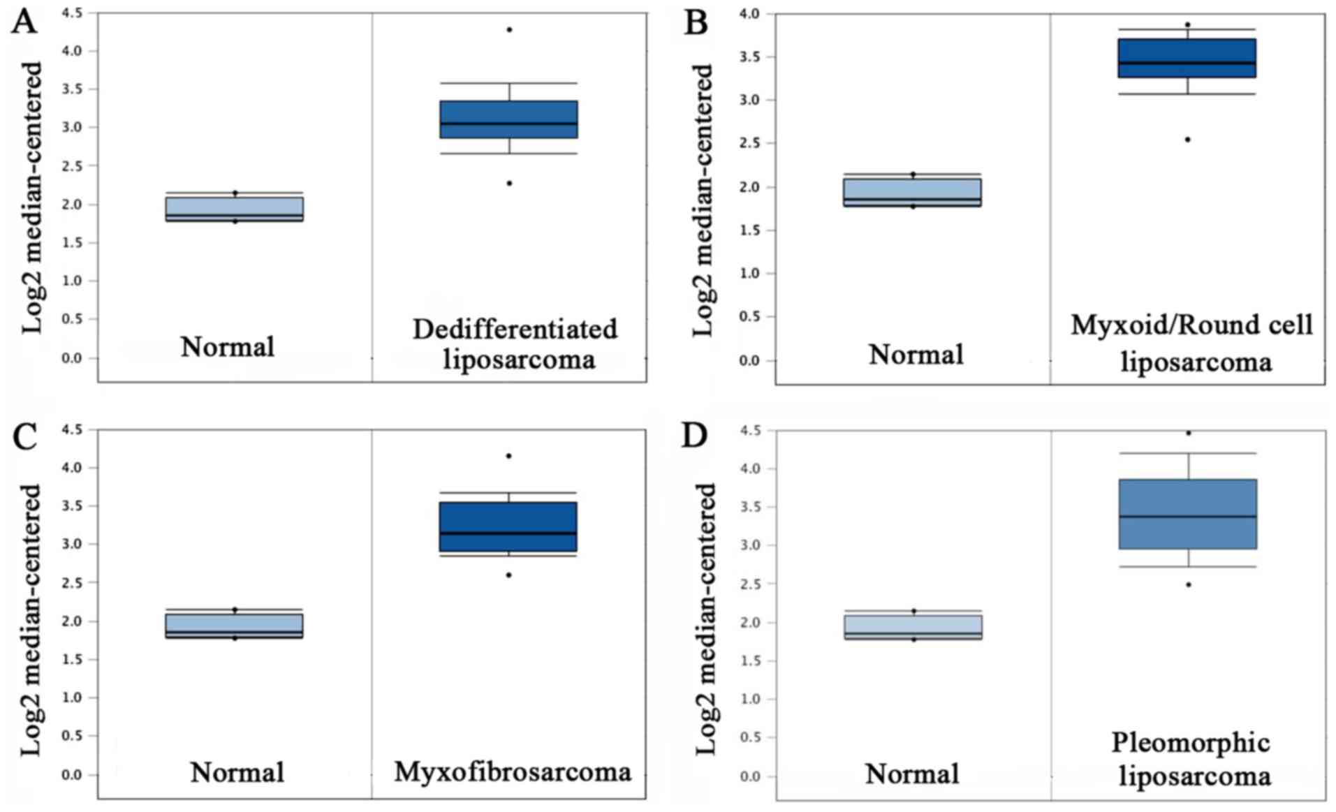
To ascertain whether CBX3 was expressed
differentially between the sarcoma tissues and the matched sarcoma
tissues, the Oncomine database was analyzed to examine the CBX3
mRNA expression levels. There were altogether 4 analyses satisfying
the inclusion standards. The statistics of Barretina sarcoma was
chosen because of the presence of the majority of the research
subjects. No statistics concerning the function of osteosarcoma is
currently available. However, it was revealed from the research
that in comparison with the normal adipose tissues, there was
markedly higher CBX3 mRNA expression level in other sarcoma types,
such as pleomorphic liposarcoma (t=12.188, P<0.001; Fig. 1D), myxofibrosarcoma (t=15.331,
P<0.001; Fig. 1C), myxoid/round
cell liposarcoma (t=15.989, P<0.001; Fig. 1B) and dedifferentiated liposarcoma
(t=2.270, P<0.001; Fig.
1A).
CBX3 is upregulated in human
osteosarcoma tissues
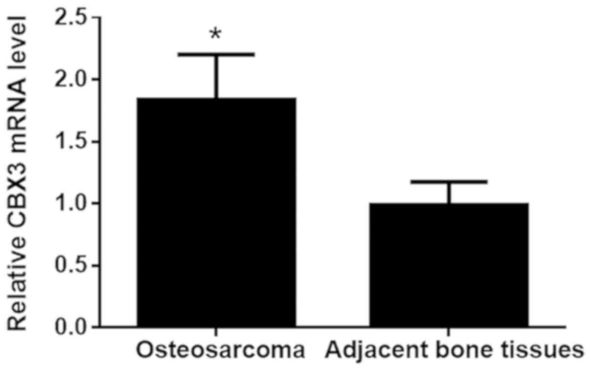
Afterwards, the qPCR assay was used for the
detection of the CBX3 expression levels in 98 osteosarcoma samples
and the corresponding non-carcinoma bone tissues. Generally,
Table I summarizes the
clinicopathological features of the osteosarcoma patients. As shown
in Fig. 2, a significantly higher
level of CBX3 mRNA was observed in the osteosarcoma tissues
compared with the level in the matched adjacent bone tissues
(t=19.21, P<0.001). It was suggested from these findings that
CBX3 may be involved in osteosarcoma progression.
 | Table I.Clinicopathological characteristics
and expression of CBX3 in the osteosarcoma patient samples. |
Table I.
Clinicopathological characteristics
and expression of CBX3 in the osteosarcoma patient samples.
| Characteristics | No of cases (%) |
|---|
| Age (years) |
|
|
<20 | 61 (62.2) |
| ≥20 | 37 (37.8) |
| Sex |
|
| Male | 56 (57.1) |
|
Female | 42 (42.9) |
| Tumor size (cm) |
|
|
<5 | 48 (49) |
| ≥5 | 50 (51) |
| Location |
|
| Upper
limb bone | 36 (36.7) |
| Lower
limb bone | 62 (63.3) |
| Clinical stage |
|
| I/II | 39 (39.8) |
|
III/IV | 59 (60.2) |
| Pathological
differentiation |
|
|
Well/Moderately | 45 (45.9) |
| Poor | 53 (54.1) |
| Response to
chemotherapy |
|
| Good | 54 (55.1) |
| Poor | 44 (44.9) |
| Lymph node
metastasis |
|
|
Absence | 58 (59.2) |
|
Presence | 40 (40.8) |
| Distant
metastasis |
|
|
Absence | 72 (73.5) |
|
Presence | 26 (26.5) |
| Recurrence |
|
|
Absence | 44 (44.9) |
|
Presence | 54 (55.1) |
| Expression of
CBX3 |
|
| Low
expression | 49 (50) |
| High
expression | 49 (50) |
Correlation of CBX3 expression with
clinicopathological characteristics in osteosarcoma
Based on the statistical analysis, osteosarcoma
patients were classified into a low CBX3 expression group (n=49)
and a high CBX3 expression group (n=49). Associations of CBX3
expression with the clinicopathological features were then
analyzed. As shown in Table II,
there was a strong association between CBX3 expression and distant
metastasis (P=0.022), clinical stage (P<0.001) and tumor size
(P=0.043), but not with recurrence (P>0.05), lymph node
metastasis, response to chemotherapy, pathological differentiation,
anatomic location, sex and age. Additionally, it was revealed from
the Spearman correlation analysis (Table III) of the correlation between
the clinicopathological features and CBX3 that there was a strong
correlation among distant metastasis (P=0.022), clinical stage
(P<0.0001) and tumor size (P=0.044).
| Table II.Correlation between CBX3 expression
and the clinicopathological characteristics of the osteosarcoma
patients. |
Table II.
Correlation between CBX3 expression
and the clinicopathological characteristics of the osteosarcoma
patients.
|
| CBX3 expression |
|
|---|
|
|
|
|
|---|
| Characteristics | Low (n=49) | High (n=49) | P-value |
|---|
| Age (years) |
|
<20 | 30 | 31 | 0.835 |
| ≥20 | 19 | 18 |
|
| Sex |
| Male | 30 | 26 | 0.414 |
|
Female | 19 | 23 |
|
| Tumor size (cm) |
| <5 | 29 | 19 | 0.043 |
| ≥5 | 20 | 30 |
|
| Location |
| Upper
limb bone | 32 | 39 | 0.675 |
| Lower
limb bone | 17 | 19 |
|
| Clinical stage |
|
I/II | 29 | 10 | <0.0001 |
|
III/IV | 20 | 39 |
|
| Pathological
differentiation |
|
Well/Moderately | 22 | 23 | 0.839 |
|
Poor | 27 | 26 |
|
| Response to
chemotherapy |
|
Good | 31 | 23 | 0.104 |
|
Poor | 18 | 26 |
|
| Lymph node
metastasis |
|
Absence | 32 | 26 | 0.218 |
|
Presence | 17 | 23 |
|
| Distant
metastasis |
|
Absence | 41 | 31 | 0.022 |
|
Presence | 8 | 18 |
|
| Recurrence |
|
Absence | 23 | 21 | 0.685 |
|
Presence | 26 | 28 |
|
| Table III.Spearman analysis of the correlation
between CBX3 expression and clinicopathological characteristics of
the osteosarcoma patients. |
Table III.
Spearman analysis of the correlation
between CBX3 expression and clinicopathological characteristics of
the osteosarcoma patients.
|
Characteristics | Spearman
correlation | P-value |
|---|
| Age | −0.21 | 0.837 |
|
Sex | 0.082 | 0.419 |
| Tumor
size | 0.204 | 0.044 |
| Location | 0.042 | 0.679 |
|
Clinical stage | 0.396 | <0.001 |
|
Pathological
differentiation | −0.02 | 0.841 |
| Response to
chemotherapy | 0.164 | 0.106 |
| Lymph
node metastasis | 0.125 | 0.222 |
| Distant
metastasis | 0.231 | 0.022 |
| Recurrence | 0.041 | 0.688 |
High CBX3 expression is associated
with poor prognosis in osteosarcoma
To investigate the prognostic value of CBX3
expression in osteosarcoma, long-rank test and Kaplan-Meier
analysis were conducted. It was shown from the results that there
was an association between high CBX3 expression and shorter OS
(P<0.0001; Fig. 3A) and DFS
(P=0.003; Fig. 3B) of the
osteosarcoma patients compared with those with low CBX3
expression.
Afterwards, multivariate and univariate Cox
regression was used for the collection and analysis of all the
clinical data. It was demonstrated that CBX3 expression serves as
an independent prognostic factor that could impact osteosarcoma
patient OS (Table IV) and DFS
(Table V).
| Table IV.Univariate and multivariate analyses
for overall survival in the osteosarcoma patients. |
Table IV.
Univariate and multivariate analyses
for overall survival in the osteosarcoma patients.
|
| Univariate
analysis | Multivariate
analysis |
|---|
|
|
|
|
|---|
| Variables | HR | 95% CI | P-value | HR | 95% CI | P-value |
|---|
| Age | 0.784 | (0.422–1.458) | 0.443 | 0.834 | (0.408–1.706) | 0.619 |
| Sex | 1.405 | (0.769–2.566) | 0.268 | 0.758 | (0.353–1.627) | 0.477 |
| Tumor size | 1.114 | (0.614–2.021) | 0.723 | 3.099 | (1.406–6.833) | 0.005 |
| Location | 0.858 | (0.458–1.608) | 0.632 | 0.876 | (0.402–1.913) | 0.741 |
| Clinical stage | 1.512 | (0.821–2.786) | 0.184 | 1.134 | (0.519–2.478) | 0.753 |
| Pathological
differentiation | 0.849 | (0.464–1.553) | 0.594 | 1.042 | (0.536–2.024) | 0.904 |
| Response to
chemotherapy | 1.101 | (0.600–2.022) | 0.756 | 0.53 | (0.242–1.158) | 0.111 |
| Lymph node
metastasis | 2.288 | (1.234–4.243) | 0.009 | 2.249 | (1.147–4.407) | 0.018 |
| Distant
metastasis | 2.006 | (1.011–3.982) | 0.046 | 1.562 | (0.694–3.517) | 0.281 |
| Recurrence | 15.06 | (3.601–62.989) | <0.001 | 27.656 |
(6.089–125.616) | <0.001 |
| Expression of
CBX3 | 3.534 | (1.808–6.909) | <0.001 | 4.259 | (1.878–9.660) | 0.001 |
| Table V.Univariate and multivariate analyses
for disease-free survival in osteosarcoma patients. |
Table V.
Univariate and multivariate analyses
for disease-free survival in osteosarcoma patients.
|
| Univariate
analysis | Multivariate
analysis |
|---|
|
|
|
|
|---|
| Variables | HR | 95% CI | P-value | HR | 95% CI | P-value |
|---|
| Age | 0.77 | (0.440–1.348) | 0.360 | 0.743 | (0.409–1.349) | 0.329 |
| Sex | 1.434 | (0.839–2.450) | 0.188 | 1.624 | (0.890–2.962) | 0.114 |
| Tumor size | 0.785 | (0.457–1.345) | 0.378 | 0.61 | (0.323–1.150) | 0.127 |
| Location | 0.573 | (0.573–1.734) | 0.996 | 0.863 | (0.452–1.647) | 0.655 |
| Clinical stage | 1.372 | (0.793–2.377) | 0.258 | 1.365 | (0.704–2.645) | 0.257 |
| Pathological
differentiation | 0.851 | (0.495–1.465) | 0.561 | 0.683 | (0.373–1.250) | 0.216 |
| Response to
chemotherapy | 1.305 | (0.763–2.230) | 0.331 | 1.156 | (0.628–2.127) | 0.641 |
| Lymph node
metastasis | 1.626 | (0.926–2.857) | 0.091 | 1.452 | (0.795–2.651) | 0.225 |
| Distant
metastasis | 1.68 | (0.922–3.061) | 0.090 | 1.327 | (0.647–2.723) | 0.441 |
| Expression of
CBX3 | 2.257 | (1.291–3.946) | 0.004 | 1.902 | (1.007–3.591) | 0.048 |
CBX3 siRNA inhibits CBX3 expression in
human osteosarcoma MG63 cells
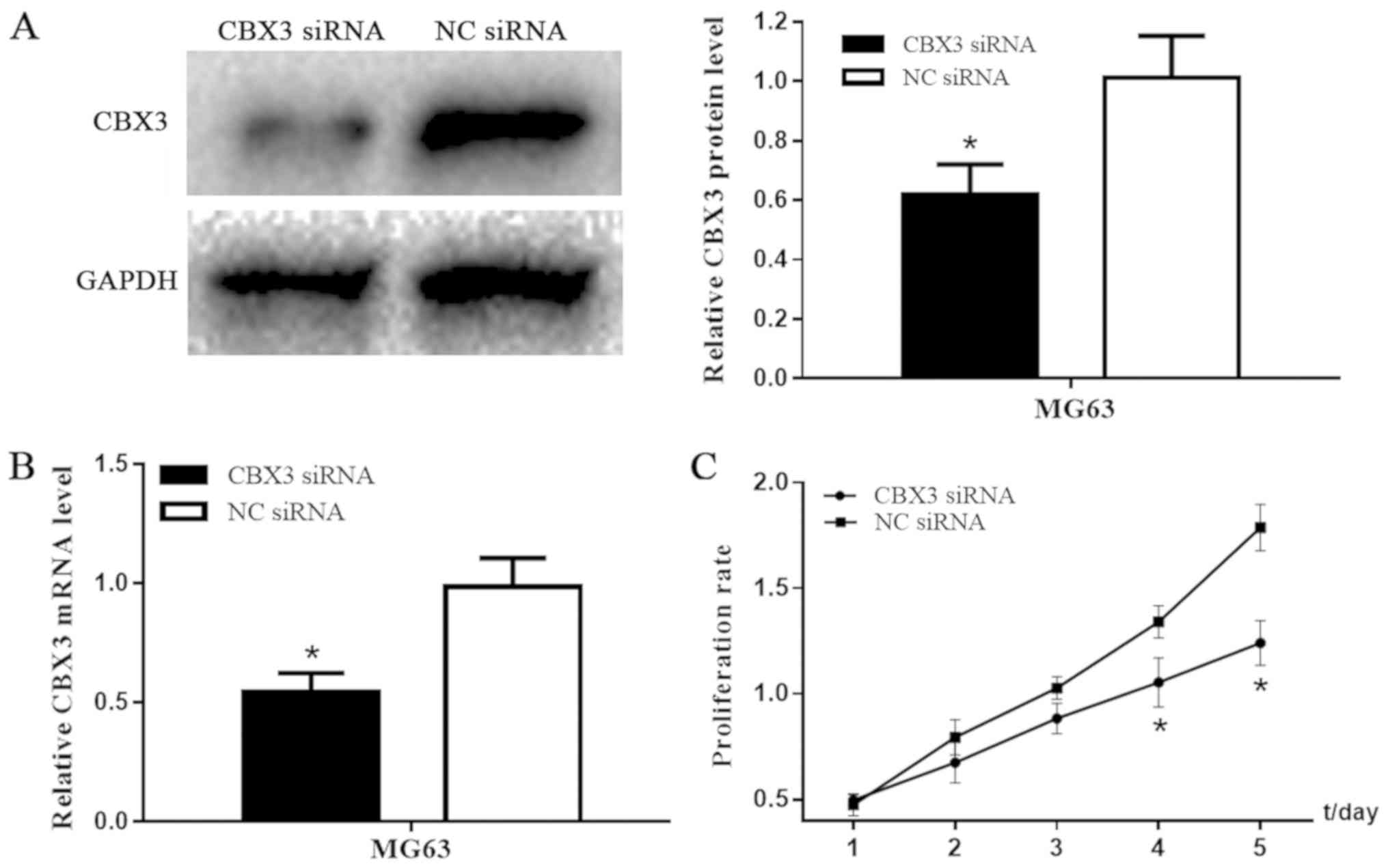
To investigate the function of CBX3 in osteosarcoma,
NC siRNA and CBX3 siRNA expressing GFP were transfected into human
osteosarcoma MG63 cells. The qPCR and western blotting were used to
analyze the knockdown efficiency. After the cells were transfected,
the expression levels of CBX3 mRNA (t=6.868, P=0.0001; Fig. 4B) and CBX3 protein (t=5.095,
P=0.0009; Fig. 4A) in the MG63
cells of the CBX3 siRNA group were significantly lower compared
with these levels in the NC siRNA group.
Downregulation of CBX3 expression
markedly inhibits the growth of osteosarcoma MG63 cells
To explore the impact of CBX3 on the growth of
osteosarcoma cells, CCK-8 assay was conducted after osteosarcoma
MG63 cells were transfected with NC siRNA or CBX3 siRNA. As shown
in Fig. 4C, the proliferation rate
of the MG63 cells was markedly inhibited by the knockdown of CBX3
expression in comparison with the NC siRNA group. Furthermore, on
day 4 (t=4.612, P=0.0017) and day 5 (t=8.017, P<0.0001), there
was an obvious reduction in the cell growth of the CBX3 siRNA group
in vitro. Thus, CBX3 knockdown prevented the growth of
osteosarcoma MG63.
Inhibition of CBX3 induces cell cycle
arrest at G0/G1 phase and increases the apoptosis of osteosarcoma
MG63 cells
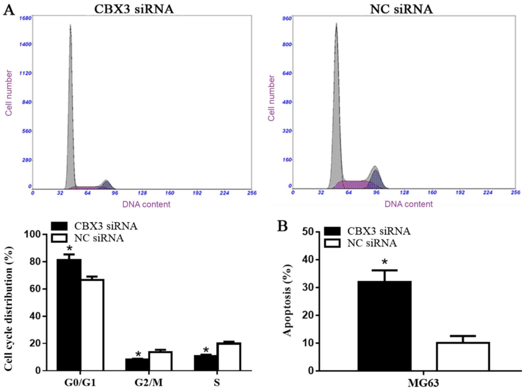
There is a direct associated between proliferation
and the distribution of the cell cycle. Therefore, we explored the
cell cycle distribution of MG63 cells after CBX3 was downregulated.
Flow cytometric analysis was used to assess the cell cycle in MG63
cells. As shown in Fig. 5A, the
number of cells in the G0 and G1 phases had an increase of 14.60%
(t=5.163, P=0.0067) in the CBX3 siRNA group while there was a
decrease by 9.30% (t=8.664, P=0.001) in the S phase and a decrease
by 5.30% (t=4.743, P=0.009) in the G2/M phase following the
knockout of CBX3. Moreover, the impact of CBX3 knockdown on the
apoptosis of osteosarcoma MG63 cells was also explored. There was
evidently higher percentage of apoptotic MG63 cells in the CBX3
siRNA group in comparison with the NC siRNA group (t=7.834,
P=0.0014; Fig. 5B).
The results demonstrated that knockdown of CBX3
expression can suppress the proliferation of MG63 cells via the
increase in the proportion of cells in the G0 and G1 phases of the
cell cycle and a decrease in the percentage of cells during the
G2/M and S phases. In addition, knockdown of CBX3 expression
induced apoptosis.
Discussion
The conserved chromatin binding proteins comprising
the heterochomatin protein 1 (HP1) family, are directly associated
with the methylated H3K9 (the methyl groups of histone H3 at lysine
9) promoter region, and therefore, take part in the heterochromatin
silencing of gene expression (7).
The aberrant expression of HP1 can result in different human
diseases, such as organism defects and cancer progression (11). Generally, chromobox protein homolog
3 (CBX3)-encoded heterochromatin protein 1γ (HP1γ) is a paralog of
HP1.
High CBX3 expression has been reported in various
types of cancers. CBX3 may be involved in the progression of
cancer. Therefore, it is a potential cancer treatment target
(12). Fan et al reported
that colon cancer cell proliferation can be improved by targeting
p21 and CDK6 in colorectal cancer (CRC) (6). Chang and colleagues found that CBX3
is overexpressed in non-small cell lung cancer (NSCLC) (13). Thus, it was hypothesized that CBX3
expression is a predictor of poor prognosis of patients suffering
from NSCLC. Slezak et al (14) found a positive correlation between
Ki-67 expression and the CBX3 expression level, which could predict
an unfavorable prognosis of prostate cancer patients. However, the
function of CBX3 in the prognosis and progression of osteosarcoma
has not yet been fully demonstrated.
Firstly, the present study investigated the function
and also the clinical significance of CBX3 in osteosarcoma patients
via the clinical data of osteosarcoma patients and public
expression profiles. The results showed that the expression levels
of CBX3 were significantly upregulated in sarcoma tissues in
comparison with those in the normal adipose tissues. Then, high
expression of CBX3 was noted in human osteosarcoma tissues in
comparison with the corresponding adjacent bone tissues. Third, it
was indicated from the statistical analysis that there was an
evident correlation between CBX3 expression levels and a high
distant metastasis rate, high clinical stage and a larger tumor
size. It was confirmed by further univariate and multivariate
analyses that CBX3 can be used as a potential prognostic biomarker
for osteosarcoma patient.
Additionally, the CBX3 siRNA was designed and used
for the transfection of MG63 cells in order to detect whether the
cell cycle distribution, apoptosis and proliferation of the
osteosarcoma cells can be impacted by downregulation of the gene
expression levels of CBX3 in vitro. It was shown that the
proliferation of MG63 cells was significantly prohibited by the
knockdown of CBX3 expression via the increase in the percentage of
cells during the G0 and G1 phases and a decrease in the proportion
of the cells in the G2/M and S phases. In addition, apoptosis was
induced. Nevertheless, further evaluation and validation must be
further conducted to evaluate the function of CBX3 in cell lines of
osteosarcoma and the explicit system implied by such
association.
In conclusion, our findings demonstrated that CBX3
is of great significance in the prognosis and progression of human
osteosarcoma. CBX3 is highly expressed in human osteosarcoma
tissues. The dismal prognosis of patients suffering from
osteosarcoma can be predicted by high CBX3 expression. In addition,
the proliferation of osteosarcoma MG63 cells was inhibited
following the downregulation of CBX3 expression. This was realized
by induction of cell apoptosis and cell cycle arrest. Thus, CBX3
can be valuable as an independent potential prognostic biomarker
for patients suffering from osteosarcoma.
Acknowledgements
Not applicable.
Funding
The present study was supported by The Youth Fund
Project of the First Affiliated Hospital of Xinxiang Medical
University (grant no. QN-2017-B018).
Availability of data and materials
The datasets used and/or analyzed during the current
study are available from the corresponding author on reasonable
request.
Authors' contributions
CM collected and analyzed the patient data and was a
major contributor in writing the manuscript. CM, XGN and YLW
performed the western blot and qPCR analyses. XHL, XL, QLZ and CM
performed the cell culture, transfection and flow cytometric
analysis. QLZ performed the statistical analysis. DPW contributed
to the conception and design of the study and revised it critically
for important intellectual content. All authors read and approved
the manuscript and agree to be accountable for all aspects of the
research in ensuring that the accuracy or integrity of any part of
the work are appropriately investigated and resolved.
Ethics approval and consent to
participate
The present study was performed according to the
Declaration of Helsinki of 1964 and all subsequent revisions, and
the Medical Ethics Committee of The First Affiliated Hospital of
Xinxiang Medical University (Xinxiang, China) approved the use of
the samples. Written informed consent for the participation in this
study was obtained from all the patients.
Patient consent for publication
Informed consent was obtained from all patients.
Competing interests
The authors declare that they have no competing
interests.
Glossary
Abbreviations
Abbreviations:
|
CBX3
|
chromobox protein homolog 3
|
|
HP1
|
heterochromatin protein 1
|
|
DMEM
|
Dulbecco's modified Eagle's medium
|
|
NBT
|
normal brain tissue
|
|
siRNA
|
small interfering RNA
|
|
qPCR
|
quantitative polymerase chain
reaction
|
|
GAPDH
|
glyceraldehyde-3-phosphate
dehydrogenase
|
|
CCK-8
|
Cell-Counting Kit-8
|
|
PBS
|
phosphate-buffered saline
|
|
OS
|
overall survival
|
|
DFS
|
disease-free survival
|
|
H3K9
|
methyl groups of histone H3 at lysine
9
|
|
HP1γ
|
heterochromatin protein 1γ
|
References
|
1
|
Zhang GY, Zhang JF, Hu XM, Luo ZP and Ma
YZ: Clinical significance of long non-coding RNA EWSAT1 as a novel
prognostic biomarker in osteosarcoma. Eur Rev Med Pharmacol Sci.
21:5337–5341. 2017.PubMed/NCBI
|
|
2
|
Fan J, Mei J, Zhang MZ, Yuan F, Li SZ, Yu
GR, Chen LH, Tang Q and Xian CJ: Clinicopathological significance
of glucose transporter protein-1 overexpression in human
osteosarcoma. Oncol Lett. 14:2439–2445. 2017. View Article : Google Scholar : PubMed/NCBI
|
|
3
|
Yoshida A, Fujiwara T, Uotani K, Morita T,
Kiyono M, Yokoo S, Hasei J, Nakata E, Kunisada T and Ozaki T:
Clinical and functional significance of intracellular and
extracellular microRNA-25-3p in osteosarcoma. Acta Med Okayama.
72:165–174. 2018.PubMed/NCBI
|
|
4
|
Longhi A, Errani C, De Paolis M, Mercuri M
and Bacci G: Primary bone osteosarcoma in the pediatric age: State
of the art. Cancer Treat Rev. 32:423–436. 2006. View Article : Google Scholar : PubMed/NCBI
|
|
5
|
Chou AJ, Geller DS and Gorlick R: Therapy
for osteosarcoma: Where do we go from here? Paediatr Drugs.
10:315–327. 2008. View Article : Google Scholar : PubMed/NCBI
|
|
6
|
Fan Y, Li H, Liang X and Xiang Z: CBX3
promotes colon cancer cell proliferation by CDK6 kinase-independent
function during cell cycle. Oncotarget. 8:19934–19946.
2017.PubMed/NCBI
|
|
7
|
Canzio D, Larson A and Narlikar GJ:
Mechanisms of functional promiscuity by HP1 proteins. Trends Cell
Biol. 24:377–386. 2014. View Article : Google Scholar : PubMed/NCBI
|
|
8
|
Esteller M: Cancer epigenomics: DNA
methylomes and histone-modification maps. Nat Rev Genet. 8:286–298.
2007. View
Article : Google Scholar : PubMed/NCBI
|
|
9
|
Chen LY, Cheng CS, Qu C, Wang P, Chen H,
Meng ZQ and Chen Z: CBX3 promotes proliferation and regulates
glycolysis via suppressing FBP1 in pancreatic cancer. Biochem
Biophys Res Commun. 500:691–697. 2018. View Article : Google Scholar : PubMed/NCBI
|
|
10
|
Livak KJ and Schmittgen TD: Analysis of
relative gene expression data using real-time quantitative PCR and
the 2(-Delta Delta C(T)) method. Methods. 25:402–408. 2001.
View Article : Google Scholar : PubMed/NCBI
|
|
11
|
Dialynas GK, Vitalini MW and Wallrath LL:
Linking heterochromatin protein 1 (HP1) to cancer progression.
Mutat Res. 647:13–20. 2008. View Article : Google Scholar : PubMed/NCBI
|
|
12
|
Zhang H, Chen W, Fu X, Su X and Yang A:
CBX3 promotes tumor proliferation by regulating G1/S phase via p21
downregulation and associates with poor prognosis in tongue
squamous cell carcinoma. Gene. 654:49–56. 2018. View Article : Google Scholar : PubMed/NCBI
|
|
13
|
Chang SC, Lai YC, Chen YC, Wang NK, Wang
WS and Lai JI: CBX3/heterochromatin protein 1 gamma is
significantly upregulated in patients with non-small cell lung
cancer. Asia Pac J Clin Oncol. Nov 10–2017.(Epub ahead of print).
doi: 10.1111/ajco.12820.
|
|
14
|
Slezak J, Truong M, Huang W and Jarrard D:
HP1γ expression is elevated in prostate cancer and is superior to
Gleason score as a predictor of biochemical recurrence after
radical prostatectomy. BMC Cancer. 13:1482013. View Article : Google Scholar : PubMed/NCBI
|