Introduction
Basaloid squamous cell carcinomas (BSCCs) in oral
lesions are extremely rare and are a variant of squamous cell
carcinoma (SCC) (1–4). Although BSCCs are reportedly
aggressive, have early lymph node metastasis, and have worse
prognosis, their biological features are similar to those of
conventional SCC (1–4). Several studies have demonstrated that
BSCCs have grossly flat or slightly elevated mucosa and
histologically small crowded cells with hyperchromatic nuclei and
stromal hyalinization (1–4). BSCC exhibits comedonecrosis and has a
palisading pattern of basal cells with squamous components in the
center, although not specifically (3,5,6). As
the histological aspects of BSCC are often similar to those of SCC,
it is difficult to distinguish BSCC from SCC for diagnosis,
especially in biopsy specimens. It has also been reported that
BSCCs exhibit high proliferation involving proliferating cell
nuclear antigen (PCNA) and Ki-67 immunoreactivity (7–9).
Interestingly, cell adhesion molecule E-cadherin and β-catenin
expression patterns reportedly differ between BSCC and SCC
(9). There are few reports that
feature tight junction molecule analysis of claudin-4 and occludin
in SCC (10,11). Their immunoreactivities were
reported to be extremely weak in cancer cells (10,11).
Whether claudin-4 and occludin are expressed in BSCC, however,
remains unknown. We hypothesized that cell proliferation, adhesion
and tight junction markers facilitate a differential diagnosis. In
the present study, the immunohistochemical detection of cell
proliferation marker PCNA, tight junction markers claudin-4 and
occludin, and stem cell marker SOX2 was conducted in BSCC and SCC.
Additionally, it was ascertained whether SOX2 overexpression
affects the expression of these factors and cell proliferation.
Materials and methods
Tissue preparation
Histological specimens collected from January 2014
to December 2018 were retrieved from our hospital's archives
according to the guidelines of the Japanese Society of Pathology.
Informed consent was provided by each patient for the use of the
clinical images. Two cases of BSCC and 20 cases of conventional SCC
were collected from craniocervical lesions, and each case included
lymph node metastasis. Specimens were obtained from the Department
of Diagnostic Pathology, Wakayama Medical University. Another BSCC
specimen was obtained from the Department of Pathology, Tsurumi
University School of Dental Medicine. Diagnosis was performed by at
least two pathologists. Clinical and pathological information is
described below. Case 1 is a female patient, 81 years of age, with
a clinically diagnosed mandibular malignant tumor; pathological
diagnosis is BSCC with abundant squamous components. Case 2 is a
male patient, 57 years of age, with a clinically diagnosed
malignant tumor in the palatine tonsil; pathological diagnosis is
BSCC with moderate squamous components and lymph node (LN)
metastasis. Case 3 is a male patient, 68 years of age, with
clinically diagnosed tongue cancer; pathological diagnosis is BSCC
without squamous components. Case 4 is a female patient, 87 years
of age, with clinically diagnosed gingival cancer; pathological
diagnosis is well-differentiated SCC. Case 5 is a male patient, 78
years of age, with clinically diagnosed tongue cancer; pathological
diagnosis is well-differentiated SCC and LN metastasis. Case 6 is a
female patient, 74 years of age, with clinically diagnosed buccal
cancer; pathological diagnosis is poorly differentiated SCC. The
clinical information regarding the other SCC cases is documented in
Table I. Partial resection and LN
dissection specimens were used in cases 2 and 5, and biopsy
specimens were used in the other cases.
 | Table I.Immunohistochemical detection of SOX2
and PCNA in BSCC and SCC specimens. |
Table I.
Immunohistochemical detection of SOX2
and PCNA in BSCC and SCC specimens.
| Case | Age | Sex | Lesion | Diagnosis | SOX2 intensity | PCNA-positive
cells |
|---|
| 1 | 81 | F | Mandibular | BSCC | 3 | 113 |
| 2 | 57 | M | Tonsil | BSCC | 3 | 358.6 |
| 3 | 68 | M | Tongue | BSCC | 3 | 224.2 |
| 4 | 87 | F | Gingiva | SCC (w) | 2 | 48.2 |
| 5 | 78 | M | Tongue | SCC (w) | 3 | 51.2 |
| 6 | 74 | F | Buccal | SCC (p) | 1 | 33.4 |
| 7 | 87 | M | Gingiva | SCC (w) | 2 | 62.4 |
| 8 | 85 | M | Gingiva | SCC (p) | 2 | 37.3 |
| 9 | 57 | M | Tongue | SCC (w) | 1 | 27.6 |
| 10 | 86 | M | Gingiva | SCC (m) | 2 | 41.4 |
| 11 | 85 | M | Gingiva | SCC (m) | 2 | 41.2 |
| 12 | 69 | M | Maxillary | SCC (p) | 2 | 50.9 |
| 13 | 64 | M | Tongue | SCC (m) | 3 | 47.6 |
| 14 | 73 | F | Tongue | SCC (m) | 2 | 73 |
| 15 | 87 | F | Oral floor | SCC (m) | 2 | 52.4 |
| 16 | 89 | F | Gingiva | SCC (w) | 3 | 56.7 |
| 17 | 69 | M | Tongue | SCC (w) | 2 | 47 |
| 18 | 90 | F | Gingiva | SCC (w) | 3 | 41.9 |
| 19 | 53 | M | Tongue | SCC (w) | 3 | 94.8 |
| 20 | 66 | F | Tongue | SCC (m) | 2 | 81.2 |
| 21 | 60 | M | Oral floor | SCC (m) | 3 | 68.4 |
| 22 | 69 | M | Oral floor | SCC (m) | 3 | 77.2 |
| 23 | 65 | M | Oral floor | SCC (p) | 3 | 77 |
Immunohistochemistry
Claudin-4, occludin, SOX2 and PCNA expression in
BSCC and SCC tissues were evaluated from serial deparaffinized
sections. Our specimens had been fixed with formalin from 24 to 48
h at room temperature and treated with routine processing as in a
previous study (12). Ten 4-mm
serial sections were prepared for staining and were incubated with
primary antibodies for 2 h. Immunohistochemistry was performed
using a Discovery Auto-Stainer with automated protocols (Ventana
Medical Systems, Inc.; Roche) as previously described (13). The average number of PCNA-positive
cells from case 1 to case 23 were counted in 10 random microscopic
fields at 400 magnification. The SOX2 intensity was determined by
qualitative assessment of three levels as weak, 1; moderate, 2; and
strong, 3. We defined a diffuse staining pattern as that positively
detected in almost all cancer cells but a dot-like staining pattern
as that positively detected in partial and marginal cancer
cells.
Cell culture and treatment
HSC2 and HSC3 human oral squamous cell carcinoma
cells, which do not have basaloid characteristics were obtained
from the Health Science Research Resources Bank (Osaka, Japan).
These cells were cultured in Dulbecco's modified Eagle's medium
(DMEM) (Sigma Chemical Co.; Merck KGaA) supplemented with 10% fetal
bovine serum and 1% penicillin streptomycin antibiotics. The
construct expression vector for SOX2 was used for transfection. The
human SOX2 plasmid (26817) was purchased from Addgene. Transient
plasmid transfection was performed as previously described
(14).
Western blot analysis
HSC2 cells were lysed using M-PER lysis buffer
(Thermo Fisher Scientific, Inc.). Protein determination was
performed using the bicinchoninic method. A total of 40 µg protein
was loaded in each lane. The total cell lysates were run on 14%
SDS-polyacrylamide gels followed by western blotting using standard
procedures. The proteins were transferred on to PVDF membrane. For
blocking, membranes were incubated with 5% skim milk for 60 min at
room temperature after protein transfer. A WesternBright Sirius kit
(Advansta) was used for antibody detection, and an AE-9300 Ez
capture MG (ATTO) was used for image data capture. We repeated the
western blot analysis three times and the results were similar.
Antibodies
The following commercial antibodies were purchased:
claudin-4 (1:100, mouse monoclonal, sc-376643; Santa Cruz
Biotechnology Inc.), occludin (1:100, mouse monoclonal, sc-133255;
Santa Cruz Biotechnology), SOX2 (1:100, rabbit polyclonal, Ab97959,
Abcam), PCNA (1:1,000, mouse monoclonal, sc-56; Santa Cruz
Biotechnology) and actin (1:10,000, mouse monoclonal, A5441; Sigma
Chemical Co.; Merck KGaA).
Real-time (quantitative) PCR
(qPCR)
We prepared three independent RNA samples from HSC3
cells. Total RNA was isolated and first-strand cDNA was synthesized
as previously described (13).
Real-time PCR was performed using SYBR Green Master Mix (Bio-Rad
Laboratories, Inc.). The thermocycling conditions were the
following: Initial denaturation at 95°C for 3 min, followed by 40
cycles of 95°C for 10 sec and 60°C for 30 sec. The
2−∆∆Cq method was used for relative quantification
(15). Amplification primer
sequences were designed as follows: SOX2-F,
5′-GAATGCCTTCATGGTGTGGT-3′ and R, 5′-TTGCTGATCTCCGAGTTGTG-3′;
claudin-4-F, 5′-ATGGCCTCCATGGGGCTACA-3′ and R,
5′-AGCGAGTCGTACACCTTGCA-3′; occludin-F,
5′-GCGGATTGGTTTATCTTGGA-3′ and R, 5′-CTGGATGACATGGCTGATTG-3′;
PCNA-F, 5′-AGGTGTTGGAGGCACTCAAG-3′ and R,
5′-AGTCCATGCTCTGCAGGTTT-3′; and 18S rRNA-F,
5′-GCGCCGCTAGAGGTGAAAT-3′ and R, 5′-GAAAACATTCTTGGCAAATGCTT-3′.
Data were normalized using 18S rRNA. qPCR was repeated three
times and the results were similar.
Cell proliferation assay
HSC2 and HSC3 cells were seeded into 96-well plates.
After transfection, 50 ml of XTT kit reaction solution was added to
each well (XTT-based) (Biological Industries) and the cells were
incubated at 37°C for an additional 2 h according to the
manufacturer's instructions. Absorbance at optical density at 480
nm (OD480) and at (OD650) was measured using
a 96-well microplate reader (SH-9000; Hitachi).
Statistical analysis
Statistical analysis with Dunnett's test was
performed using JMP Pro software version 13.0 (SAS Institute Japan,
Tokyo, Japan). The data are expressed as the mean value ± SE (bars)
of three independent samples. *P<0.05 was considered to indicate
a statistically significant result, and ANOVA was used followed by
Dunnett's test.
Results
Macroscopic and radioscopic aspects in
BSCC
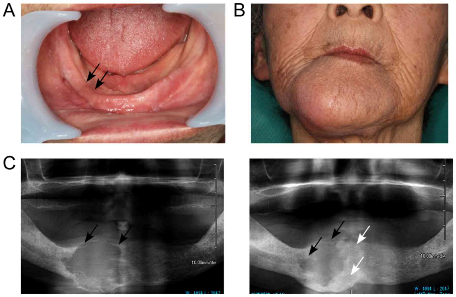
Representative macroscopic and radioscopic aspects
in case 1 of BSCC are shown in Fig.
1. Fig. 1A shows flat
appearance of gingival mucosa in a right anterior lesion. Fig. 1B shows the huge mandibular swelling
that was found. Diffuse swelling was found in the palatine tonsil
or tongue in case 2 and 3, respectively (data not shown). An
orthopantomograph of the BSCC at first medical examination showed a
multilocular cyst surrounded by well-defined radiolucent borders
(Fig. 1C, left panel). The patient
was treated with radiotherapy and the orthopantomograph at 8 months
after the first examination showed a decreased multilocular cyst
with calcification (Fig. 1C, right
panel). This indicated that radiotherapy was effective. The other
two patients with BSCC were not treated with radiotherapy.
Histological aspects of BSCC and
SCC
Fig. 2A shows
representative histological hematoxylin and eosin (H&E)-stained
images from case 1, 2 and 3 of BSCC. Fig. 2B shows representative histological
images from case 4, 5 and 6 of SCC.
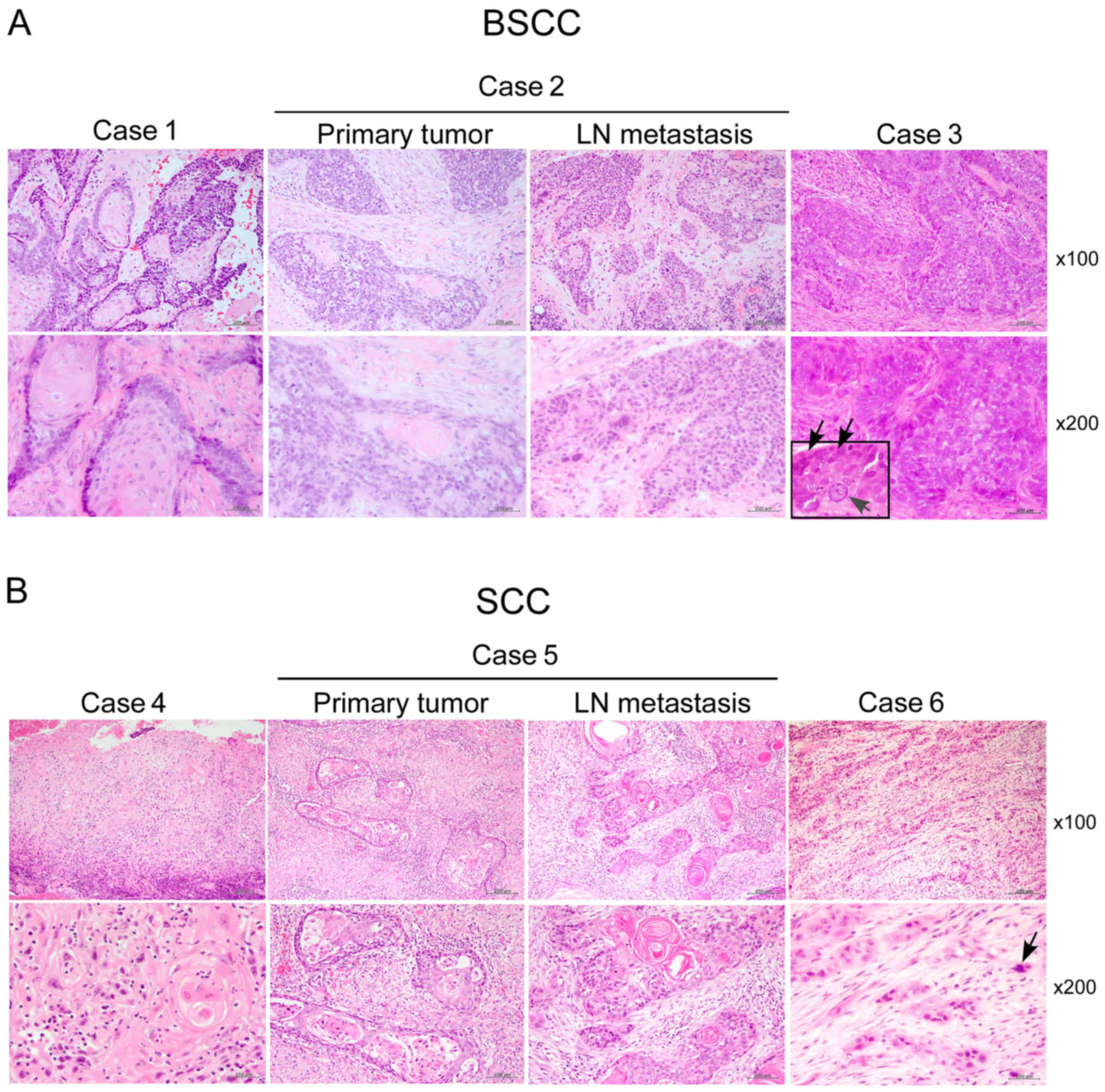 | Figure 2.Histological features of BSCC and SCC.
(A) Representative H&E images of case 1, 2 and 3 of BSCC. Case
1, BSCC with abundant squamous components. Case 2, BSCC with
moderate squamous components. Case 2 includes primary tumor and
lymph node (LN) metastasis. Case 3, BSCC without squamous
components. Top panels shows ×100 magnification and bottom panels
show ×200 magnification (scale bars, 200 mm). Square inset shows a
larger image. Black arrows show a palisading pattern in the basal
cells, and the gray arrow shows large atypical nucleus. (B)
Representative H&E images of case 4, 5, and 6 of SCC. Case 4,
SCC with abundant squamous components. Case 5, SCC with abundant
squamous components of primary tumor and LN metastasis. Case 6, SCC
without squamous components. There are no palisading pattern in
case 6, but a large atypical nucleus is observed (black arrow). Top
panel shows ×100 magnification and bottom panel shows ×200
magnification (scale bars, 200 mm). BSCC, basaloid squamous cell
carcinoma; SCC, squamous cell carcinoma; H&E, hematoxylin and
eosin. |
Immunohistochemical detection of
claudin-4, occludin, SOX2 and PCNA in BSCC and SCC
Representative images of claudin-4, occludin, SOX2
and PCNA immunoreactivities for case 1, 2 and 3 of BSCC are shown
in Fig. 3. Claudin-4
immunoreactivity in case 1 was strongly detected in the cell
membrane of squamous components, whereas no obvious detection was
observed in cancer cells. Claudin-4 immunoreactivity in case 2 of
the primary tumor was weakly detected in the membrane of squamous
components, whereas no obvious detection was observed in the cancer
cells. No obvious detection of claudin-4 immunoreactivity in case 2
was observed in cancer cells of LN metastasis. Claudin-4
immunoreactivity in case 3 was not detected in the cancer cells.
Occludin immunoreactivity was similar to claudin-4 as above, but in
case 1 it was weakly detected when compared to claudin-4. We
defined diffuse staining patterns as positively detected in almost
all cancer cells but dot-like staining patterns as positively
detected in partial and marginal cancer cells. SOX2 in case 1 had a
diffuse staining pattern in the nucleus, cytoplasm and in the
membrane of squamous components. SOX2 immunoreactivity in cases 2
and 3 of the primary tumor and LN metastasis exhibited a diffuse
staining pattern in the nucleus and some positive staining in
squamous components was observed. The PCNA immunoreactivity of the
primary tumor and LN metastasis in case 1, 2 and 3 also had diffuse
staining in the nucleus, but there was no obvious detection
observed in squamous components.
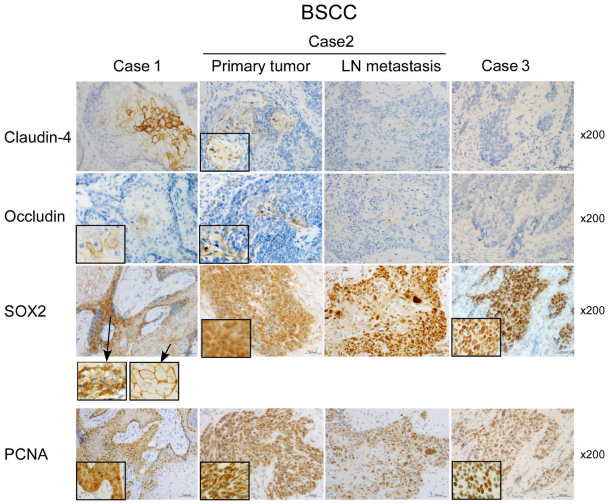 | Figure 3.Immunoreactivities of claudin-4,
occludin, SOX2 and PCNA in BSCC. Representative images for the
immunoreactivities of claudin-4, occludin, SOX2 and PCNA in case 1,
2 and 3 of BSCC. Each panel shows ×200 magnification (scale bars,
200 mm). Each square inset shows larger immunostaining images. The
square in SOX2 shows larger immunostaining images in the nucleus
and cytoplasm, and in the membrane, respectively. The square in
PCNA shows larger immunostaining images in the nucleus. BSCC,
basaloid squamous cell carcinoma; SOX2, SRY-box 2; PCNA,
proliferating cell nuclear antigen. |
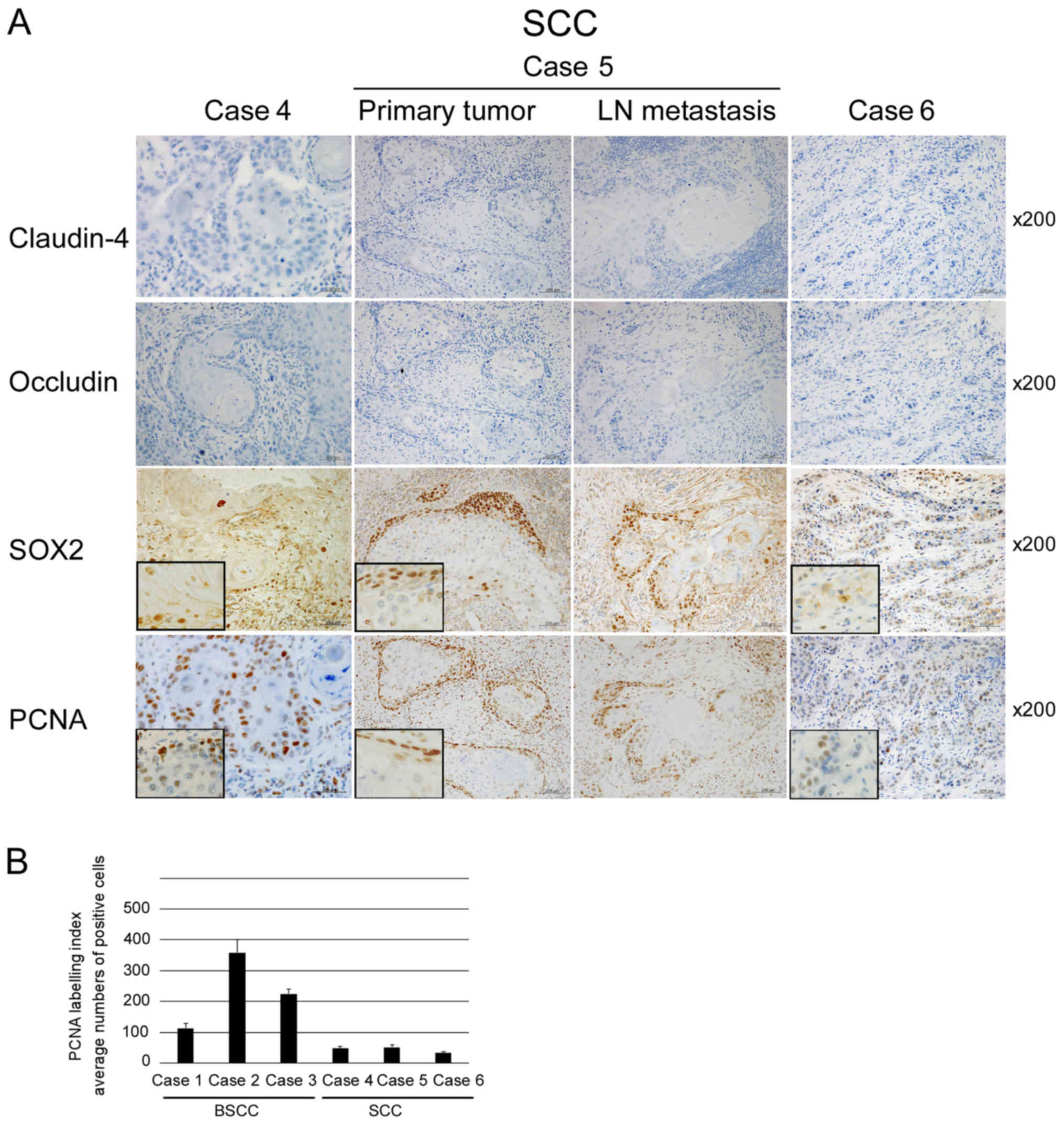
Representative images of claudin-4, occludin, SOX2
and PCNA immunoreactivities in SCC are shown in Fig. 4A. No positive immunoreactivities
for claudin-4 and occludin in the primary tumor and LN metastasis
from cases 4, 5 and 6 were detected in either cancer cells or
squamous components. SOX2 immunoreactivities from case 4, 5 and 6
were dot-like staining patterns in the nucleus of cancer cells. No
obvious detection was observed in squamous components. PCNA
immunoreactivity from case 4, 5 and 6 of the primary tumor was
similar to that of SOX2. There appeared to be weaker detection of
PCNA immunoreactivity in case 6 compared with case 4 and 5. SOX2
intensities and PCNA-positive cells of BSCC and SCC cases are
provided in Table I and Fig. 4B. The numbers of PCNA-positive
cells were higher in BSCC compared to SCC. SOX2 intensities in BSCC
were all strong, but they exhibited a variation in SCC.
SOX2 overexpression exhibits little
effect on the expression of claudin-4, occludin and PCNA
SOX2 expression was strongly detected in all cases
of BSCC. Therefore, it was ascertained whether SOX2 overexpression
affects expression of claudin-4, occludin and PCNA using oral SCC
cancer HSC2 cells. SOX2 overexpression had little effect on the
protein expression of occludin and PCNA (Fig. 5A). Endogenous protein expression of
claudin-4 was not detected in HSC2 cells, thus expression levels
were further examined by qPCR in HSC3 cells. SOX2 overexpression
had little effect on the mRNA expression of claudin-4, occludin and
PCNA in HSC3 cells (Fig. 5B).
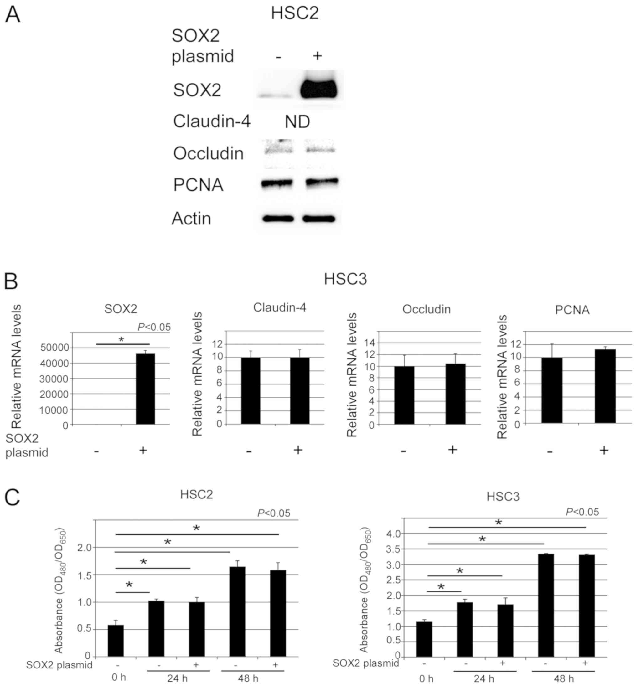 | Figure 5.SOX2 overexpression in oral cancer
HSC2 and HSC3 cells. (A) Representative western blotting images of
SOX2, occludin, PCNA and actin in HSC2 cells with (+) or without
(−) SOX2 overexpression. ND for claudin-4 means not detected. The
western blot analysis was repeated three times and similar results
were obtained. (B) Relative SOX2, claudin-4, occludin and PCNA mRNA
expression in HSC3 cells with (+) or without (−) SOX2
overexpression. Data are expressed as mean values ± SE (bars) of
three independent samples. *P<0.05, as determined using t-test.
qPCR was repeated three times and similar results were obtained.
(C) Cell proliferation of HSC2 and HSC3 cells with (+) or without
(−) SOX2 overexpression was determined. The absorbance
(OD480/OD650) at 0, 24, and 48 h is shown.
Data are expressed as mean values ± SE (bars) of three independent
samples. *P<0.05, as determined using Dunnett's test. SOX2,
SRY-box 2; PCNA, proliferating cell nuclear antigen. |
SOX2 overexpression exhibits little
effect on cell proliferation
Abnormalities in SOX2 expression affect
proliferation, migration and apoptosis in various types of cancer
cells. It was thus ascertained whether SOX2 overexpression affects
cell proliferation of oral cancer HSC2 and HSC3 cells. SOX2
overexpression had little effect on the cell proliferation of HSC2
and HSC3 cells at 24 and 48 h (Fig.
5C).
Discussion
Basaloid squamous cell carcinoma (BSCC) is regarded
as a high-grade malignant tumor, yet it remains unclear how to
distinguish it from squamous cell carcinoma (SCC). Oral BSCC is
extremely rare, thus it is important to understand both the
clinical and histological aspects. At our university, a diagnosed
of oral BSCC has been made only in 2 cases during the past 20
years. Moreover, in another university, only 1 case of BSCC was
found during the past 20 years. Therefore, more than 3 cases of
BSCC could not be obtained, making additional cases difficult to
find. Articles reporting on oral BSCC are quite rare, and there are
certain studies that have used only 2 BSCC cases (16–18).
We will attempt use more BSCC cases, collaborating with other
universities in future research. Clinically, BSCC is highly
sensitive to radiotherapy (4,19).
SCC, on the other hand, is not sensitive. In case 1, radiotherapy
induced the suppression of tumor growth and calcification in the
mandible. This clinical aspect is compatible with BSCC.
The histological aspects of BSCC are similar to SCC.
Takubo et al (20) reported
that a hyaline substance positively stained with periodic
acid-Shiff (PAS) staining had been observed in BSCC of the
esophagus. In the present study, however, we did not obviously
observe positive staining of PAS in BSCC (data not shown). In
addition, there are no critical markers for distinguishing between
the two. Therefore, immunohistochemical detection of claudin-4,
occludin, SOX2 and PCNA was conducted in BSCC compared to SCC.
Overexpression of claudins has been reported in several types of
cancer, and the overexpression or downregulation of claudins and
occludin are associated with tumor progression and recurrence
(21–24). Therefore, whether claudin-4 and
occludin are candidate markers for BSCC was ascertained. It is
worth noting that the immunoreactivities of claudin-4 and occludin
in BSCC were detected in the membrane of squamous components. They
were not observed in cancer cells of BSCC without squamous
components in case 3. This implies that claudin-4 and occludin play
important roles in squamous components of BSCC. The squamous
components are created by keratinization. Claudin-4 and occludin
may therefore regulate the adhesion of keratinization in BSCC. On
the contrary, no obvious detection of claudin-4 and occludin
immunoreactivities in SCC were observed in either squamous
components or cancer cells. It has been reported that claudin-4
immunoreactivity in SCC is weakly or not significantly detected in
the membrane of cancer cells. Considering this, it was speculated
that the functions of claudin-4 and occludin in squamous components
of BSCC differ from those of SCC. The differences in
immunoreactivities between BSCC and SCC are valuable for
differential diagnosis. However, these differences are not useful
in BSCC without squamous components. Future research must clarify
this possibility by using claudins with more cases of BSCC.
The staining patterns of PCNA immunoreactivity
between BSCC and SCC were also found to be different. The PCNA
labeling index is over 50% in BSCC and is associated with the
malignant feature of BSCC (25).
In SCC, the PCNA immunoreactivities exhibited dot-like staining
patterns in nuclei, whereas in BSCC they appeared as diffuse
staining patterns in the nuclei. Sampaio-Góes et al
(7) and Yu et al (25) also reported findings that
corroborate ours. Our specimens were fixed with formalin from 24 to
48 h. It has been reported that formalin fixation for over 24 and
48 h ensures better results for immunoreactivity (26). Thus, our samples used for
immunoreactivity analysis were maintained in optimum
conditions.
SOX2 overexpression is a poor prognostic factor and
is associated with tumor progression (27–31).
SOX2 regulates matrix metalloproteinase (MMP)-2, MMP-3, vimentin,
slug, E-cadherin and β-catenin (31–33).
Whether SOX2 plays a significant role in oral cancer, however
remains unclear. We found that SOX2 immunoreactivity in SCC was
presented in a dot-like staining pattern, but in BSCC this was
exhibited as a diffuse staining pattern. SOX2 immunoreactivity in
BSCC in case 1 was also observed in the membrane of squamous
components. We therefore examined whether SOX2 overexpression
affects the expression of claudin-4 and occludin, but it had little
effect. This implies that claudin-4 and occludin overexpression may
occur independently of SOX2. Finally, the effect of SOX2
overexpression on cell proliferation was determined, involving PCNA
expression. No significant effect was observed implying that other
factors, such as c-Myc, may be associated with cell proliferation
of oral cancer.
Human papillomavirus (HPV) affects the progression,
recurrence and radiotherapy sensitivity of tonsillar and posterior
tongue SCC (34,35). In addition, the protein expression
of angiogenesis-related proteins and TNF-receptors differ between
HPV-positive and -negative tissues (34). Although our BSCC sample from case 2
was from the tonsil, the p16 immunoreactivity was negative. It is
possible that the observed immunoreactivities of case 2 may not be
due to HPV infection. Our tongue SCC cases were from a side margin
lesion; however, future studies needs to clarify whether the HPV
status affects progression and immunoreactivities.
Poorly differentiated SCC has no squamous
components, thus it is extremely difficult to distinguish poorly
differentiated SCC from BSCC without squamous components. In this
study, it was demonstrated that the staining patterns of SOX2 and
PCNA immunoreactivities in BSCC and SCC were different. SOX2 and
PCNA presented as dot-like staining, appearing as partially
marginal staining in cancer cells of SCC, whereas they were diffuse
staining in almost all cancer cells of BSCC. On the other hand,
there were no differences in claudin-4 and occludin
immunoreactivities. These diffuse staining patterns of SOX2 and
PCNA suggest that BSCC has more aggressive and proliferative
potential than SCC. It has been reported that the diffuse staining
pattern of NOTCH1 is correlated with mutation status and poor
prognosis in adenoid cystic carcinoma (36). Therefore, diffuse staining patterns
of SOX2 and PCNA may correlate with BSCC mutation and
progression.
This observation may be useful for additional
diagnosis. In conclusion, claudin-4 and occludin immunoreactivities
and staining patterns of SOX2 and PCNA may be utilized to
potentially carry out a differential diagnosis between BSCC and
SCC
Acknowledgements
The authors would like to thank Dr Yumi Ito from the
Department of Pathology, Tsurumi University School of Dental
Medicine, who supplied a BSCC specimen.
Funding
The present study was supported by JSPS KAKENHI
grant no. 16K09624 (FS), and by Nihon University Multidisciplinary
Research Grant for 2018 (UKB).
Availability of data and materials
The datasests used and/or analyzed during the
current study are available from the corresponding author on
reasonable request.
Authors' contributions
FS and UKB performed experiments, carried out the
pathological diagnosis and completed the draft. FS, SIM and YM
performed the pathological diagnosis. IT and SF performed the
clinical diagnosis and submitted clinical images and materials.
UKB, IT, SF, SIM and YM revised manuscript. All authors read and
approved the final manuscript.
Ethics approval and consent to
participate
This study was approved by the Wakayama Medical
University Research Ethics Committee (Protocol no. 1715) and
histological specimens were retrieved from our hospital archives.
Informed consent was provided by each patient for the use of the
clinical images.
Patient consent for publication
The patient provided written informed consent for
the publication of clinical images.
Competing interests
The authors declare that they have no competing
interests.
References
|
1
|
Fritsch VA, Gerry DR and Lentsch EJ:
Basaloid squamous cell carcinoma of the oral cavity: An analysis of
92 cases. Laryngoscope. 124:1573–1578. 2014. View Article : Google Scholar : PubMed/NCBI
|
|
2
|
Thariat J, Badoual C, Faure C, Butori C,
Marcy PY and Righini CA: Basaloid squamous cell carcinoma of the
head and neck: Role of HPV and implication in treatment and
prognosis. J Clin Pathol. 63:857–866. 2010. View Article : Google Scholar : PubMed/NCBI
|
|
3
|
Ereño C, Gaafar A, Garmendia M,
Etxezarraga C, Bilbao FJ and López JI: Basaloid squamous cell
carcinoma of the head and neck: A clinicopathological and follow-up
study of 40 cases and review of the literature. Head Neck Pathol.
2:83–91. 2008. View Article : Google Scholar : PubMed/NCBI
|
|
4
|
Shen W, Sakamoto N and Yang L:
Cause-specific mortality prediction model for patients with
basaloid squamous cell carcinomas of the head and neck: A competing
risk analysis. J Cancer. 9:4009–4017. 2018. View Article : Google Scholar : PubMed/NCBI
|
|
5
|
Wain SL, Kier R, Vollmer RT and Bossen EH:
Basaloid-squamous carcinoma of the tongue, hypopharynx, and larynx:
Report of 10 cases. Hum Pathol. 17:1158–1166. 1986. View Article : Google Scholar : PubMed/NCBI
|
|
6
|
Luna MA, el Naggar A, Parichatikanond P,
Weber RS and Batsakis JG: Basaloid squamous carcinoma of the upper
aerodigestive tract. Clinicopathologic and DNA flow cytometric
analysis. Cancer. 66:537–542. 1990. View Article : Google Scholar : PubMed/NCBI
|
|
7
|
Sampaio-Góes FC, Oliveira DT, Dorta RG,
Nonogaki S, Landman G, Nishimoto IN and Kowalski LP: Expression of
PCNA, p53, Bax, and Bcl-X in oral poorly differentiated and
basaloid squamous cell carcinoma: Relationships with prognosis.
Head Neck. 27:982–989. 2005. View Article : Google Scholar : PubMed/NCBI
|
|
8
|
Coletta RD, Cotrim P, Vargas PA, Villalba
H, Pires FR, de Moraes M and de Almeida OP: Basaloid squamous
carcinoma of the oral cavity: Report of 2 cases and study of AgNOR,
PCNA, p53, and MMP expression. Oral Surg Oral Med Oral Pathol Oral
Radiol Endod. 91:563–569. 2001. View Article : Google Scholar : PubMed/NCBI
|
|
9
|
Pereira CH, Morais MO, Martins AF, Soares
MQ, Alencar Rde C, Batista AC, Leles CR and Mendonça EF: Expression
of adhesion proteins (E-cadherin and β-catenin) and cell
proliferation (Ki-67) at the invasive tumor front in conventional
oral squamous cell and basaloid squamous cell carcinomas. Arch Oral
Biol. 61:8–15. 2016. View Article : Google Scholar : PubMed/NCBI
|
|
10
|
Sappayatosok K and Phattarataratip E:
Overexpression of claudin-1 is associated with advanced clinical
stage and invasive pathologic characteristics of oral squamous cell
carcinoma. Head Neck Pathol. 9:173–180. 2015. View Article : Google Scholar : PubMed/NCBI
|
|
11
|
Phattarataratip E and Sappayatosok K:
Expression of claudin-5, claudin-7 and occludin in oral squamous
cell carcinoma and their clinico-pathological significance. J Clin
Exp Dent. 8:e299–e306. 2016.PubMed/NCBI
|
|
12
|
Wu Y, Sato F, Yamada T, Bhawal UK,
Kawamoto T, Fujimoto K, Noshiro M, Seino H, Morohashi S, Hakamada
K, et al: The BHLH transcription factor DEC1 plays an important
role in the epithelial-mesenchymal transition of pancreatic cancer.
Int J Oncol. 41:1337–1346. 2012. View Article : Google Scholar : PubMed/NCBI
|
|
13
|
Sato F, Kohsaka A, Takahashi K, Otao S,
Kitada Y, Iwasaki Y and Muragaki Y: Smad3 and Bmal1 regulate p21
and S100A4 expression in myocardial stromal fibroblasts via TNF-α.
Histochem Cell Biol. 148:617–624. 2017. View Article : Google Scholar : PubMed/NCBI
|
|
14
|
Sato F, Muragaki Y and Zhang Y: DEC1
negatively regulates AMPK activity via LKB1. Biochem Biophys Res
Commun. 467:711–716. 2015. View Article : Google Scholar : PubMed/NCBI
|
|
15
|
Livak KJ and Schmittgen TD: Analysis of
relative gene expression data using real-time quantitative PCR and
the 2(-Delta Delta C(T)) method. Methods. 25:402–408. 2001.
View Article : Google Scholar : PubMed/NCBI
|
|
16
|
Peddapell K, Rao GV, Sravya T and Ravipati
S: Basaloid squamous cell carcinoma: Report of two rare cases and
review of literature. J Oral Maxillofac Pathol. 22:2852018.
View Article : Google Scholar
|
|
17
|
Heera R, Ayswarya T, Padmakumar SK and
Ismayil P: Basaloid squamous cell carcinoma of oral cavity: Report
of two cases. J Oral Maxillofac Pathol. 20:5452016. View Article : Google Scholar : PubMed/NCBI
|
|
18
|
Patel PN, Mutalik VS, Rehani S and
Radhakrishnan R: Basaloid squamous cell carcinoma of oral cavity
with incongruent clinical course. BMJ Case Rep. 2013.bcr 2013200441
2013. View Article : Google Scholar
|
|
19
|
Maebayashi T, Ishibashi N, Aizawa T,
Sakaguchi M, Taku H, Ohhara M, Takimoto T and Tanaka Y: A
long-surviving patient with advanced esophageal basaloid squamous
cell carcinoma treated only with radiotherapy: Case report and
literature review. BMC Gastroenterol. 17:1512017. View Article : Google Scholar : PubMed/NCBI
|
|
20
|
Takubo K, Mafune K, Tanaka Y, Miyama T and
Fujita K: Basaloid-squamous carcinoma of the esophagus with marked
deposition of basement membrane substance. Acta Pathol Jpn.
41:59–64. 1991.PubMed/NCBI
|
|
21
|
Resnick MB, Konkin T, Routhier J, Sabo E
and Pricolo VE: Claudin-1 is a strong prognostic indicator in stage
II colonic cancer: A tissue microarray study. Mod Pathol.
18:511–518. 2005. View Article : Google Scholar : PubMed/NCBI
|
|
22
|
Sheehan GM, Kallakury BV, Sheehan CE,
Fisher HA, Kaufman RP Jr and Ross JS: Loss of claudins-1 and −7 and
expression of claudins-3 and −4 correlate with prognostic variables
in prostatic adenocarcinomas. Hum Pathol. 38:564–569. 2007.
View Article : Google Scholar : PubMed/NCBI
|
|
23
|
Lourenço SV, Coutinho-Camillo CM, Buim ME,
Pereira CM, Carvalho AL, Kowalski LP and Soares FA: Oral squamous
cell carcinoma: Status of tight junction claudins in the different
histopathological patterns and relationship with clinical
parameters. A tissue-microarray-based study of 136 cases. J Clin
Pathol. 63:609–614. 2010. View Article : Google Scholar : PubMed/NCBI
|
|
24
|
Martin TA, Mansel RE and Jiang WG: Loss of
occludin leads to the progression of human breast cancer. Int J Mol
Med. 26:723–734. 2010. View Article : Google Scholar : PubMed/NCBI
|
|
25
|
Yu GY, Gao Y, Peng X, Chen Y, Zhao FY and
Wu MJ: A clinicopathologic study on basaloid squamous cell
carcinoma in the oral and maxillofacial region. Int J Oral
Maxillofac Surg. 37:1003–1008. 2008. View Article : Google Scholar : PubMed/NCBI
|
|
26
|
Patil S, Rao RS, Ganavi BS and Majumdar B:
Natural sweeteners as fixatives in histopathology: A longitudinal
study. J Nat Sci Biol Med. 6:67–70. 2015. View Article : Google Scholar : PubMed/NCBI
|
|
27
|
Wang H, Zhou Y, Liu Q, Xu J and Ma Y:
Prognostic value of SOX2, Cyclin D1, p53, and ki-67 in patients
with esophageal squamous cell carcinoma. Onco Targets Ther.
11:5171–5181. 2018. View Article : Google Scholar : PubMed/NCBI
|
|
28
|
Wuebben EL, Wilder PJ, Cox JL, Grunkemeyer
JA, Caffrey T, Hollingsworth MA and Rizzino A: SOX2 functions as a
molecular rheostat to control the growth, tumorigenicity and drug
responses of pancreatic ductal adenocarcinoma cells. Oncotarget.
7:34890–34906. 2016. View Article : Google Scholar : PubMed/NCBI
|
|
29
|
Chung JH, Jung HR, Jung AR, Lee YC, Kong
M, Lee JS and Eun YG: SOX2 activation predicts prognosis in
patients with head and neck squamous cell carcinoma. Sci Rep.
8:16772018. View Article : Google Scholar : PubMed/NCBI
|
|
30
|
Yang N, Hui L, Wang Y, Yang H and Jiang X:
SOX2 promotes the migration and invasion of laryngeal cancer cells
by induction of MMP-2 via the PI3K/Akt/mTOR pathway. Oncol Rep.
31:2651–2659. 2014. View Article : Google Scholar : PubMed/NCBI
|
|
31
|
Girouard SD, Laga AC, Mihm MC, Scolyer RA,
Thompson JF, Zhan Q, Widlund HR, Lee CW and Murphy GF: SOX2
contributes to melanoma cell invasion. Lab Invest. 92:362–370.
2012. View Article : Google Scholar : PubMed/NCBI
|
|
32
|
Liu X, Qiao B, Zhao T, Hu F, Lam AK and
Tao Q: Sox2 promotes tumor aggressiveness and
epithelial-mesenchymal transition in tongue squamous cell
carcinoma. Int J Mol Med. 42:1418–1426. 2018.PubMed/NCBI
|
|
33
|
Han X, Fang X, Lou X, Hua D, Ding W, Foltz
G, Hood L, Yuan Y and Lin B: Silencing SOX2 induced
mesenchymal-epithelial transition and its expression predicts liver
and lymph node metastasis of CRC patients. PLoS One. 7:e413352012.
View Article : Google Scholar : PubMed/NCBI
|
|
34
|
Ramqvist T, Näsman A, Franzén B, Bersani
C, Alexeyenko A, Becker S, Haeggblom L, Kolev A, Dalianis T and
Munck-Wikland E: Protein expression in tonsillar and base of tongue
cancer and in relation to human papillomavirus (HPV) and clinical
outcome. Int J Mol Sci. 19:E9782018. View Article : Google Scholar : PubMed/NCBI
|
|
35
|
Näsman A, Bersani C, Lindquist D, Du J,
Ramqvist T and Dalianis T: Human papillomavirus and potentially
relevant biomarkers in tonsillar and base of tongue squamous cell
carcinoma. Anticancer Res. 37:5319–5328. 2017.PubMed/NCBI
|
|
36
|
Sajed DP, Faquin WC, Carey C, Severson EA,
H Afrogheh A, A Johnson C, Blacklow SC, Chau NG, Lin DT, Krane JF,
et al: Diffuse staining for activated NOTCH1 correlates with NOTCH1
mutation status and is associated with worse outcome in adenoid
cystic carcinoma. Am J Surg Pathol. 41:1473–1482. 2017. View Article : Google Scholar : PubMed/NCBI
|