Introduction
Gastric cancer is the fourth most common type of
cancer worldwide and the second leading cause of cancer-associated
mortalities after lung cancer. The majority of patients with early
gastric cancer have no obvious symptoms, being pain and weight loss
the most common clinical symptoms of advanced gastric cancer
patients. The diagnostic methods for gastric cancer include
laboratory tests, endoscopy and pathological observation, while the
treatment methods include surgery, chemotherapy and radiation
therapy. The prognosis depends on the type of gastric cancer
(1,2).
Forkhead box protein 3 (FOXP3) is a member of the
forkhead transcription factor family, which is considered as the
molecular marker of regulatory T cells (3). Mutation of the FOXP3 gene can
cause severe autoimmune diseases. Therefore, FOXP3 is considered
critical for the regulation of immunological homeostasis (4,5). FOXP3, as
a transcriptional regulatory factor, regulates the activity of
regulatory T cells via the direct regulation of certain genes
(3). Regulatory T cells are a subset
of T lymphocytes with a suppressive function, which are involved in
the maintenance of immunological self-tolerance (6,7).
Regulatory T cells exhibit high expression of cluster of
differentiation 25, and exhibit an immunosuppressive function via
direct contact with cells or secretion of transforming growth
factor-β and interleukin-10 (8).
Regulatory T cells are important for immunological tolerance, tumor
immunity and transplantation immunity (9). Regulatory T cells effectively inhibit
the antitumor immune response and are involved in the process of
tumor immune escape (10). The number
of regulatory T cells in the peripheral blood of patients with
tumors is significantly increased, and thus their presence markedly
inhibits the antitumor immune response (11).A previous study has demonstrated that
the number of regulatory T cells in the peripheral blood is closely
associated with the survival and prognosis of gastric cancer
patients (12). Depletion of
regulatory T cells improves the specific immune response of tumor
bearing mice to tumors and dendritic cell vaccines to low
antigenicity tumors (13). Recently,
the association between FOXP3 expression and gastric cancer has
gained increasing attention (12,13);
however, few studies have investigated the correlation between
FOXP3 expression and various gastric cancer lesion grades (14).
In the present study, sarcosine ethyl ester
hydrochloride and sodium nitrate were used to induce gastric cancer
in a rat model to investigate the correlation between FOXP3
expression and different gastric cancer lesion types.
Materials and methods
Experimental animals
A total of 92 adult male Wistar rats (weight,
250–3000 g; age, 8–12 weeks) were purchased from the Animal
Experimental Center of Fudan University (Shanghai, China) and
maintained in separate cages (n=4/cage) at the vivarium of Henan
University Huaihe Hospital (Henan, China) on a 12 h light/dark
cycle. The rats were then weighed after 12 h of fasting and
randomly divided into an experimental group (n=46) and a normal
control group (n=46). The present study was performed in accordance
with the Guide for the Care and Use of Laboratory Animals of the
National Institutes of Health (Bethesda, MD, USA) (8th edition)
(15). The animal use protocol in the
present study was reviewed and approved by the Institutional Animal
Care and Use Committee of Henan University Huaihe Hospital
(Kaifeng, China).
Experimental method
To establish a gastric cancer model in rats, the
experimental group was administered a mixture of sarcosine ethyl
ester hydrochloride (2.5 mg/kg; Sigma-Aldrich, St. Louis, MO, USA)
and sodium nitrite (2.5 mg/kg; Sangon Biotech Co., Ltd., Shanghai,
China) by lavage, daily for 6 months. The rats of the control group
were administered saline (2.5 mg/kg) by lavage, daily for 6 months.
Anti-FOXP3 monoclonal antibody (catalogue number, SAB4700610;
dilution, 1:100), anti-β-actin monoclonal antibody (catalogue
number, A2228; dilution, 1:100) and anti-FOXP3 polyclonal antibody
(catalogue number, AV32564; dilution, 1:200) were purchased from
Sigma-Aldrich. PEF FOXP3 eukaryotic expression vector, RPMI 1640
medium and the pSV2neo plasmid were purchased from Invitrogen
(Thermo Fisher Scientific, Inc., Waltham, MA, USA). Lipofectamine
2000, 3-(4,5-dimethylthiazol-2-yl)-2,5-diphenyltetrazolium bromide,
immunohistochemical SP kits and 3,3′-diaminobenzidine chromogenic
reagent kit were purchased from OriGene Technologies, Inc.
(Beijing, China).
The time period for establishing the gastric cancer
model in rats was 6 months, and the model was evaluated via the
grading of gastric lesions. Once the model was established, rats
were sacrificed using anesthesia with chloral hydrate (Sangon
Biotech Co., Ltd.). Serum was stored at −80°C for use, while
gastrointestinal tissues were weighed and observed. A section of
gastrointestinal tissue was stored at −80°C for use, while the
other part of gastrointestinal tissue was fixed in 4% formaldehyde
solution, and the pieces (4-mm thick tissue sections) were then
embedded in paraffin wax. The expression of FOXP3 in
gastrointestinal tissues was determined by immunohistochemistry
with anti-FOXP3 antibodies, which were incubated at room
temperature for 30 min.
Grading of gastric lesions
The grades of gastrointestinal tissue lesions in the
experimental group were classified as follows: Grade I, hyperplasia
(simple hyperplasia and papillary hyperplasia); grade II,
precancerous lesions (papilloma, endogenous epithelioma and
dysplasia); grade III, early cancer (basal cell carcinoma,
carcinoma in situ and early invasive carcinoma); grade IV,
invasive carcinoma (12). Gastric
cancer lesions in the experimental group rats were assessed
according to the Borrmann classification (16): Type I (nodular); type II
(circumscribed ulcerative type), tumor with larger ulcers,
infiltrating into the surrounding tissues; type III (infiltrating
ulcerative type) tumor with major ulcers, with evident edge uplift,
unclear boundaries and evident infiltration into the surrounding
tissues; and type IV (diffuse infiltration type), tumor with
diffuse invasive growth and unclear boundaries (13).
The intensity of FOXP3 expression was divided into
four categories according to the number of positive cells in the
gastrointestinal tract: negative (−), <5% positive cells; weak
positive (+), 5–19% positive cells; positive (++), 20–60% positive
cells; and strong positive (+++), >60% positive cells (17).
Gene transfection
The experiment was divided into three groups as
follows: Group A, PEF FOXP3 and pSV2neo co-transfection; group B,
no transfection; and group C, PEF and pSV2neo co-transfection
group. Groups B and C served as the control groups. Transfection
was performed according to the manufacturer's protocol. The
positive clone group was formed after 3 weeks, then amplified,
cultured and reserved (18).
TriPure Isolation reagent (Roche Diagnostics, Basel,
Switzerland) was used to extract total RNA from the gastric cells
obtained from the rats. A total of 3 µg total RNA was obtained from
each group to perform reverse transcription-polymerase chain
reaction (RT-PCR). A total of 8 µl RT product was then used to
perform PCR. FOXP3 fragment specific primers (Da An Gene Co., Ltd.,
Guangzhou, China) were synthesized according to the FOXP3 gene RNA
sequence, and β-actin was used as the reference gene. The primer
sequences were as follows: Forward, 5′-CACAACATGCGACCCCCTTTCACC-3′
and reverse, 5′-AGGTTGTGGCGGATGGCGTTCTTC-3′ for FOXP3 (synthetic
product, 167 bp); forward, 5′-GGCACCACACCTTCTACA-3′ and reverse,
5′-AGGAAGGCTGGAAGAGTG-3′ for β-actin (synthetic product, 540 bp).
The PCR was conducted according to the following conditions:
pre-denaturation at 94°C for 30 sec; 30 cycles of denaturation at
94°C for 30 sec, annealing at 63°C for 30 sec and extension at 72°C
for 40 sec; and terminal extension at 72°C for 3 min. FOXP3
expression levels were analyzed by 1.5% agarose gel
electrophoresis. The results were analyzed by gel imaging and
Quantity One version 4.6.2 software (Bio-Rad Laboratories, Inc.,
Hercules, CA, USA).
Statistical analysis
All data was analyzed using SPSS 14.0 software (SPSS
Inc., Chicago, IL, USA). Categorical data were analyzed by t-test,
while continuous data were analyzed by analysis of variance.
Results of immunohistochemistry and RT-PCR were analyzed by
χ2 test. The correlation between the expression of FOXP3
and the lesion classification of gastric tissue was analyzed by
Cochran-Mantel-Haenszel test. P<0.05 was considered to indicate
a statistically significant difference.
Results
Modeling results and classification of
gastrointestinal lesions
Of the 46 rats in the experimental group, 6 rats
developed hyperplastic lesions (grade I), 8 rats developed
precancerous lesions (grade II), 17 rats developed early gastric
cancer (grade III) and 14 rats developed infiltrative gastric
carcinoma (grade IV). In all gastric tissues obtained from the
experimental group, the whole gastrointestinal tract exhibited
ulcerative infiltration. Part of the marginal ulcer was infiltrated
and destructed, the boundary of which became not clear, and a
number of ulcer-surrounding tissues were infiltrated. Microscopy
revealed gastric carcinoma cancer cells of various sizes, arranged
in nests, with decreased mesenchyme, hyperchromatic nuclei and
increased mitotic activity, indicating that gastric cancer was
successfully induced in the rat model by sarcosine ethyl ester
hydrochloride and sodium nitrite.
FOXP3 transcription in gastric tissues
of the experimental and control groups
RT-PCR revealed that the transcription levels of
FOXP3 were different in grade IV gastric lesions compared with the
control group. The results revealed that, in the experimental
group, FOXP3 transcription levels were positively correlated with
gastric lesion grade. However, no FOXP3 expression was observed in
the gastric mucosa of the control group (Figs. 1 and 2).
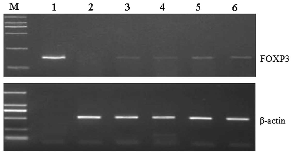 | Figure 1.Reverse transcription-polymerase chain
reaction analysis of FOXP3 gene transcription revealed high
levels of FOXP3 in advanced gastric cancer. Lane M, DL2000
DNA marker; lane 1, FOXP3 expression plasmid (control group); lane
2, control group; lane 3, experimental group (grade I); lane 4,
experimental group (grade II); lane 5, experimental group (grade
III); lane 6, experimental group (grade IV). Sizes: β-actin, 540
bp; FOXP3, 167 bp. FOXP3, forkhead box protein 3. |
FOXP3 expression in gastric cancer
cells
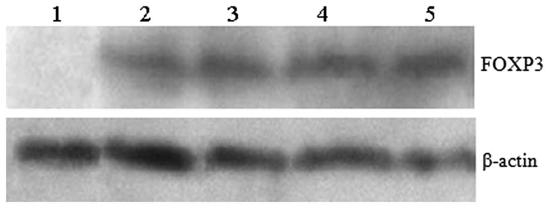
Western blot analysis revealed that FOXP3 protein
expression levels were different in grade IV gastric lesions
compared with the control group. Furthermore, in the experimental
group, FOXP3 protein levels were positively correlated with gastric
lesion grade. However, no FOXP3 protein expression was identified
in gastric mucosa of the control group (P<0.05) (Figs. 2 and 3).
FOXP3 protein distribution and
immunohistochemical staining in gastric cancer lesions of the
experimental group
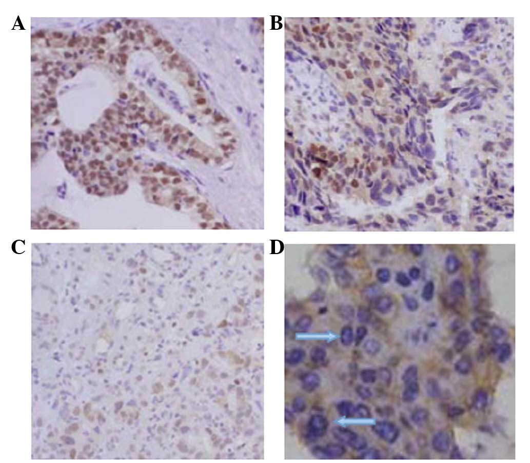
In the experimental group, FOXP3 protein expression
was identified in the gastric tissues, which was mainly distributed
in the nuclei of gastric cancer cells. Infiltrative cancer (grade
III and IV lesions) exhibited strong FOXP3 staining, whereas early
cancer and precancerous lesions (grades I and II) exhibited weak
FOXP3 staining (Fig. 4).
Of the 32 gastric cancer pathological sections
obtained (which included grade III, early cancer and grade IV,
invasive carcinoma.), 20 cases (62.5%) exhibited positive FOXP3
staining, of which, 4 cases exhibited positive nuclear and
cytoplasmic staining and 16 cases exhibited positive nuclear
staining. Of the 8 precancerous lesion sections, 4 cases (50.00%)
exhibited positive FOXP3 staining. Of the 6 proliferative lesion
sections, 2 cases (33.33%) exhibited positive FOXP3 staining. No
positive FOXP3 staining was observed in any of the 46 control
tissue sections. In the experimental group, FOXP3 was predominantly
distributed in the nuclei of gastric cancer cells, and the
expression of FOXP3 positively correlated with the gastric lesion
grade. These results indicated that FOXP3 may exhibit an important
function in the formation and development of gastric cancer.
Discussion
The incidence rate of gastric cancer continues to be
the highest among all malignancies of the digestive system, despite
having decreased in recent years (19). The prognosis of gastric cancer is
closely associated with cancer stage and treatment, and >90% of
patients with early gastric cancer survive for >5 years and may
be cured following adequate treatment (20). However, the 5-year survival rate of
advanced gastric cancer patients is <5% following treatment
(21). Therefore, early diagnosis is
critical to improve patient prognosis and increase the survival
rate. Gastric cancer patients usually present with symptoms at a
late stage, and the incidence and mortality rates of the disease in
China are two-fold higher than the global incidence (15.86/100,000)
and mortality rates (7.20/100,000) (22). Therefore, constructing animal models
of gastric cancer to simulate the pathological state and
pathophysiological processes of gastric cancer patients represents
a useful approach to investigate the underlying mechanism and to
identify novel treatments for the disease (23,24).
It has been previously demonstrated that the level
and function of regulatory T cells in the majority of gastric
cancer patients may be useful to predict patient survival and
prognosis (25). FOXP3 is an
important protein marker of regulatory T cells, which exhibits
important functions in development and maturation of regulatory T
cells (3). In recent years, FOXP3
expression in gastric cancer tissue has become a research focus
(26). However, few studies have
investigated the association between FOXP3 expression and different
types of gastric cancer lesions, and the potential underlying
mechanism remains unknown (27,28).
In the present study, a gastric cancer rat model was
established using sarcosine ethyl ester hydrochloride and sodium
nitrite. FOXP3 expression and localization was investigated using
RT-PCR, western blot analysis and immunohistochemistry. The
transcription levels of FOXP3 were different in grade IV gastric
cancer lesions compared with the control group. The levels of FOXP3
transcription were positively correlated with gastric cancer stage.
The transcription level of FOXP3 in normal gastric tissue was not
significantly different from that in gastric cancer. Notably, the
level of FOXP3 expression was closely associated with the gastric
cancer lesion grade. Previous studies have revealed that FOXP3
expression was localized to the nucleus and cytoplasm in pancreatic
and colon cancer cells (29,30), which is consistent with the results of
the present study, which demonstrated that FOXP3 expression in
gastric cancer tissues was mainly localized to the nuclei and
cytoplasm. However, in the present study, no FOXP3 expression was
identified in normal gastric tissue by immunohistochemical
analysis. In addition, the expression level of FOXP3 was positively
correlated with gastric lesion grade. Of the 32 gastric cancer
tissue sections, 20 cases (62.50%) exhibited positive FOXP3 protein
expression, including 4 cases with positive nuclear and cytoplasmic
FOXP3 staining and 16 cases with positive nuclear staining. Of the
8 precancerous gastric tissues, 4 cases (50.00%) exhibited positive
FOXP3 protein expression. Of the 6 proliferative lesion tissue
sections, 2 cases (33.33%) exhibited positive FOXP3 protein
expression. These results indicated that FOXP3 may exhibit a
modulatory function in the formation and development of gastric
carcinoma.
A previous study has reported that gastric cancer
cells with increased FOXP3 expression demonstrate increased drug
resistance, indicating that FOXP3 may be involved in the signal
transduction pathways that are associated with drug resistance, and
may interact with and key molecules of these pathways (31). The combined application of various
chemotherapeutic drugs was commonly used as a method to inhibit the
growth of tumor cells in the clinic. However, gastric cancer, as
one of the most common tumors in China, exhibits obvious
characteristics of drug resistance, and the recurrence rate of
gastric cancer following surgical resection is high (32). Therefore, detailed investigation
regarding the generation and development of gastric carcinoma
resistance may improve chemotherapy and immunotherapy of gastric
cancer. The factors that influence the development and prognosis of
gastric cancer are complex, including tumor growth pattern, tumor
types, cancer gene expression, clinical stage and lymph node
metastasis (20). Advances in
molecular biology and immunohistochemistry have increased the
understanding of the close association between gastric cancer,
pathology and immunology (33).
In conclusion, in the present study, FOXP3
expression was identified in gastric cancer cell lines and tissues
by establishing a gastric cancer rat model, and performing RT-PCR,
western blot analysis and immunohistochemistry. In addition, the
results revealed that the expression of FOXP3 was associated with
gastric lesion grade, indicating that the expression of FOXP3 may
exhibit an important function in the formation, development and
prognosis of gastric cancer. In present study, the mechanism
responsible for the effect of FOXP3 on gastric cancer has not been
completely clarified due to certain limitations on the experimental
conditions. Therefore, the functions and detailed mechanism of
FOXP3 in gastric carcinoma requires further investigation.
References
|
1
|
Sugano K: Screening of gastric cancer in
Asia. Best Pract Res Clin Gastroenterol. 29:895–905. 2015.
View Article : Google Scholar : PubMed/NCBI
|
|
2
|
Smid D, Skalicky T, Dolezal J, Kubackova D
and Fichtl J: Surgical treatment of gastric cancer. Bratisl Lek
Listy. 116:666–670. 2015.PubMed/NCBI
|
|
3
|
de Reuver PR, Mehta S, Gill P, Andrici J,
D'Urso L, Clarkson A, Mittal A, Hugh TJ, Samra JS and Gill AJ:
Immunoregulatory forkhead box protein p3-positive lymphocytes are
associated with overall survival in patients with pancreatic
neuroendocrine tumors. J Am Coll Surg. 222:281–287. 2016.
View Article : Google Scholar : PubMed/NCBI
|
|
4
|
Kasprowicz DJ, Smallwood PS, Tyznik AJ and
Ziegler SF: Scurfin (FoxP3) controls T-dependent immune responses
in vivo through regulation of CD4+ T cell effector function. J
Immunol. 171:1216–1223. 2003. View Article : Google Scholar : PubMed/NCBI
|
|
5
|
Gambineri E, Torgerson TR and Ochs HD:
Immune dysregulation, polyendocrinopathy, enteropathy, and X-linked
inheritance (IPEX), a syndrome of systemic autoimmunity caused by
mutations of FOXP3, a critical regulator of T cell homeostasis.
Curr Opin Rheumatol. 15:430–435. 2003. View Article : Google Scholar : PubMed/NCBI
|
|
6
|
Lopes JE, Torgerson TR, Schubert LA,
Anover SD, Ocheltree EL, Ochs HD and Ziegler SF: Analysis of FOXP3
reveals multiple domains required for its function as a
transcriptional repressor. J Immunol. 177:3133–3142. 2006.
View Article : Google Scholar : PubMed/NCBI
|
|
7
|
Buckner JH and Ziegler SF: Functional
analysis of FOXP3. Ann N Y Acad Sci. 1143:151–169. 2008. View Article : Google Scholar : PubMed/NCBI
|
|
8
|
Zheng L, Wang X, Xu L, Wang N, Cai P,
Liang T and Hu L: Foxp3 gene polymorphisms and haplotypes associate
with susceptibility of Graves' disease in Chinese Han population.
Int Immunopharmacol. 25:425–431. 2015. View Article : Google Scholar : PubMed/NCBI
|
|
9
|
Safinia N, Scotta C, Vaikunthanathan T,
Lechler RI and Lombardi G: Regulatory T cells: Serious contenders
in the promise for immunological tolerance in transplantation.
Front Immunol. 6:4382015. View Article : Google Scholar : PubMed/NCBI
|
|
10
|
Bettelli E, Dastrange M and Oukka M: Foxp3
interacts with nuclear factor of activated T cells and NF-kappa B
to repress cytokine gene expression and effector functions of T
helper cells. Proc Natl Acad Sci USA. 102:5138–5143. 2005.
View Article : Google Scholar : PubMed/NCBI
|
|
11
|
Klabusay M: The role of regulatory T-cells
in antitumor immune response. Klin Onkol. 28(Suppl 4): 4S23–4S27.
2015.(In Czech). View Article : Google Scholar : PubMed/NCBI
|
|
12
|
Hou J, Yu Z, Xiang R, Li C and Wang L,
Chen S, Li Q, Chen M and Wang L: Correlation between infiltration
of FOXP3+ regulatory T cells and expression of B7-H1 in the tumor
tissues of gastric cancer. Exp Mol Pathol. 96:284–291. 2014.
View Article : Google Scholar : PubMed/NCBI
|
|
13
|
Li B, Samanta A, Song X, Iacono KT, Bembas
K, Tao R, Basu S, Riley JL, Hancock WW, Shen Y, et al: FOXP3
interactions with histone acetyltransferase and class II histone
deacetylases are required for repression. Proc Natl Acad Sci USA.
104:4571–4576. 2007. View Article : Google Scholar : PubMed/NCBI
|
|
14
|
Yuan XL, Shen DF, Lu J, Dong P, Wang J, Li
MX and Shen LS: The populations, distribution of regulatory T
cells, Foxp3 mRNA expression gastric cancer patients and the
association with malignant stage. Chinese Journal of Laboratory
Medicine. 31:378–383. 2008.(In Chinese).
|
|
15
|
Mühler MR, Clément O, Salomon LJ, Balvay
D, Autret G, Vayssettes C, Cuénod CA and Siauve N: Maternofetal
pharmacokinetics of a gadolinium chelate contrast agent in mice.
Radiology. 258:455–460. 2011. View Article : Google Scholar : PubMed/NCBI
|
|
16
|
Chen BB, Liang PC, Liu KL, Hsiao JK, Huang
JC, Wong JM, Lee PH, Shun CT and Ming-Tsang Y: Preoperative
diagnosis of gastric tumors by three-dimensional multidetector row
CT and double contrast barium meal study: Correlation with surgical
and histologic results. J Formos Med Assoc. 106:943–952. 2007.
View Article : Google Scholar : PubMed/NCBI
|
|
17
|
Stengel A, Goebel M, Wang L, Rivier J,
Kobelt P, Mönnikes H, Lambrecht NW and Taché Y: Central nesfatin-1
reduces dark-phase food intake and gastric emptying in rats:
Differential role of corticotropin-releasing factor2 receptor.
Endocrinology. 150:4911–4919. 2009. View Article : Google Scholar : PubMed/NCBI
|
|
18
|
Xiong Y, Svingen PA, Sarmento OO, Smyrk
TC, Dave M, Khanna S, Lomberk GA, Urrutia RA and Faubion WA Jr:
Differential coupling of KLF10 to Sin3-HDAC and PCAF regulates the
inducibility of the FOXP3 gene. Am J Physiol Regul Integr Comp
Physiol. 307:R608–R620. 2014. View Article : Google Scholar : PubMed/NCBI
|
|
19
|
Merchant SJ, Kim J, Choi AH, Sun V, Chao J
and Nelson R: A rising trend in the incidence of advanced gastric
cancer in young Hispanic men. Gastric Cancer. Feb 29–2016.(Epub
ahead of print). View Article : Google Scholar : PubMed/NCBI
|
|
20
|
Hacker U and Lordick F: Current standards
in the treatment of gastric cancer. Dtsch Med Wochenschr.
140:1202–1205. 2015.(In German). PubMed/NCBI
|
|
21
|
Kohno D, Nakata M, Maejima Y, Shimizu H,
Sedbazar U, Yoshida N, Dezaki K, Onaka T, Mori M and Yada T:
Nesfatin1 neurons in paraventricular and supraoptic nuclei of the
rat hypothalamus coexpress oxytocin and vasopressin and are
activated by refeeding. Endocrinology. 149:1295–1301. 2008.
View Article : Google Scholar : PubMed/NCBI
|
|
22
|
Yanai H, Matsumoto Y, Harada T, Nishiaki
M, Tokiyama H, Shigemitsu T, Tada M and Okita K: Endoscopic
ultrasonography and endoscopy for staging depth of invasion in
early gastric cancer: A pilot study. Gastrointest Endosc.
46:212–216. 1997. View Article : Google Scholar : PubMed/NCBI
|
|
23
|
Levi E, Sochacki P, Khoury N, Patel BB and
Majumdar AP: Cancer stem cells in Helicobacter pylori infection and
aging: Implications for gastric carcinogenesis. World J
Gastrointest Pathophysiol. 5:366–372. 2014.PubMed/NCBI
|
|
24
|
Ke X, Wang J, Li L, Chen IH, Wang H and
Yang XF: Roles of CD4+CD25 (high) FOXP3+ Tregs in lymphomas and
tumors are complex. Front Biosci. 13:3986–4001. 2008.PubMed/NCBI
|
|
25
|
Kakinuma T, Nadiminti H, Lonsdorf AS,
Murakami T, Perez BA, Kobayashi H, Finkelstein SE, Pothiawala G,
Belkaid Y and Hwang ST: Small numbers of residual tumor cells at
the site of primary inoculation are critical for anti-tumor
immunity following challenge at a secondary location. Cancer
Immunol Immunother. 56:1119–1131. 2007. View Article : Google Scholar : PubMed/NCBI
|
|
26
|
Hao Q, Zhang C, Gao Y, Wang S, Li J, Li M,
Xue X, Li W, Zhang W and Zhang Y: FOXP3 inhibits NF-κB activity and
hence COX2 expression in gastric cancer cells. Cell Signal.
26:564–569. 2014. View Article : Google Scholar : PubMed/NCBI
|
|
27
|
Zhou Z, Song X, Li B and Greene MI: FOXP3
and its partners: Structural and biochemical insights into the
regulation of FOXP3 activity. Immunol Res. 42:19–28. 2008.
View Article : Google Scholar : PubMed/NCBI
|
|
28
|
Mizukami Y, Kono K, Kawaguchi Y, Akaike H,
Kamimura K, Sugai H and Fujii H: Localisation pattern of Foxp3+
regulatory T cells is associated with clinical behaviour in gastric
cancer. Br J Cancer. 98:148–153. 2008. View Article : Google Scholar : PubMed/NCBI
|
|
29
|
Allan SE, Passerini L, Bacchetta R,
Crellin N, Dai M, Orban PC, Ziegler SF, Roncarolo MG and Levings
MK: The role of 2 FOXP3 isoforms in the generation of human CD4+
Tregs. J Clin Invest. 115:3276–3284. 2005. View Article : Google Scholar : PubMed/NCBI
|
|
30
|
Karanikas V, Speletas M, Zamanakou M,
Kalala F, Loules G, Kerenidi T, Barda AK, Gourgoulianis KI and
Germenis AE: Foxp3 expression in human cancer cells. J Transl Med.
6:192008. View Article : Google Scholar : PubMed/NCBI
|
|
31
|
Zuo T, Wang L, Morrison C, Chang X, Zhang
H, Li W, Liu Y, Wang Y, Liu X, Chan MW, et al: FOXP3 is an X-linked
breast cancer suppressor gene and an important repressor of the
HER-2/ErbB2 oncogene. Cell. 129:1275–1286. 2007. View Article : Google Scholar : PubMed/NCBI
|
|
32
|
Durães C, Almeida GM, Seruca R, Oliveira C
and Carneiro F: Biomarkers for gastric cancer: Prognostic,
predictive or targets of therapy? Virchows Arch. 464:367–378. 2014.
View Article : Google Scholar : PubMed/NCBI
|
|
33
|
Zheng Y, Josefowicz SZ, Kas A, Chu TT,
Gavin MA and Rudensky AY: Genome-wide analysis of Foxp3 target
genes in developing and mature regulatory T cells. Nature.
445:936–940. 2007. View Article : Google Scholar : PubMed/NCBI
|