Introduction
Cholangiocarcinoma (CCA) is a cancer of the bile
duct caused by the malignant transformation of cholangiocytes, the
epithelial cells lining the bile duct. CCA is the second most
common type of primary hepatic malignancy (1,2). Due to
late diagnosis, resistance to chemo- and radiotherapy, and the high
rates of local invasion and distant metastasis, the prognosis for
CCA is very poor. The median survival time of patients with CCA is
<2 years (3), and the 5-year
survival rate of patients receiving curative resection is 0–40%
(4). The prevalence of CCA is the
highest in Asia, particularly in the northeastern region of
Thailand (5). Opisthorchis
viverrini infection is hypothesized to be a causal factor of
CCA in Thailand, as it is markedly associated with the incidence of
CCA (6).
In total, >90% of cancer-associated mortality is
due to the local or distant metastasis of cancer cells (7). The epithelial-mesenchymal transition
(EMT) is an important process in cancer metastasis, characterized
by alterations in the gene expression and morphology of cells,
which leads to a reduction of intercellular adhesion, and an
increase in cell motility (8–11). This process is associated with a
reduction of E-cadherin expression (12,13) and an
increase in the expression of vimentin, an intermediate filament
protein, leading to increased cell motility and promoting tumor
metastasis (14,15).
Growth arrest and DNA damage-inducible-β (Gadd45β)
is a stress-response protein; its expression is induced by
physiological or environmental stress. The aberrant expression of
Gadd45β in various types of cancer has implicated its involvement
in tumorigenesis (16). Gadd45β
belongs to the Gadd45 protein family (Gadd45α, Gadd45β, Gadd45γ)
(17). Gadd45β may form a homodimer
or heterodimer with the other Gadd45 proteins, or interact with a
variety of other proteins, including proliferating cell nuclear
antigen, cyclin-dependent kinase 1, p21, mitogen-activated protein
kinase kinase kinase 4, mitogen-activated protein kinase kinase 7
and p38 mitogen activated protein kinase (MAPK). The function of
Gadd45β differs depending on the interacting molecules, including
cell cycle control, DNA repair, survival and death control, and
stress signaling (18–22).
Cancer cells must survive and propagate in a
strenuous environment of hypoxia, nutrient competition and
oxidative stress (23). It is
essential for cells to acquire the ability to thrive in these
stressful conditions. The function of Gadd45β as a stress-response
protein in cancer is paradoxical; the downregulation of Gadd45β via
promoter methylation in hepatocellular carcinoma suggests that
Gadd45β may act as a tumor suppressor (24), whereas the upregulation of Gadd45β in
colorectal cancer was associated with recurrence and mortality of
patients with colorectal cancer (25), suggesting a tumor-promoting role. When
the Gadd45β gene from normal adjacent tissue was over-expressed in
colorectal cancer cell lines, apoptotic cell death was induced
(25). In addition, Gadd45β was
identified as upregulated in the metastasis of uveal melanoma to
the liver (26), and the silencing of
Gadd45β in human embryonic carcinoma cells decreased viability and
invasiveness (27), suggesting that
Gadd45β may contribute to the malignant phenotypes of cancer.
However, the function of Gadd45β in metastasis and EMT is not yet
fully characterized.
In the present study, it was identified that
patients with CCA exhibit increased Gadd45β expression in tumor
tissue, and that a high level of Gadd45β expression was associated
with metastasis. Gadd45β expression in a CCA cell line, HuCCA-1,
was suppressed using siRNA-mediated gene silencing, and the effects
on cell viability, survival and death signaling pathways,
migration, invasiveness, and the EMT pathway were studied. The data
of the present study thus indicated that Gadd45β expression
promoted the viability, migration and invasion of the HuCCA-1
cells, traits required for successful metastasis.
Materials and methods
Ethical approval
All procedures performed in the present study
involving human participants were performed in accordance with the
1964 Declaration of Helsinki and its later amendments, or
comparable ethical standards. The study was conducted with the
approval of the Ethical Committee of Rajavithi Hospital (Bangkok,
Thailand).
Immunohistochemical (IHC)
staining
Paraffinized tissue samples from 28 patients with
CCA who had undergone surgical treatment at Rajavithi Hospital
between January 2010 and October 2010 were selected for
retrospective analysis. Standard IHC technique was used for the
detection of Gadd45β in paraffinized sections on glass slides.
Polyclonal Gadd45β antibody (HPA029816-100UL; 1:500 dilution;
Sigma-Aldrich; Merck KGaA, Darmstadt, Germany) was hybridized with
the sections overnight at 4°C, followed by incubation with
biotinylated goat anti-rabbit IgG at RT for 1 h (E0432; dilution
1:500; Dako; Agilent Technologies, Inc., Santa Clara, CA, USA).
Subsequently avidin-biotin-peroxidase conjugate was added (ABC
Elite; Vector Laboratories, Burlingame, CA, USA) for 30 min at room
temperature and the immunohistochemical reaction was developed with
freshly prepared reagents from a Histofine SAB-PO kit (Nichirei,
Inc., Tokyo, Japan). The samples were visualized at ×400
magnification using an Olympus BX53 microscope (Olympus
Corporation, Tokyo, Japan) and categorized into four grades
according to the intensity and percentage of positively stained
cells: Negative or <5%, grade 0; weak or 5–25%, grade 1;
moderate or 25–50%, grade 2; and strong or >50%, grade 3. IHC
grade 0 (3 cases) and grade 1 (4 cases), which exhibited weak
and/or <25% staining, were grouped as low expression, whereas
grade 2 (5 cases) and grade 3 (16 cases), which exhibited moderate
to strong and/or >25% staining, were grouped as high expression
of Gadd45β for the purpose of statistical analysis.
Cell line and culture condition
The human CCA cell line, HuCCA-1 (28), developed from a Thai patient with CCA,
was used for the study. The cells were provided by Professor
Satitaya Sirisinha (Mahidol University, Bangkok, Thailand). These
cells were grown in HAM's F-12 medium supplemented with 10% fetal
bovine serum (Gibco; Thermo Fisher Scientific, Inc., Waltham, MA,
USA) at 37°C in 5% CO2 humidified atmosphere.
Small interfering (si)RNA-mediated
Gadd45β gene silencing
Commercial Gadd45β siRNA [GADD45β siRNA(h):
sc-37416], a pool of 3 different siRNA duplexes to the Gadd45β gene
(NCBI RefSeq. 12759252), was purchased from Santa Cruz
Biotechnology, Inc. (Dallas, TX, USA). Silencer®
Negative control siRNA no. 1 (cat. no. 4404021; Ambion; Thermo
Fisher Scientific, Inc.) was used as negative control siRNA. The
cells were washed with PBS and detached using 0.25% Trypsin/EDTA
(Gibco; Thermo Fisher Scientific, Inc.). Cells were seeded at
2×10⁵ cells/well into a 6 well plate for 24 h, then
transfected with 40 nM siRNA using Lipofectamine®
RNAiMax reagent (Invitrogen; Thermo Fisher Scientific, Inc.)
according to the manufacturer's instructions. The medium containing
transfection reagents was changed for 10% FBS supplemented
HAM's-F12 at 24 h after transfection.
Western blot analysis
At 48 h post-transfection, total cellular protein
was extracted and prepared following a protocol described
previously (29). Each protein sample
of 40 µg was separated via SDS-PAGE (12% gel; 120 V for 2 h)
followed by an electroblotting transfer of the protein to a
nitrocellulose membrane at 30 V at 4°C for 15 h. The membranes were
blocked in 1.5% skimmed milk at room temperature for 1 h, the
membranes were incubated with the primary antibodies at 4°C
overnight on a rocking platform. The next day, the membranes were
washed with TBS with Tween-20 prior to incubation with horseradish
peroxidase-conjugated secondary antibodies for 1 h at room
temperature. Following 3 washes with TBST for 10 min, ECL Plus
Western Blotting Detection system (Bio-Rad Laboratories, Inc.,
Hercules, CA, USA) was used to visualize the immunoreactive bands
in G:Box ChemiXL 1.4 (Syngene; Synoptics, Cambridge, UK).
Rabbit polyclonal Gadd45β antibodies were purchased
from Abcam (Cambridge, UK) and goat polyclonal GAPDH antibodies
from Santa Cruz Biotechnology, Inc. The rest of the antibodies used
in the study were purchased from Cell Signaling Technology, Inc.
(Danvers, MA, USA); the details regarding all antibodies used in
western blotting are included in Table
I. Densitometry analysis was performed using ImageJ software
(version 1.49v; National Institutes of Health, Bethesda, MD, USA)
(30). The density of the protein of
interest relative to GAPDH was determined in HuCCA-1 cells
transfected with Gadd45β or negative control siRNA. The activation
of Akt, p38 MAPK and extracellular signal-regulated kinase (ERK)1/2
signaling pathways was examined using phospho-specific antibodies
at 48 h after siRNA transfection. The densitometry measurements for
total Akt, ERK1/2 and p38 MAPK were used for the normalization of
protein loading. The phospho-protein/total protein density ratio
was determined in HuCCA-1 cells transfected with Gadd45β siRNA or
negative control siRNA. The phospho-protein/total protein density
ratio of cells transfected with negative control siRNA was
designated as 100%.
 | Table I.The primary antibodies used for
western blotting in the present study. |
Table I.
The primary antibodies used for
western blotting in the present study.
| Antibody | Cat. no. | Clone no. | Type of
antibody | Organism | Company | Dilution |
|---|
| Gadd45β | ab105060 |
| Polyclonal | Rabbit | Abcam | 1:2,000 |
| GAPDH | sc-48166 | I-19 | Polyclonal | Goat | Santa Cruz
Biotechnology, Inc. | 1:2,000 |
| Cleaved PARP
(Asp214) | #5625 | D64E10 | Monoclonal | Rabbit | Cell Signaling
Technology, Inc. | 1:1,000 |
| Total PARP | #9542 |
| Monoclonal | Rabbit | Cell Signaling
Technology, Inc. | 1:1,000 |
| Cleaved caspase-3
(Asp175) | #9664 | 5A1E | Monoclonal | Rabbit | Cell Signaling
Technology, Inc. | 1:1,000 |
| Full-length caspase
3 | #9662 |
| Monoclonal | Rabbit | Cell Signaling
Technology, Inc. | 1:1,000 |
| Phospho-p38 MAPK
(Thr180/Tyr182) | #9211 |
| Monoclonal | Rabbit | Cell Signaling
Technology, Inc. | 1:1,000 |
| p38 MAPK | #9212 |
| Monoclonal | Rabbit | Cell Signaling
Technology, Inc. | 1:1,000 |
| Phospho-Akt
(Ser473) | #4058 | 193H12 | Monoclonal | Rabbit | Cell Signaling
Technology, Inc. | 1:1,000 |
| Akt | #9272 |
| Monoclonal | Rabbit | Cell Signaling
Technology, Inc. | 1:1,000 |
| Phospho-ERK1/2
(Thr202/Tyr204) | #9101 |
| Monoclonal | Rabbit | Cell Signaling
Technology, Inc. | 1:1,000 |
| ERK1/2 | #9102 |
| Monoclonal | Rabbit | Cell Signaling
Technology, Inc. | 1:1,000 |
| Vimentin | #5741 | D21H3 | Monoclonal | Rabbit | Cell Signaling
Technology, Inc. | 1:1,000 |
| Claudin-1 | #13255 | D5H1D | Monoclonal | Rabbit | Cell Signaling
Technology, Inc. | 1:1,000 |
| β-catenin | #8480 | D10A8 | Monoclonal | Rabbit | Cell Signaling
Technology, Inc. | 1:1,000 |
| ZO-1 | #8193 | D7D12 | Monoclonal | Rabbit | Cell Signaling
Technology, Inc. | 1:1,000 |
| Slug | #9585 | C19G7 | Monoclonal | Rabbit | Cell Signaling
Technology, Inc. | 1:1,000 |
| E-cadherin | #3195 | 24E10 | Monoclonal | Rabbit | Cell Signaling
Technology, Inc. | 1:1,000 |
Cell viability assays
Live/Dead® cell viability
assay
HuCCA-1 cells were grown on coverslips prior to
transfection with siRNA. At 48 h post-transfection, cell viability
was assessed using the Live/Dead® Viability/Cytotoxicity
kit for mammalian cells (Invitrogen; Thermo Fisher Scientific,
Inc.) according to the manufacturer's instructions. Briefly, a
working solution, containing 2 µM calcein AM and 4 µM ethidium
homodimer (EthD-1), was prepared in PBS. The cells were washed with
pre-warmed PBS, and 150 µl of Live/Dead solution was applied to
each cover slip. Following incubation at 37°C in a 5%
CO2 humidified atmosphere for 30 min, the reagents were
removed and the cover slips were washed with PBS, mounted on the
glass-slides, and viewed using a Nikon Eclipse TE2000-U microscope
(Nikon Corporation, Tokyo, Japan) using B-2A (green, calcein) and
G-2A (red, EthD-1) excitation filters. Images were recorded at ×100
total magnification using ACT-1C software (version 1.02; Nikon
Corporation). Live cells were stained with calcein-AM (green),
whereas dead cells were stained with ethidium homodimer (red).
WST-1 assay
A total of 5,000 HuCCA-1 cells were seeded in
96-well plates for 24 h prior to siRNA transfection. At 24, 48 and
72 h post-transfection, 10 µl of WST-1 reagent (Roche Diagnostics,
Basel, Switzerland) was added into each well. The plates were
incubated at 37°C in a humidified atmosphere containing 5%
CO2 for 2 h. Then the absorbance at 450 nm was
measured.
Hoechst staining
At 48 h post-transfection, the culture media was
carefully removed to avoid the loss of detached cells. HuCCA-1
cells were then fixed in 2% w/v paraformaldehyde solution for 30
min at room temperature prior to permeabilization using 0.03% v/v
Triton X-100 for 30 min. Nuclear staining was performed with
Hoechst 333258 dye at room temperature (5 µg/ml; Thermo Fisher
Scientific, Inc.) for 2 h, followed by visualization at ×400 total
magnification with an Olympus IX83 live cell fluorescence
microscope (Olympus Corporation).
Gelatin zymography
To analyze the activity of MMPs, the gelatinase
activity of the enzymes secreted into the medium was determined
using gelatin zymography assay, following a previously described
protocol (29). The gelatinolytic
activity of MMPs was observed as clear bands on the blue background
of the Coomassie-stained gel. Densitometry analysis was performed
using ImageJ software (30).
Cell invasion and migration
assays
Cells were harvested at 48 h post-transfection for
in vitro invasion and migration assays using transwell
chambers, as described by Li et al (31). Briefly, HuCCA-1 cells
(1×10⁵ cells/well) transfected with Gadd45β siRNA or
negative control siRNA were seeded into the upper chamber of the
transwells coated with Matrigel for the invasion assay and with no
coating for the migration assay, and analyzed after 6 h of
incubation at 37°C. HAM's F-12 supplemented with 10% FBS, used as
chemoattractant, was added to the lower chamber. Cells were seeded
in the upper chamber in serum free medium. Three independent
experiments were performed in duplicate transwells.
Statistical analysis
Data is presented as the mean ± standard deviation
and every experiment was performed at least three times. The
χ2 test was used for clinicopathological data analysis,
and experimental data analysis was performed using a paired t-test.
P<0.05 was considered to indicate a statistically significant
difference.
Results
High levels of Gadd45β expression are
associated with metastatic incidence in CCA
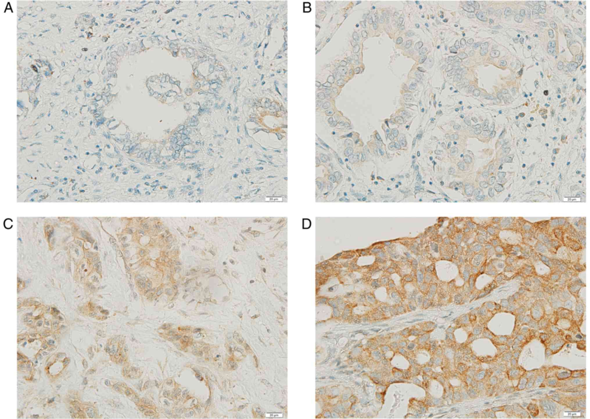
Gadd45β expression in the histological specimens of
CCA tissue from 28 patients was examined with IHC staining. The
expression of Gadd45β was detected in all tumor tissues (n=28), and
75% (n=21) exhibited higher Gadd45β expression than the surrounding
stroma (Fig. 1A-D). Although Gadd45β
expression was previously reported to be moderate in the liver,
gall bladder and pancreas (32), the
expression level in cholangiocytes or CCA tissue has not previously
been reported, to the best of our knowledge. A high level of
Gadd45β expression was associated with the incidence of metastasis
in patients (P=0.035), although no statistically significant
association was observed between the level of Gadd45β expression
and age, sex, lymph node involvement or differentiation status
(Table I).
Gadd45β knockdown reduces the
viability of HuCCA-1 cells
To investigate the functional importance of Gadd45β
in CCA, Gadd45β expression was suppressed using siRNA-mediated
silencing in HuCCA-1, a CCA cell line that expresses moderate
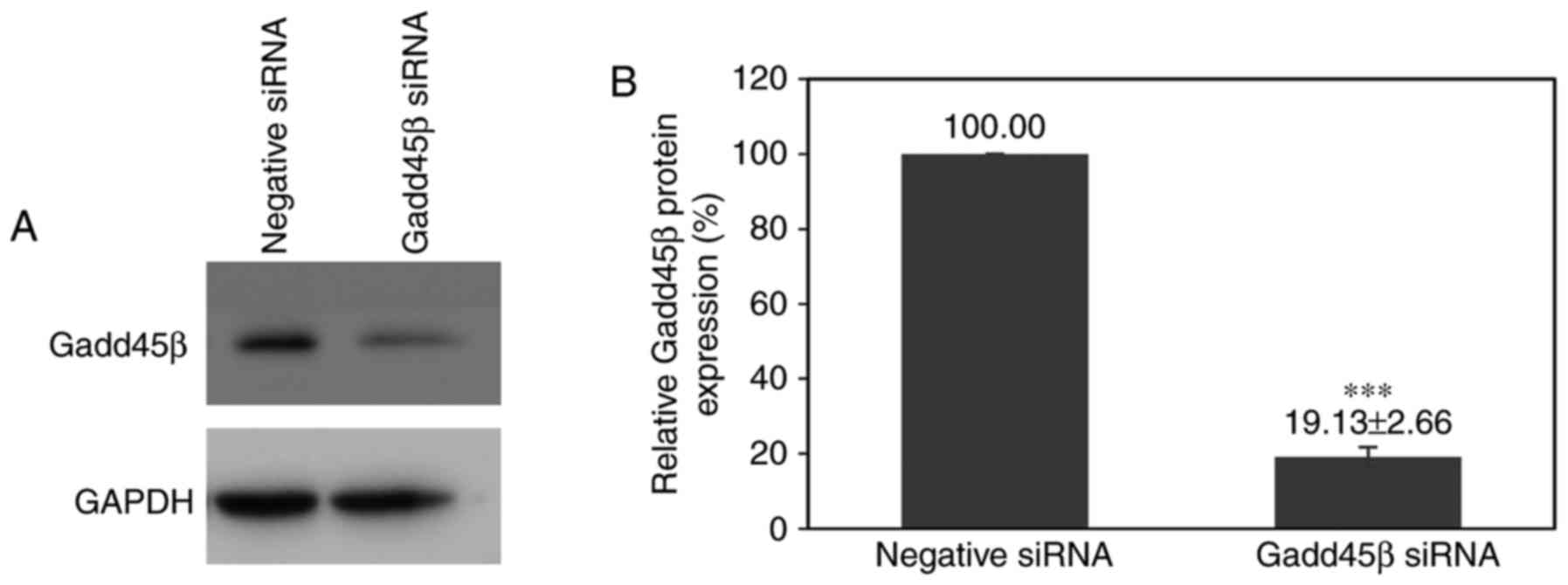
levels of Gadd45β. Gadd45β siRNA transfection decreased the
expression of Gadd45β in HuCCA-1 to 19.13±2.66% relative to
negative control siRNA transfection (Fig.
2A and B).
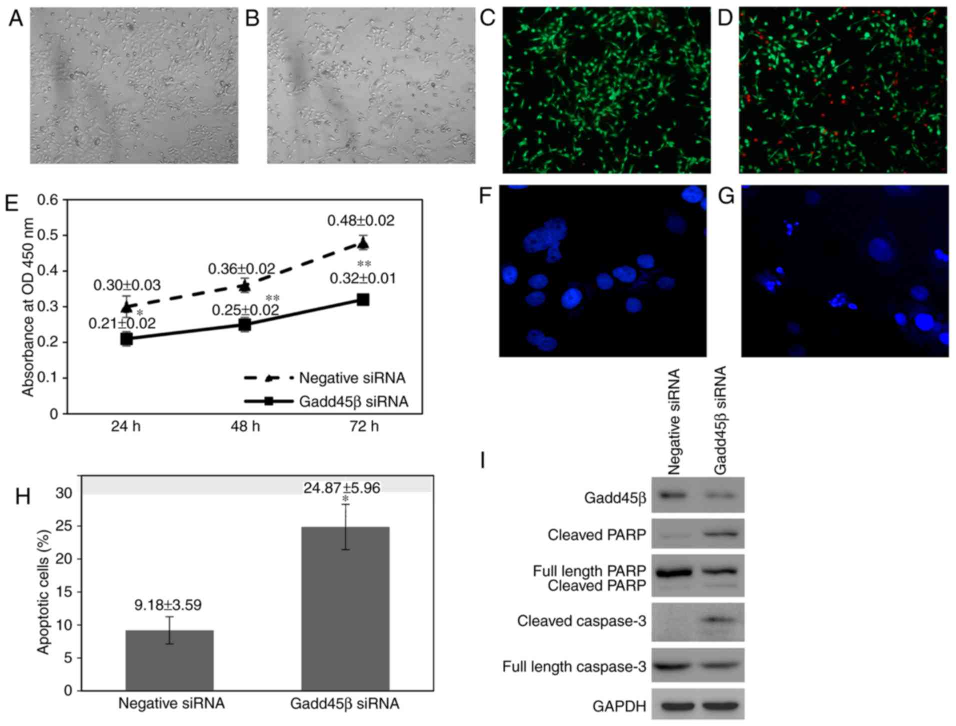
During Gadd45β silencing experiments, a decrease in
the confluence of Gadd45β-silenced HuCCA-1 cells was observed
compared with negative control siRNA-transfected cells (Fig. 3A and B). Furthermore, there was an
increase in the number of cells floating in the medium of
Gadd45β-silenced HuCCA-1 cells, indicating that silencing of
Gadd45β perturbed the viability of HuCCA-1 cells (data not shown).
A Live/Dead® cell viability assay confirmed that there
was an increase in the number of dead cells in HuCCA-1 cells when
Gadd45β was silenced (Fig. 3C and D).
Cell viability was assessed using a WST-1 assay, in which the WST-1
reagent was added directly into the cells without removing the
culture medium, preventing the loss of detached viable cells.
Consistent with visual observation, Gadd45β silencing significantly
decreased the viability of HuCCA-1 cells (Fig. 3E).
Gadd45β silencing kills HuCCA-1 cells
via apoptosis
The mode of cell death in HuCCA-1 cells upon Gadd45β
silencing was examined by staining the nuclear DNA with Hoechst
333258. Upon silencing of Gadd45β, HuCCA-1 cells were observed to
exhibit nuclear condensation and DNA fragmentation, characteristics
of apoptotic cells (33). The nuclei
of live cells were homogenously stained in blue, whereas those of
apoptotic cells appeared condensed and fragmented. Gadd45β-silenced
HuCCA-1 cells exhibited an increased rate of apoptotic cells
compared with negative control cells (24.87±5.96% vs. 9.18±3.59%;
Fig. 3F-H). Consistently, western
blot analysis revealed that poly (ADP-ribose) polymerase (PARP)
cleavage and caspase-3 cleavage were observed, indicating that
HuCCA-1 cells died via apoptosis when Gadd45β was silenced
(Fig. 3I) (34).
Gadd45β silencing reduces Akt activity
in HuCCA-1
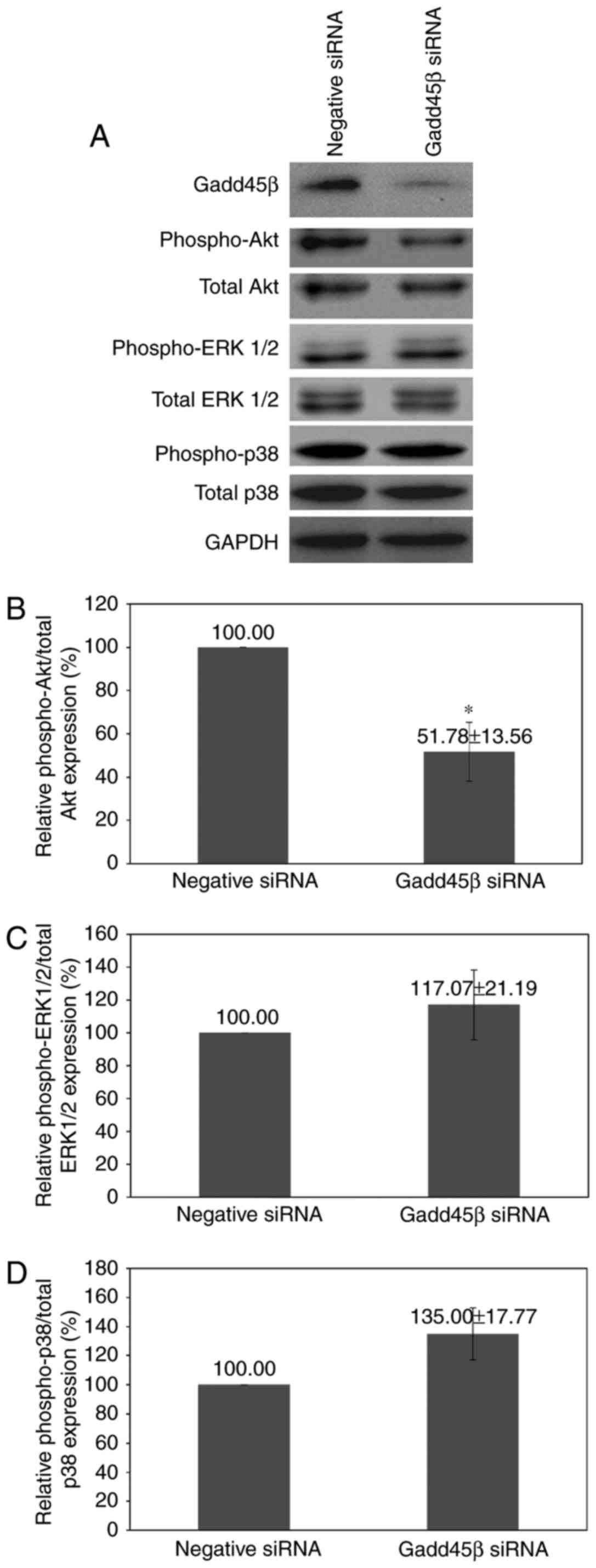
To determine which signaling pathways were involved,
three of the principal pathways involved in the regulation of cell
viability and proliferation, the Akt, ERK and p38 MAPK pathways,
were studied. Akt is associated with cell proliferation, survival
and apoptosis inhibition (35). p38
MAPK is a stress-activated protein kinase pathway important for
cell proliferation, differentiation, survival and migration
(36,37). ERK1/2 activation has been demonstrated
to protect cells from apoptosis (38).
Gadd45β silencing resulted in a marked reduction of
Akt phosphorylation (P=0.0237), whereas no significant alteration
in p38 MAPK or ERK1/2 phosphorylation was observed (Fig. 4A-D). The decrease in Akt activity may
be responsible for the reduction of viability in HuCCA-1 upon
Gadd45β silencing.
Gadd45β silencing reduces the invasion
and migration of HuCCA-1 cells
A hallmark of malignancy is the ability to invade
local tissues or to spread to distant sites (39). During this process, the cancer cells
must acquire the ability to invade and migrate through connective
tissue barriers, including the basement membrane, surrounding
matrix and existing blood vessels (40). IHC data analysis of patient samples
revealed that there was an association between the high expression
of Gadd45β and metastasis (Table
II). Furthermore, Gadd45β was demonstrated to be important for
the viability of HuCCA-1 cells in the present study; viability is
required by the cancer cells for successful metastasis (41). Therefore, the effect of Gadd45β
silencing on the migration and invasion abilities of HuCCA-1 cells
was investigated using in vitro transwell migration and
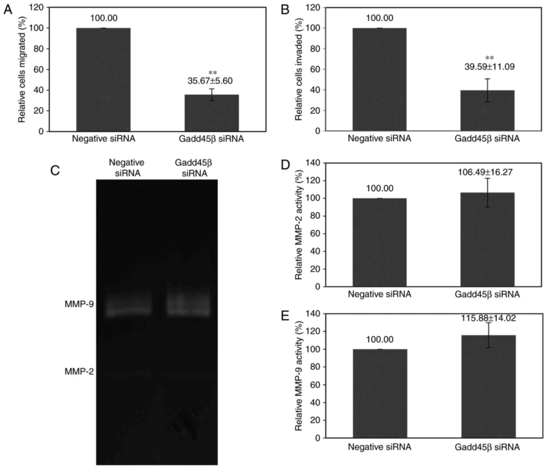
invasion assays. Gadd45β silencing decreased the migration of
HuCCA-1 cells to 35.67±5.60%, and invasion to 39.59±11.09%,
compared with the negative control siRNA (Fig. 5A and B).
| Table II.Association between Gadd45β
expression and the clinicopathological features of patients with
cholangiocarcinoma. |
Table II.
Association between Gadd45β
expression and the clinicopathological features of patients with
cholangiocarcinoma.
|
|
| Gadd45β
expression |
|
|---|
|
|
|
|
|
|---|
| Clinicopathological
parameter | Total | Low | High | P-value |
|---|
| Total, n (%) | 28 | 7 (25.0) | 21 (75.0) |
|
| Age, years |
|
|
| 0.378 |
| Mean ±
standard deviation | 59.68±10.41 |
|
|
|
| <60,
n (%) | 12 (42.9) | 4 (14.3) | 8 (28.6) |
|
| ≥60, n
(%) | 16 (57.1) | 3 (10.7) | 13 (46.4) |
|
| Sex, n (%) |
|
|
| 0.512 |
|
Male | 13 (46.4) | 4 (14.3) | 9 (32.1) |
|
|
Female | 15 (53.6) | 3 (10.7) | 12 (42.9) |
|
| Tumor size, cm |
|
|
| 0.385a |
| Mean ±
standard deviation | 2.90±2.4 |
|
|
|
| <5,
n (%) | 25 (92.6) | 7 (25.9) | 18 (66.7) |
|
| ≥5, n
(%) | 2 (7.4) | 0 (0) | 2 (7.4) |
|
| Lymph node
metastasis, n (%) |
|
|
| 0.190 |
|
Absent | 14 (50.0) | 5 (17.9) | 9 (32.1) |
|
|
Present | 14 (50.0) | 2 (7.1) | 12 (42.9) |
|
| Distant metastasis,
n (%) |
|
|
| 0.035c |
|
Absent | 19 (67.9) | 7 (25.0) | 12 (42.9) |
|
|
Present | 9 (32.1) | 0 (0) | 9 (32.1) |
|
| Differentiation
status, n (%) |
|
|
| 0.696b |
|
Well | 11 (52.4) | 3 (14.3) | 8 (38.1) |
|
|
Moderate | 10 (47.6) | 2 (9.5) | 8 (38.1) |
|
| Type of surgery, n
(%) |
|
|
| 0.076 |
| R0 | 14 (50.0) | 6 (21.0) | 8 (28.6) |
|
| R1 | 7 (25.0) | 1 (3.6) | 6 (21.4) |
|
| R2 | 7 (25.0) | 0 (0) | 7 (25.0) |
|
Cancer cells may secrete matrix metalloproteinases
(MMPs) or stimulate surrounding stromal cells to secrete MMPs,
consequently leading to digestion of the extracellular matrix,
promoting local invasion and metastasis (42,43).
However, gelatin zymography analysis revealed that Gadd45β
silencing did not significantly alter the activity of MMP-2
(P=0.5609) and MMP-9 (P=0.1886) secreted by HuCCA-1 cells (Fig. 5C-E).
Gadd45β silencing reverses the EMT
changes in HuCCA-1
Epithelial-mesenchymal transition is an important
process during cancer metastasis, denoted by acquisition of
mesenchymal phenotypes, including increased motility and
invasiveness (8,9,44–46). Since Gadd45β silencing decreased the
invasion and migration of HuCCA-1 cells, properties associated with
EMT, including morphological changes and EMT marker expression,
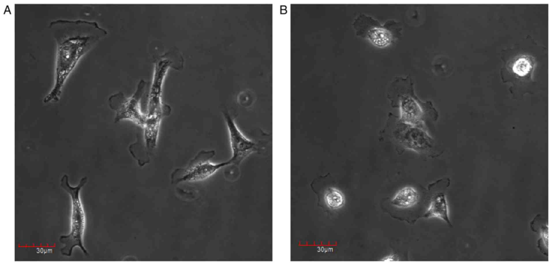
were examined. It was demonstrated that Gadd45β silencing may have
induced HuCCA-1 cells to undergo morphological changes, from
spindle-shaped and fibroblast-like in appearance, to a flattened
epithelial-like phenotype (Fig. 6A and
B). The expression patterns of the proteins involved in the EMT
pathway were further investigated with western blotting. It was
identified that Gadd45β silencing affected the expression of
multiple EMT markers (Fig. 7A-G).
Epithelial marker E-cadherin expression was increased (P=0.0430),
whereas the expression of the mesenchymal markers, Vimentin
(P=0.0085) and Slug (P=0.0399), was decreased by Gadd45β silencing.
However, there was no change in β-catenin expression (P=0.6006;
Fig. 7A and D).
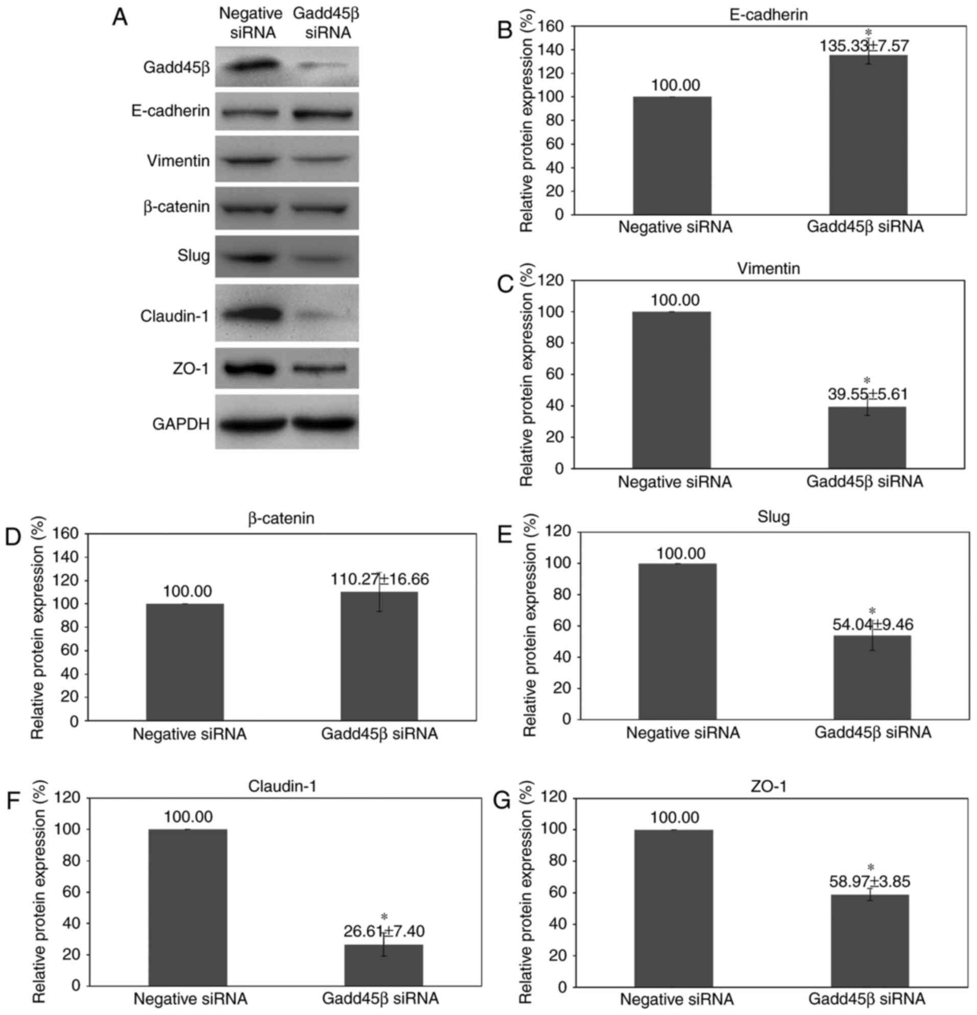 | Figure 7.Effect of Gadd45β silencing upon EMT
marker expression. (A) Representative images of western blot
analysis for the expression of E-cadherin, vimentin, β-catenin,
Slug, claudin-1 and ZO-1 at 48 h after Gadd45β silencing in
HuCCA-1. Densitometry analysis of (B) E-cadherin, (C) vimentin, (D)
β-catenin, (E) Slug, (F) claudin-1 and (G) ZO-1. Columns, mean of
three independent experiments; bars, standard error of the mean.
*P<0.05. Gadd45β, growth arrest and DNA damage-inducible-β; EMT,
epithelial-mesenchymal transition; ZO-1, zona occludens protein 1;
siRNA, small interfering RNA. |
Notably, proteins associated with tight junctions
were also affected by Gadd45β silencing. Claudin-1 and zona
occludens protein 1 (ZO-1) constitute the tight junctions between
cells (47,48); one of the mechanisms involved in
cancer invasion is collective migration, in which the expression
levels of tight-junction proteins are important to allow cells to
move as a cluster (49). There was a
decrease in claudin-1 (P=0.0100; Fig. 7A
and F) and the ZO-1 expression (P=0.0087; Fig. 7A and G) upon Gadd45β silencing.
Discussion
The IHC staining of paraffin embedded tissue samples
from patients with CCA revealed that the majority of CCA tissue
samples expressed increased Gadd45β relative to non-tumor tissue,
and that the high level of Gadd45β in CCA was associated with an
increased incidence of metastasis in patients. These findings
suggest that Gadd45β may be of functional importance in CCA. As
cancer cells are continuously exposed to stressful environments
including hypoxia, competition for nutrients and oxidative stress
during growth (23), Gadd45β appears
to enable CCA cells to cope with stress and to thrive in the harsh
tumor microenvironment.
In the present study, the functional importance of
Gadd45β in CCA was determined using siRNA-mediated gene silencing
in HuCCA-1, a cell line established from a patient with CCA.
Gadd45β silencing decreased the proliferation and induced apoptosis
in HuCCA-1 cells, as demonstrated by nuclear condensation and
fragmentation, together with the activation of caspase-3 and PARP
cleavage. The impairment of cell proliferation together with an
increased rate of apoptosis in Gadd45β-silenced cells suggests that
Gadd45β serves a pro-survival role in CCA. These data are in
agreement with previous reports on genotoxic stress-induced
apoptosis in hematopoietic cells of Gadd45β-deficient mice
(50). Additionally, Gadd45β has been
demonstrated to promote the survival of mouse embryo fibroblasts in
response to tumor necrosis factor-α (51) and of B cells during Fas-induced
apoptosis (52).
To study the underlying molecular mechanisms by
which Gadd45β promotes viability in HuCCA-1, key cellular survival
and death signaling pathways were studied, since the activation of
death signals or the reduction of survival signals reduces the
viability of cells. The results of the present study demonstrated
that Gadd45β silencing significantly decreased the Akt activity,
although p38 MAPK and ERK1/2 activity was unchanged. Activation of
the Akt/PI3K pathway has been associated with growth and metastasis
(53); therefore, the decrease in
p-Akt may be responsible for the reduced growth and induction of
apoptosis in HuCCA-1 cells upon Gadd45β silencing. The altered cell
signaling activities appeared to shift the balance from
pro-survival to pro-apoptotic following the silencing of
Gadd45β.
EMT is an important biological process that allows
malignant tumor cells to acquire migratory and invasive phenotypes;
prerequisites for cancer invasion and metastasis. It is indicated
by the acquisition of fibroblast-like morphology, with a reduction
in intercellular adhesion and increased cell motility (11–18). The
IHC data from the present study indicated that Gadd45β may be
involved in the metastasis of CCA. Gadd45β silencing of HuCCA-1
cells induced a marked reduction in invasiveness and migration,
although the activity of MMPs was not affected. A number of reports
have implicated Gadd45β in the cell migration ability, an important
characteristic of metastatic cancer cells. For example Salerno
et al (54) demonstrated that
the granulocytes of Gadd45β-deficient mice displayed the impairment
of lipopolysacchiride-stimulated chemotactic migration.
Furthermore, Kodama and Negishi (55)
demonstrated that the ectopic expression of Gadd45β increased,
whereas siRNA-mediated silencing of Gadd45β decreased, pregnane X
receptor-induced cell migration in hepatocellular carcinoma
cells.
Examination of the expression of EMT markers
revealed that Gadd45β silencing resulted in reduction in Slug
expression, with a consequent increase in E-cadherin and decrease
in vimentin expression. High Slug expression has been demonstrated
to induce EMT in cancer cells (56)
and the effects are orchestrated by an increase in vimentin
expression (57). As Slug expression
is negatively regulated by GS3Kβ (58), which in turn is inactivated by pAkt
(59), the decrease in Akt activity
following Gadd45β silencing may have led to the downregulation of
Slug expression, with the consequent suppression of vimentin
expression and an increase in the invasiveness and migration of
HuCCA-1 cells.
Furthermore, a decrease in the expression of the
tight junction proteins, claudin-1 and ZO-1, was induced by Gadd45β
silencing. Claudins and ZO-1 are integral proteins of the tight
junctions that seal adjacent epithelial cells (47,48).
Although the conventional roles of claudins and ZO-1 are to control
paracellular ion flux and maintain cell polarity, accumulating
evidence has demonstrated that these proteins also exhibit
non-junctional functions, including in cell motility, and may
function in the EMT, apoptosis resistance, invasion and metastasis
of cancer cells (60–62). Claudins have been demonstrated to
promote collective migration, where groups of cells move in a
coordinated manner and remain connected via cell-cell junctions.
Additionally, claudin expression has been associated with increased
MMP activity and cell survival (62).
Claudin-1 expression has been demonstrated to be associated with
invasive and metastatic phenotypes in a range of types of cancer
including colon, liver, oral squamous cell carcinoma and melanoma
(63). ZO-1 is a component of the
‘junctional plaque’ at the cytoplasmic surface of the tight
junctions, which links integral membrane proteins with the
cytoskeleton (64). It was identified
in a previous study that ZO-1 can translocate from the cell
membrane to the nucleus and may be involved in cell signaling
(65). The upregulation of ZO-1 has
been reported in melanoma, and the silencing of ZO-1 led to a
marked reduction in the rate of cell invasion (66). Thus, the identified association
between a decrease in claudin-1 and ZO-1 expression with impaired
cell invasion and migration in the present study appears to be
consistent with these studies.
In conclusion, the silencing of Gadd45β
significantly decreased the viability, metastatic phenotypes and
EMT markers of HuCCA-1 cells, suggesting that Gadd45β serves a
function in the metastatic process in CCA. This is in accord with
the IHC data of patients with CCA, in which Gadd45β expression was
associated with metastasis. However, a limitation of this study was
that the experiments were performed in only one CCA cell line.
Further studies should be performed in more CCA cell lines with
more stable knock down systems, as transient siRNA transfection has
a limited gene silencing duration. The study of Gadd45β expression,
function and Gadd45β-mediated signaling pathways in CCA will be
important in understanding cancer biology, in which the cancer
cells are dynamically evolving with accumulated mutations. Since
the cancer cells must thrive in a competitive environment, a stress
response protein like Gadd45β may be critical. The understanding or
manipulation of Gadd45β-dependent survival signaling and EMT
pathways may be beneficial as a potential therapeutic option,
adjuvant to the conventional therapies for CCA.
Acknowledgements
The authors thank Professor Satitaya Sirisinha
(Mahidol University, Bangkok, Thailand) for providing the HuCCA-1
cell line and the Central Instrument Facility, Center of
Nanoimaging, Faculty of Science of Mahidol University. The authors
express their thanks to Professor Tavan Janvilisri and Ms. Phorutai
Pearngam for assistance in the study of EMT pathways. The present
study was supported by Mahidol University (grant no. 18/2555).
Glossary
Abbreviations
Abbreviations:
|
CCA
|
cholangiocarcinoma
|
|
ZO-1
|
zona occludens protein 1
|
|
MMP
|
matrix metalloproteinase
|
|
EMT
|
epithelial-mesenchymal transition
|
|
siRNA
|
small interfering RNA
|
References
|
1
|
Blechacz B and Gores GJ:
Cholangiocarcinoma: Advances in pathogenesis, diagnosis, and
treatment. Hepatology. 48:308–321. 2008. View Article : Google Scholar : PubMed/NCBI
|
|
2
|
Welzel TM, McGlynn KA, Hsing AW, O'Brien
TR and Pfeiffer RM: Impact of classification of hilar
cholangiocarcinomas (Klatskin tumors) on the incidence of intra-
and extrahepatic cholangiocarcinoma in the United States. J Natl
Cancer Inst. 98:873–875. 2006. View Article : Google Scholar : PubMed/NCBI
|
|
3
|
Farley DR, Weaver AL and Nagorney DM:
‘Natural history’ of unresected cholangiocarcinoma: Patient outcome
after noncurative intervention. Mayo Clin Proc. 70:pp. 425–429.
1995; View
Article : Google Scholar : PubMed/NCBI
|
|
4
|
Anderson CD, Pinson CW, Berlin J and Chari
RS: Diagnosis and treatment of cholangiocarcinoma. Oncologist.
9:43–57. 2004. View Article : Google Scholar : PubMed/NCBI
|
|
5
|
Sripa B, Brindley PJ, Mulvenna J, Laha T,
Smout MJ, Mairiang E, Bethony JM and Loukas A: The tumorigenic
liver fluke Opisthorchis viverrini-multiple pathways to cancer.
Trends Parasitol. 28:395–407. 2012. View Article : Google Scholar : PubMed/NCBI
|
|
6
|
Sripa B, Kaewkes S, Sithithaworn P,
Mairiang E, Laha T, Smout M, Pairojkul C, Bhudhisawasdi V, Tesana
S, Thinkamrop B, et al: Liver fluke induces cholangiocarcinoma.
PLoS Med. 4:e2012007. View Article : Google Scholar : PubMed/NCBI
|
|
7
|
Weigelt B, Peterse JL and van't Veer LJ:
Breast cancer metastasis: Markers and models. Nat Rev Cancer.
5:591–602. 2005. View
Article : Google Scholar : PubMed/NCBI
|
|
8
|
Heerboth S, Housman G, Leary M, Longacre
M, Byler S, Lapinska K, Willbanks A and Sarkar S: EMT and tumor
metastasis. Clin Transl Med. 4:62015. View Article : Google Scholar : PubMed/NCBI
|
|
9
|
Nieto MA: Epithelial plasticity: A common
theme in embryonic and cancer cells. Science. 342:12348502013.
View Article : Google Scholar : PubMed/NCBI
|
|
10
|
Moreno-Bueno G, Portillo F and Cano A:
Transcriptional regulation of cell polarity in EMT and cancer.
Oncogene. 27:6958–6969. 2008. View Article : Google Scholar : PubMed/NCBI
|
|
11
|
Peinado H, Olmeda D and Cano A: Snail, Zeb
and bHLH factors in tumour progression: An alliance against the
epithelial phenotype? Nat Rev Cancer. 7:415–428. 2007. View Article : Google Scholar : PubMed/NCBI
|
|
12
|
Adhikary A, Chakraborty S, Mazumdar M,
Ghosh S, Mukherjee S, Manna A, Mohanty S, Nakka KK, Joshi S, De A,
et al: Inhibition of epithelial to mesenchymal transition by
E-cadherin up-regulation via repression of slug transcription and
inhibition of E-cadherin degradation: Dual role of scaffold/matrix
attachment region-binding protein 1 (SMAR1) in breast cancer cells.
J Biol Chem. 289:25431–25444. 2014. View Article : Google Scholar : PubMed/NCBI
|
|
13
|
Techasen A, Loilome W, Namwat N, Khuntikeo
N, Puapairoj A, Jearanaikoon P, Saya H and Yongvanit P: Loss of
E-cadherin promotes migration and invasion of cholangiocarcinoma
cells and serves as a potential marker of metastasis. Tumour Biol.
35:8645–8652. 2014. View Article : Google Scholar : PubMed/NCBI
|
|
14
|
Mendez MG, Kojima S and Goldman RD:
Vimentin induces changes in cell shape, motility, and adhesion
during the epithelial to mesenchymal transition. FASEB J.
24:1838–1851. 2010. View Article : Google Scholar : PubMed/NCBI
|
|
15
|
Chaw SY, Majeed AA, Dalley AJ, Chan A,
Stein S and Farah CS: Epithelial to mesenchymal transition (EMT)
biomarkers-E-cadherin, beta-catenin, APC and Vimentin-in oral
squamous cell carcinogenesis and transformation. Oral Oncol.
48:997–1006. 2012. View Article : Google Scholar : PubMed/NCBI
|
|
16
|
Qiu W, David D, Zhou B, Chu PG, Zhang B,
Wu M, Xiao J, Han T, Zhu Z, Wang T, et al: Down-regulation of
growth arrest DNA damage-inducible gene 45beta expression is
associated with human hepatocellular carcinoma. Am J Pathol.
162:1961–1974. 2003. View Article : Google Scholar : PubMed/NCBI
|
|
17
|
Zumbrun SD, Hoffman B and Liebermann DA:
Distinct mechanisms are utilized to induce stress sensor gadd45b by
different stress stimuli. J Cell Biochem. 108:1220–1231. 2009.
View Article : Google Scholar : PubMed/NCBI
|
|
18
|
Liebermann DA and Hoffman B: Gadd45 in
stress signaling. J Mol Signal. 3:152008. View Article : Google Scholar : PubMed/NCBI
|
|
19
|
Vairapandi M, Balliet AG, Fornace AJ Jr,
Hoffman B and Liebermann DA: The differentiation primary response
gene MyD118, related to GADD45, encodes for a nuclear protein which
interacts with PCNA and p21WAF1/CIP1. Oncogene. 12:2579–2594.
1996.PubMed/NCBI
|
|
20
|
Smith ML, Ford JM, Hollander MC, Bortnick
RA, Amundson SA, Seo YR, Deng CX, Hanawalt PC and Fornace AJ Jr:
p53-mediated DNA repair responses to UV radiation: Studies of mouse
cells lacking p53, p21, and/or gadd45 genes. Mol Cell Biol.
20:3705–3714. 2000. View Article : Google Scholar : PubMed/NCBI
|
|
21
|
Jónsson ZO and Hübscher U: Proliferating
cell nuclear antigen: More than a clamp for DNA polymerases.
Bioessays. 19:967–975. 1997. View Article : Google Scholar : PubMed/NCBI
|
|
22
|
Kelman Z and Hurwitz J: Protein-PCNA
interactions: A DNA-scanning mechanism? Trends Biochem Sci.
23:236–238. 1998. View Article : Google Scholar : PubMed/NCBI
|
|
23
|
Leprivier G, Rotblat B, Khan D, Jan E and
Sorensen PH: Stress-mediated translational control in cancer cells.
Biochim Biophys Acta. 1849:845–860. 2015. View Article : Google Scholar : PubMed/NCBI
|
|
24
|
Qiu W, Zhou B, Zou H, Liu X, Chu PG, Lopez
R, Shih J, Chung C and Yen Y: Hypermethylation of growth arrest DNA
damage-inducible gene 45 beta promoter in human hepatocellular
carcinoma. Am J Pathol. 165:1689–1699. 2004. View Article : Google Scholar : PubMed/NCBI
|
|
25
|
Wang L, Xiao X, Li D, Chi Y, Wei P, Wang
Y, Ni S, Tan C, Zhou X and Du X: Abnormal expression of GADD45B in
human colorectal carcinoma. J Transl Med. 10:2152012. View Article : Google Scholar : PubMed/NCBI
|
|
26
|
Meir T, Dror R, Yu X, Qian J, Simon I,
Pe'er J and Chowers I: Molecular characteristics of liver
metastases from uveal melanoma. Invest Ophthalmol Vis Sci.
48:4890–4896. 2007. View Article : Google Scholar : PubMed/NCBI
|
|
27
|
Inowa T, Hishikawa K, Matsuzaki Y, Isagawa
T, Takeuchi T, Aburatani H, Kitamura T and Fujita T: GADD45β
determines chemoresistance and invasive growth of side population
cells of human embryonic carcinoma. Stem Cells Int.
2010:7829672010. View Article : Google Scholar : PubMed/NCBI
|
|
28
|
Sirisinha S, Tengchaisri T, Boonpucknavig
S, Prempracha N, Ratanarapee S and Pausawasdi A: Establishment and
characterization of a cholangiocarcinoma cell line from a Thai
patient with intrahepatic bile duct cancer. Asian Pac J Allergy
Immunol. 9:153–157. 1991.PubMed/NCBI
|
|
29
|
Pongcharoen P, Jinawath A and Tohtong R:
Silencing of CD44 by siRNA suppressed invasion, migration and
adhesion to matrix, but not secretion of MMPs, of
cholangiocarcinoma cells. Clin Exp Metastasis. 28:827–839. 2011.
View Article : Google Scholar : PubMed/NCBI
|
|
30
|
Schneider CA, Rasband WS and Eliceiri KW:
NIH Image to ImageJ: 25 years of image analysis. Nat Methods.
9:671–675. 2012. View Article : Google Scholar : PubMed/NCBI
|
|
31
|
Li YH and Zhu C: A modified Boyden chamber
assay for tumor cell transendothelial migration in vitro.
Clin Exp Metastasis. 17:423–429. 1999. View Article : Google Scholar : PubMed/NCBI
|
|
32
|
Uhlen M, Fagerberg L, Hallström BM,
Lindskog C, Oksvold P, Mardinoglu A, Sivertsson Å, Kampf C,
Sjöstedt E, Asplund A, et al: Proteomics. Tissue-based map of the
human proteome. Science. 347:12604192015. View Article : Google Scholar : PubMed/NCBI
|
|
33
|
Kerr JF, Wyllie AH and Currie AR:
Apoptosis: A basic biological phenomenon with wide-ranging
implications in tissue kinetics. Br J Cancer. 26:239–257. 1972.
View Article : Google Scholar : PubMed/NCBI
|
|
34
|
Tewari M, Quan LT, O'Rourke K, Desnoyers
S, Zeng Z, Beidler DR, Poirier GG, Salvesen GS and Dixit VM:
Yama/CPP32 beta, a mammalian homolog of CED-3, is a
CrmA-inhibitable protease that cleaves the death substrate
poly(ADP-ribose) polymerase. Cell. 81:801–809. 1995. View Article : Google Scholar : PubMed/NCBI
|
|
35
|
Kumar A, Rajendran V, Sethumadhavan R and
Purohit R: Akt kinase pathway: A leading target in cancer research.
ScientificWorldJournal. 2013:7561342013. View Article : Google Scholar : PubMed/NCBI
|
|
36
|
Nebreda AR and Porras A: p38 MAP kinases:
Beyond the stress response. Trends Biochem Sci. 25:257–260. 2000.
View Article : Google Scholar : PubMed/NCBI
|
|
37
|
Kyriakis JM and Avruch J: Mammalian
mitogen-activated protein kinase signal transduction pathways
activated by stress and inflammation. Physiol Rev. 81:807–869.
2001. View Article : Google Scholar : PubMed/NCBI
|
|
38
|
Erhardt P, Schremser EJ and Cooper GM:
B-Raf inhibits programmed cell death downstream of cytochrome c
release from mitochondria by activating the MEK/Erk pathway. Mol
Cell Biol. 19:5308–5315. 1999. View Article : Google Scholar : PubMed/NCBI
|
|
39
|
Hanahan D and Weinberg RA: Hallmarks of
cancer: The next generation. Cell. 144:646–674. 2011. View Article : Google Scholar : PubMed/NCBI
|
|
40
|
Talmadge JE and Fidler IJ: AACR centennial
series: The biology of cancer metastasis: Historical perspective.
Cancer Res. 70:5649–5669. 2010. View Article : Google Scholar : PubMed/NCBI
|
|
41
|
Mehlen P and Puisieux A: Metastasis: A
question of life or death. Nat Rev Cancer. 6:449–458. 2006.
View Article : Google Scholar : PubMed/NCBI
|
|
42
|
Davies KJ: The complex interaction of
matrix metalloproteinases in the migration of cancer cells through
breast tissue Stroma. Int J Breast Cancer. 2014:8390942014.
View Article : Google Scholar : PubMed/NCBI
|
|
43
|
Roomi MW, Monterrey JC, Kalinovsky T, Rath
M and Niedzwiecki A: Patterns of MMP-2 and MMP-9 expression in
human cancer cell lines. Oncol Rep. 21:1323–1333. 2009.PubMed/NCBI
|
|
44
|
Micalizzi DS, Farabaugh SM and Ford HL:
Epithelial-mesenchymal transition in cancer: Parallels between
normal development and tumor progression. J Mammary Gland Biol
Neoplasia. 15:117–134. 2010. View Article : Google Scholar : PubMed/NCBI
|
|
45
|
Powell DR, Blasky AJ, Britt SG and
Artinger KB: Riding the crest of the wave: Parallels between the
neural crest and cancer in epithelial-to-mesenchymal transition and
migration. Wiley Interdiscip Rev Syst Biol Med. 5:511–522. 2013.
View Article : Google Scholar : PubMed/NCBI
|
|
46
|
Wallerand H, Cai Y, Wainberg ZA, Garraway
I, Lascombe I, Nicolle G, Thiery JP, Bittard H, Radvanyi F and
Reiter RR: Phospho-Akt pathway activation and inhibition depends on
N-cadherin or phospho-EGFR expression in invasive human bladder
cancer cell lines. Urol Oncol. 28:180–188. 2010. View Article : Google Scholar : PubMed/NCBI
|
|
47
|
Furuse M, Fujita K, Hiiragi T, Fujimoto K
and Tsukita S: Claudin-1 and −2: Novel integral membrane proteins
localizing at tight junctions with no sequence similarity to
occludin. J Cell Biol. 141:1539–1550. 1998. View Article : Google Scholar : PubMed/NCBI
|
|
48
|
Stevenson BR, Siliciano JD, Mooseker MS
and Goodenough DA: Identification of ZO-1: A high molecular weight
polypeptide associated with the tight junction (zonula occludens)
in a variety of epithelia. J Cell Biol. 103:755–766. 1986.
View Article : Google Scholar : PubMed/NCBI
|
|
49
|
Rørth P: Collective cell migration. Annu
Rev Cell Dev Biol. 25:407–429. 2009. View Article : Google Scholar : PubMed/NCBI
|
|
50
|
Gupta M, Gupta SK, Balliet AG, Hollander
MC, Fornace AJ, Hoffman B and Liebermann DA: Hematopoietic cells
from Gadd45a- and Gadd45b-deficient mice are sensitized to
genotoxic-stress-induced apoptosis. Oncogene. 24:7170–7179. 2005.
View Article : Google Scholar : PubMed/NCBI
|
|
51
|
De Smaele E, Zazzeroni F, Papa S, Nguyen
DU, Jin R, Jones J, Cong R and Franzoso G: Induction of gadd45beta
by NF-kappaB downregulates pro-apoptotic JNK signalling. Nature.
414:308–313. 2001. View Article : Google Scholar : PubMed/NCBI
|
|
52
|
Zazzeroni F, Papa S, Algeciras-Schimnich
A, Alvarez K, Melis T, Bubici C, Majewski N, Hay N, De Smaele E,
Peter ME and Franzoso G: Gadd45 beta mediates the protective
effects of CD40 costimulation against Fas-induced apoptosis. Blood.
102:3270–3279. 2003. View Article : Google Scholar : PubMed/NCBI
|
|
53
|
Yothaisong S, Dokduang H, Techasen A,
Namwat N, Yongvanit P, Bhudhisawasdi V, Puapairoj A, Riggins GJ and
Loilome W: Increased activation of PI3K/Akt signaling pathway is
associated with cholangiocarcinoma metastasis and PI3K/mTOR
inhibition presents a possible therapeutic strategy. Tumour Biol.
34:3637–3648. 2013. View Article : Google Scholar : PubMed/NCBI
|
|
54
|
Salerno DM, Tront JS, Hoffman B and
Liebermann DA: Gadd45a and Gadd45b modulate innate immune functions
of granulocytes and macrophages by differential regulation of p38
and JNK signaling. J Cell Physiol. 227:3613–3620. 2012. View Article : Google Scholar : PubMed/NCBI
|
|
55
|
Kodama S and Negishi M: Pregnane X
receptor PXR activates the GADD45beta gene, eliciting the p38 MAPK
signal and cell migration. J Biol Chem. 286:3570–3578. 2011.
View Article : Google Scholar : PubMed/NCBI
|
|
56
|
Medici D, Hay ED and Olsen BR: Snail and
Slug promote epithelial-mesenchymal transition through
beta-catenin-T-cell factor-4-dependent expression of transforming
growth factor-beta3. Mol Biol Cell. 19:4875–4887. 2008. View Article : Google Scholar : PubMed/NCBI
|
|
57
|
Vuoriluoto K, Haugen H, Kiviluoto S,
Mpindi JP, Nevo J, Gjerdrum C, Tiron C, Lorens JB and Ivaska J:
Vimentin regulates EMT induction by Slug and oncogenic H-Ras and
migration by governing Axl expression in breast cancer. Oncogene.
30:1436–1448. 2011. View Article : Google Scholar : PubMed/NCBI
|
|
58
|
Kim JY, Kim YM, Yang CH, Cho SK, Lee JW
and Cho M: Functional regulation of Slug/Snail2 is dependent on
GSK-3β-mediated phosphorylation. FEBS J. 279:2929–2939. 2012.
View Article : Google Scholar : PubMed/NCBI
|
|
59
|
Cross DA, Alessi DR, Vandenheede JR,
McDowell HE, Hundal HS and Cohen P: The inhibition of glycogen
synthase kinase-3 by insulin or insulin-like growth factor 1 in the
rat skeletal muscle cell line L6 is blocked by wortmannin, but not
by rapamycin: Evidence that wortmannin blocks activation of the
mitogen-activated protein kinase pathway in L6 cells between Ras
and Raf. Biochem J. 303:21–26. 1994. View Article : Google Scholar : PubMed/NCBI
|
|
60
|
Bezdekova M, Brychtova S, Sedlakova E,
Langova K, Brychta T and Belej K: Analysis of Snail-1, E-cadherin
and claudin-1 expression in colorectal adenomas and carcinomas. Int
J Mol Sci. 13:1632–1643. 2012. View Article : Google Scholar : PubMed/NCBI
|
|
61
|
Stebbing J, Filipovic A and Giamas G:
Claudin-1 as a promoter of EMT in hepatocellular carcinoma.
Oncogene. 32:4871–4872. 2013. View Article : Google Scholar : PubMed/NCBI
|
|
62
|
Oliveira SS and Morgado-Diaz JA: Claudins:
Multifunctional players in epithelial tight junctions and their
role in cancer. Cell Mol Life Sci. 64:17–28. 2007. View Article : Google Scholar : PubMed/NCBI
|
|
63
|
Kwon MJ: Emerging roles of claudins in
human cancer. Int J Mol Sci. 14:18148–18180. 2013. View Article : Google Scholar : PubMed/NCBI
|
|
64
|
Bauer H, Zweimueller-Mayer J, Steinbacher
P, Lametschwandtner A and Bauer HC: The dual role of zonula
occludens (ZO) proteins. J Biomed Biotechnol. 2010:4025932010.
View Article : Google Scholar : PubMed/NCBI
|
|
65
|
Gottardi CJ, Arpin M, Fanning AS and
Louvard D: The junction-associated protein, zonula occludens-1,
localizes to the nucleus before the maturation and during the
remodeling of cell-cell contacts. Proc Natl Acad Sci USA. 93:pp.
10779–10784. 1996; View Article : Google Scholar : PubMed/NCBI
|
|
66
|
Smalley KS, Brafford P, Haass NK, Brandner
JM, Brown E and Herlyn M: Up-regulated expression of zonula
occludens protein-1 in human melanoma associates with N-cadherin
and contributes to invasion and adhesion. Am J Pathol.
166:1541–1554. 2005. View Article : Google Scholar : PubMed/NCBI
|