Introduction
Osteosarcoma (OS), one of the most frequent types of
primary malignant bone neoplasm, originates from the metaphysis of
the long bones in children and adolescents (1). The estimated morbidity rate for OS is
4.4 per million worldwide, with a peak incidence at 15–19 years old
(2). Phenotypically, OS is relatively
homogeneous, but multiple genetic alterations and environmental
factors have been demonstrated to be closely associated with
carcinogenesis, and progression of OS (3,4).
Presently, the main therapeutic strategies for patients with OS are
orthopedic surgical intervention, chemotherapy and occasionally
radiotherapy (5). Although progress
in treatments has occurred, the 5-year overall survival rates for
OS patients without metastatic disease are ~50-60%, and is
significantly low for those with metastasis (6,7). The
majority of patients with OS eventually develop metastasis,
particularly pulmonary metastasis, which is the major cause of
treatment failures (8). Therefore, it
is necessary to understand the molecular mechanism underlying
metastasis of OS, and investigate novel therapeutics treatments
that target the molecular pathway regulating the metastasis of
OS.
MicroRNA (miRNA/miR) are a family of endogenous,
non-coding, single stranded and short RNAs of 18–25 nucleotides in
length. They modulate gene expression through interactions with
complementary sequences in the 3′untranslated regions (3′UTRs) of
target mRNAs in a sequence-specific manner, resulting in
degradation or translational repression of target mRNAs (9–12). Growing
evidence has demonstrated that miRNAs serve important functions in
a wide range of physiological and pathological processes, including
cell growth, development, differentiation, apoptosis, survival,
migration and invasion (13,14). Notably, accumulated evidence has
indicated that the aberrant expression of miRNAs is required to
maintaining the malignant phenotype of cancer cells, and that they
function as tumor suppressors or oncogenes as a result of the
diversity of their target mRNAs (15). Particularly, numerous miRNAs have been
reported to serve essential functions in carcinogenesis and
progression of OS, and may be independent prognostic markers or
therapeutic targets for patients with OS (16–18).
Therefore, it is essential to further investigate the expression
and roles of miRNAs in OS, and provide insight into identifying the
novel therapeutic treatment for patients with OS.
Previous studies demonstrated that miR-302a
expression is aberrantly expressed in multiple tumor types
(19–21). However, miR-302a expression pattern
and its biological roles remains to be investigated in OS. The
present study aimed to investigate the expression level of miR-302a
in OS tissues and cell lines, and the biological roles of miR-302a
in OS cells. Furthermore, the molecular mechanism underlying its
tumor suppressive roles was evaluated.
Material and methods
Ethics statement and OS tissue
samples
The present study was approved by the Ethics
Committee of Tianjin Hospital (Tianjin, China) and written informed
consent was obtained from all patients involved in the present
study. A total of 34 paired OS tissues and matched normal adjacent
tissues (NATs) were obtained from patients with OS (20 male and 14
female; age range, 17–63 years; mean age, 38 years) between June
2013 and March 2015. Patients with OS who received other
therapeutic treatments prior to surgery were excluded from this
study. Upon resection, the OS tissues and matched NATs were
immediately snap-frozen in liquid nitrogen and stored at −80°C.
Cell lines and cell culture
The OS HOS, MG63, SAOS2 and U2OS cell lines and
human normal osteoblastic hFOB 1.19 cell line were obtained from
the American Type Culture Collection (Manassas, VA, USA). All cells
were cultured in Dulbecco's modified Eagle's medium supplemented
with 10% fetal bovine serum (FBS), 100 U/ml penicillin and 100 U/ml
streptomycin (all Gibco; Thermo Fisher Scientific, Inc., Waltham,
MA, USA) in a humidified incubator with 5% CO2 at
37°C.
Oligonucleotide transfection
miR-302a mimics and negative control (NC) were
purchased from Shanghai GenePharma Co., Ltd. (Shanghai, China).
IGF-1R and control siRNAs were synthesized and purified by
Guangzhou RiboBio Co., Ltd. (Guangzhou, China). The sequence of the
miR-302a mimic was 5′UAAGUGCUUCCAUGUUUUGGUGA3′. The sequence of the
NC mimic was 5′UUCUCCGAACGUGUCACGUTT3′. The sequence of the IGF-1R
siRNA was 5′CACCGCGGCTGGAAACTCTTCTACACGAATGTAGAAGAGTTTCCAGCCGC3′.
The sequence of the control siRNA was
5′CACCGCTCACCGGCTCCAGATTTATCGAAATAAATCTGGAGCCGGTGAGC3′.
Oligonucleotide transfection was conducted using Lipofectamine 2000
(Invitrogen; Thermo Fisher Scientific, Inc.) according to the
manufacturer's protocol.
Reverse transcription-quantitative
polymerase chain reaction (RT-qPCR)
Total RNA was extracted using TRIzol reagent
(Invitrogen; Thermo Fisher Scientific, Inc.), and then reverse
transcribed into cDNA using the PrimeScript RT-PCR kit (Takara Bio,
Inc., Otsu, Japan). The expression levels of miR-302a were
determined using SYBR Premix Ex Taq TM II kit (Takara Bio, Inc.),
following the manufacturer's protocol. RNU48 was used as the
internal control for quantification of miR-302a expression. A total
of 200 ng cDNA was used in the qPCR reactions using an ABI7500
Real-time PCR system (Applied Biosystems; Thermo Fisher Scientific,
Inc.), and the thermocycler conditions of qPCR were: 5 min at 95°C,
followed by 40 cycles of 95°C for 30 sec and 65°C for 45 sec. The
expression levels of IGF-1R mRNA were measured using SYBR Green PCR
Master Mix (Applied Biosystems; Thermo Fisher Scientific, Inc.),
and GAPDH was used to normalize the data for quantification of
IGF-1R mRNA expression. The thermocycler conditions of qPCR were as
follows: 95°C for 10 min; 40 cycles of 95°C for 15 sec; and 60°C
for 1 min. The primer sequences used for qPCR were: miR-302a:
forward, 5′CGTGGATGTACTTGCTTTGAA3′; reverse, 5′TCACCAAAACATGGAAGC
AC3′; RNU48 forward, 5′CTCGCTTCGGCAGCACATATACT3′; reverse,
5′ACGCTTCACGAATTTGCGTGTC3′; IGF-1R forward,
5′AGGATATTGGGCTTTACAACCTG3′; reverse, 5′GAGGTAACAGAGGTCAGCATTTT3′;
GAPDH forward, 5′TGCACCACCAACTGCTTA3′; reverse,
5′GGATGCAGGGATGATGTTC3′. Each sample was examined in triplicate.
Data was analyzed using the 2−ΔΔCq method (22).
Transwell migration and Matrigel
invasion assay
Transwell migration and Matrigel invasion assays
were performed to investigate the effects of miR-302a on the
migration and invasion capacity of OS cells. For Matrigel invasion
assays, the Transwell chambers (8 mm pore filter) were coated with
100 µl Matrigel (5 mg/ml) (both BD Biosciences, San Jose, CA, USA).
For Transwell migration and invasion assays, transfected MG63 and
U2OS cells were collected and resuspended in serum-free culture
medium. A total of 1×10⁵ transfected cells in 300 µl
serum-free culture medium were seeded into the upper chamber, while
500 µl culture medium supplemented with 20% FBS was added to the
lower chamber. Following 12 (for migration) or 24 h (for invasion)
of incubation at 37°C, the nonmigrated and noninvaded cells were
carefully scraped off using a cotton swab. Migrated and invaded
cells were fixed, stained with 0.5% crystal violet and washed with
PBS, followed by counting using a CKX41 inverted microscope
(Olympus Corporation, Tokyo, Japan) at magnification, ×200 in five
randomized fields.
Target prediction of miRNAs
miRanda (http://www.microrna.org) and TargetScan version 7.0
(http://www.targetscan.org/) were used to
predict the target genes of miR-302a.
Dual-Luciferase report assay
The pGL3-IGF-1R-3′UTR wild-type (Wt) and
pGL3-IGF-1R-3′UTR mutant (Mut) were purchased from Shanghai
GenePharma Co., Ltd. For the Luciferase report assay, OS cells were
transfected with miR-302a mimics or NC, and luciferase reporter
vector using Lipofectamine 2000. At 48 h following transfection,
cells were collected and luciferase activities were measured using
Dual-Luciferase Reporter Assay system (Promega Corporation,
Madison, WI, USA), according to the manufacturer's protocol. The
Renilla luciferase activities were detected as an internal
control for relative firefly luciferase activities.
Western blot analysis
MG63 and U2OS cells were lysed in ice-cold lysis
buffer (50 mM Tris-HCl, pH 6.8, 32 mM 2-ME, 2% w/v SDS, 10%
glycerol) along with protease inhibitor. Protein concentration was
determined and 20 µg protein was separated by 10% SDS-PAGE and
transferred to polyvinylidene difluoride membranes (EMD Millipore,
Billerica, MA, USA). Subsequent to blocking at 37°C with 5% non-fat
milk in Tris-buffered saline (TBS) for 30 min, the membranes were
incubated with primary antibodies directed against IGF-1R (1:1,000
dilution; cat. no., ab39398) and GADPH (1:1,000 dilution; cat. no.,
ab8245; both Abcam, Cambridge, UK) overnight at 4°C. Subsequently,
the membranes were washed with TBS-Tween 20, and incubated with the
corresponding horseradish peroxidase-conjugated secondary
antibodies (1:1,000 dilution; cat. no., ab6785 and ab150077; Abcam)
for 1 h at room temperature. An enhanced chemiluminescent system
(EMD Millipore) was used to visualize signal bands, and analyzed
with Image Lab software (version 3.0; Bio-Rad Laboratories, Inc.,
Hercules, CA, USA).
Statistical analysis
Data are presented as the mean ± standard deviation.
The differences between groups were compared with Student's t test
or one-way analysis of variance using SPSS statistical software
(version 13.0; SPSS Inc., Chicago, IL, USA). P<0.05 was
considered to indicate a statistically significant difference.
Results
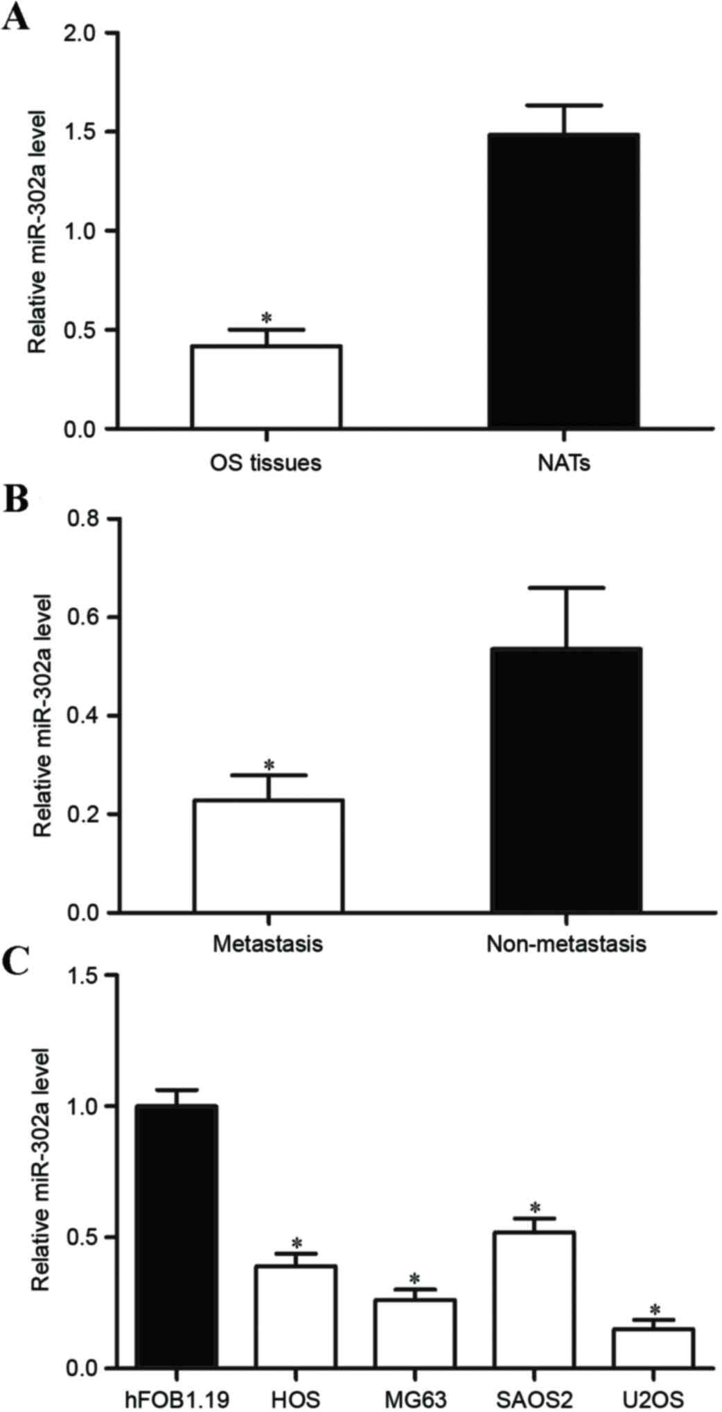
miR-302a mRNA expression is
downregulated in OS tissues and cell lines
miR-302a has been studied in numerous human cancer
types; however, its expression and functions in OS have not been
investigated previously. In the present study, the expression
levels of miR-302a were measured in OS tissue samples. It was
revealed that the expression of miR-302a were significantly lower
in OS tissue compared with that in matched NATs (Fig. 1A; P<0.05). In addition, the
expression of miR-302a in OS tissue with metastasis was
significantly lower compared with that in OS tissue without
metastatic disease (Fig. 1B;
P<0.05). miR-302a expression was determined in OS cell lines and
the human normal osteoblastic hFOB 1.19 cell line. As demonstrated
in Fig. 1C, miR-302a was
significantly downregulated in OS cell lines compared with that in
hFOB 1.19 cells (P<0.05). These results indicate that miR-302a
is downregulated in OS and may be involved in the metastasis of
OS.
miR-302a inhibits the migration and
invasion of OS cells in vitro
To investigate the effects of miR-302a in the
metastasis of OS, OS cells were transfected with miR-302a mimics or
NC, and then subjected to Transwell migration and Matrigel invasion
assays. In the four OS cell lines, MG63 and U2OS cell lines were
selected for functional experiments due to lower miR-302a
expression levels. Following transfection, miR-302a expression
relative to GAPDH was assessed using RT-qPCR. As demonstrated in
Fig. 2A, miR-302a expression was
significantly upregulated in MG63 and U2OS cells transfected with
miR-302a mimics compared with those transfected with the negative
control (P<0.05). Transwell migration and invasion assays
revealed that the overexpression of miR-302a inhibited migration
and invasion capacity of MG63 and U2OS cells (Fig. 2B and C; both P<0.05). These results
indicate that miR-302a acts as a tumor metastasis suppressor in
OS.
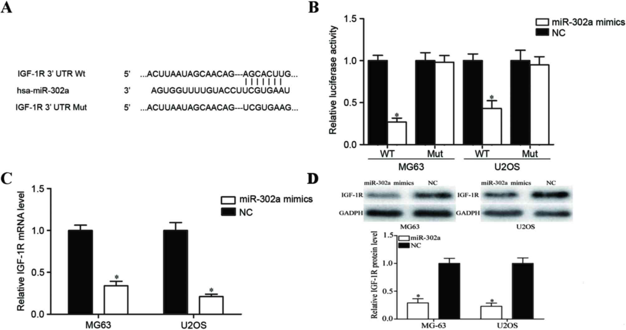
IGF-1R is a direct target gene of
miR-302a in OS
To determine the molecular mechanism underlying
miR-302a in the metastasis of OS, miRanda and TargetScan databases
were used to predict potential target genes of miR-302a. IGF-1R was
predicted to be one of its targets (Fig.
3A). To confirm this predication, a Dual-Luciferase reporter
assay was performed. The results demonstrated that the
overexpression of miR-302a decreased the luciferase activities of
pGL3-IGF-1R-3′UTR Wt; however, no significant differences in the
luciferase activities of pGL3-IGF-1R-3′UTR Mut were observed when
compared to the negative control groups (Fig. 3B; P<0.05). Furthermore, RT-qPCR and
western blot analyses were performed to investigate the regulatory
effects of miR-302a on IGF-1R mRNA and protein expression. As
illustrated in Fig. 3C and D, IGF-1R
was significantly downregulated at mRNA and protein levels in
miR-302a mimics-transfected MG63 and U2OS cells (all P<0.05).
These results suggest that IGF-1R is a direct target gene of
miR-302a in OS.
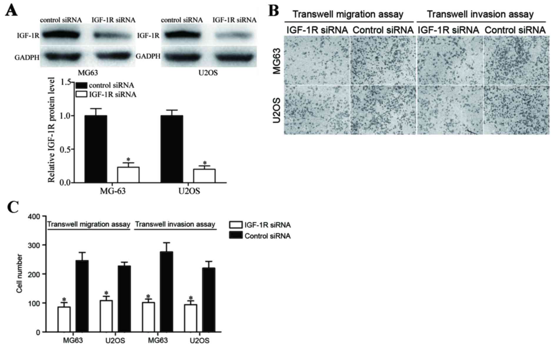
Knockdown IGF-1R mimics functions with
miR-302a overexpression of OS cells
To investigate the effects of IGF-1R in OS cells,
MG63 and U2OS cells were transfected with IGF-1R siRNA or control
siRNA, and then Transwell migration and Matrigel invasion assays
were performed. As illustrated in Fig.
4A, IGF-1R siRNA reduced IGF-1R expression at the protein level
compared with the control siRNA treated group. In addition,
downregulation of IGF-1R in MG63 and U2OS cells significantly
inhibited the migration and invasion abilities of cells compared
with the control group (Fig. 4B and
C; P<0.05). These findings indicate that IGF-1R silencing
and miR-302a overexpression possess similar functions resulting in
cell migration and invasion suppression.
Discussion
Metastasis serves an important role in OS
progression, and the majority of mortalities of patients with OS
are primarily due to complications arising from metastasis. The
molecular mechanisms underlying the carcinogenesis and progression
of OS are apparent, however the molecular basis of the presence of
metastasis remains unclear. Therefore, studies investigating the
molecular mechanism underlying the metastasis of OS are warranted,
in order to develop more efficient therapeutic strategies to treat
patients with OS and prevent metastasis. Increasing evidences have
indicated that miRNAs perform essential regulatory functions in
numerous biological events, including metastasis (13,14). In
the current study, the expression levels of miR-302a were
determined in OS tissue and cell lines using RT-qPCR. The results
demonstrated that miR-302a was significantly downregulated in OS
tissues and cell lines. In addition, the downregulation of miR-302a
in OS tissues was significantly associated with metastasis.
Furthermore, enforced miR-302a expression inhibited metastasis of
OS cells, suggesting its involvement in OS metastasis. To the best
of our knowledge, this is the first study to investigate the
expression and function of miR-302a in OS.
miR-302a has been primarily studied in ovarian
(19), colorectal (20), prostate (21) and breast (23) cancer. Guo et al (19) reported that miR-302a was downregulated
in ovarian cancer, and upregulation of miR-302a significantly
inhibited cancer cell proliferation and improved apoptosis through
targeting SDC1. In addition, expression of miR-302a was lower in
colorectal cancer cells compared with that in normal colon
epithelium cells. Overexpression of miR-302a suppressed growth and
invasion capacity of colorectal cancer cells via regulation of
mitogen-activated protein kinase, and phosphoinositide
3-kinase/protein kinase B (Akt) signaling pathways (20). In prostate cancer, miR-302a expression
levels were demonstrated to be decreased in cancer tissue samples,
particularly in tissues with a Gleason score of ≥8. Enforced
miR-302a expression in prostate cancer cells resulted in a
significant suppression in cell growth in vitro and in
vivo, and enhanced G1/S cell cycle arrest by
targeting Akt (21). Liang et
al (23) revealed that miR-302a
was downregulated in metastatic breast cancer cells and tumor
tissue samples. Ectopic expression of miR-302a suppressed invasion
and metastasis of breast cancer cell in vitro, and in
vivo via blockade of C-X-C chemokine receptor 4 (23). Furthermore, miR-302a was demonstrated
to contribute to radioresistance of breast cancer cells. Lower
miR-302a expression was revealed in irradiated breast cancer cells,
and sensitized radioresistant breast cancer cells to radiation
therapy in vitro and in vivo through negative
regulation of AKT1 and RAD52 homolog DNA repair protein (24). These findings indicate that miR-302a
serves important functions in the tumorigenesis and progression of
these cancer types, and is a promising molecular target for the
treatment of such diseases.
In the present study, IGF-1R was demonstrated to be
a direct target gene of miR-302a in OS. To validate the molecular
mechanism underlying the suppressive functions of miR-302a in the
metastasis of OS, miRanda and TargetScan was used to predict the
potential target genes of miR-302a. The analysis revealed that
IGF-1R was one of the targets of miR-302a. To confirm this
prediction, luciferase reporter assays were performed. The results
indicated that the overexpression of miR-302a decreased the
luciferase activities of IGF-1R-3′UTR Wt; however, no significant
differences in the luciferase activities of IGF-1R-3′UTR Mut were
observed. Furthermore, RT-qPCR and western blot analysis
demonstrated that IGF-1R was downregulated in OS cells at mRNA and
protein levels following transfection with miR-302a. The results of
the current study also indicate that miR-302a expression levels are
associated with metastatic features of OS. Taken together, it is
reasonable to suggest that alterations in miR-302a expression
modulate the metastasis of OS cells via directly targeting
IGF-1R.
IGF-1R, a transmembrane tyrosine kinase receptor of
the insulin receptor family, has been reported to serve essential
functions in carcinogenesis and tumor progression, including
malignant transformation, proliferation, anti-apoptosis,
vascularization, and metastasis (25,26). It
has been demonstrated to be upregulated in various human cancer
types, such as hepatocellular carcinoma, non-small lung cancer and
prostate cancer (27–29). Furthermore, in OS, IGF-1R was revealed
to be upregulated in OS tissues, and expression levels of IGF-1R
were correlated with poor prognosis of patients with OS. In
functional studies, the knockdown of IGF-1R resulted in a decrease
in the adhesion, motility and metastasis of OS (28). In the present study, it was revealed
that the downregulation of IGF-1R inhibited the migration and
invasion capacity of OS cells. Therefore, regarding its
cancer-associated functions, it is warranted that IGF-1R is
investigated as a potential target for the treatment of patients
with OS.
In conclusion, the results of the present study
demonstrated that miR-302a is downregulated in OS, and acts as a
tumor metastasis suppressor in human OS through targeting IGF-1R.
The capacity of miR-302a to suppress metastasis of OS may provide a
novel approach to preventing metastasis in patients with OS.
References
|
1
|
Geller DS and Gorlick R: Osteosarcoma: A
review of diagnosis, management, and treatment strategies. Clin Adv
Hematol Oncol. 8:705–718. 2010.PubMed/NCBI
|
|
2
|
Liu J, Xue H, Zhang J, Suo T, Xiang Y,
Zhang W, Ma J, Cai D and Gu X: MicroRNA-144 inhibits the metastasis
of gastric cancer by targeting MET expression. J Exp Clin Cancer
Res. 34:352015. View Article : Google Scholar : PubMed/NCBI
|
|
3
|
Kansara M and Thomas DM: Molecular
pathogenesis of osteosarcoma. DNA Cell Biol. 26:1–18. 2007.
View Article : Google Scholar : PubMed/NCBI
|
|
4
|
Gorlick R: Current concepts on the
molecular biology of osteosarcoma. Cancer Treat Res. 152:467–478.
2009. View Article : Google Scholar : PubMed/NCBI
|
|
5
|
Wang L, Jin F, Qin A, Hao Y, Dong Y, Ge S
and Dai K: Targeting Notch1 signaling pathway positively affects
the sensitivity of osteosarcoma to cisplatin by regulating the
expression and/or activity of Caspase family. Mol Cancer.
13:1392014. View Article : Google Scholar : PubMed/NCBI
|
|
6
|
Osaki M, Takeshita F, Sugimoto Y, Kosaka
N, Yamamoto Y, Yoshioka Y, Kobayashi E, Yamada T, Kawai A, Inoue T,
et al: MicroRNA-143 regulates human osteosarcoma metastasis by
regulating matrix metalloprotease-13 expression. Mol Ther.
19:1123–1130. 2011. View Article : Google Scholar : PubMed/NCBI
|
|
7
|
Bramer JA, van Linge JH, Grimer RJ and
Scholten RJ: Prognostic factors in localized extremity
osteosarcoma: A systematic review. Eur J Surg Oncol. 35:1030–1036.
2009. View Article : Google Scholar : PubMed/NCBI
|
|
8
|
Marina N, Gebhardt M, Teot L and Gorlick
R: Biology and therapeutic advances for pediatric osteosarcoma.
Oncologist. 9:422–441. 2004. View Article : Google Scholar : PubMed/NCBI
|
|
9
|
Nikiforova MN, Gandhi M, Kelly L and
Nikiforov YE: MicroRNA dysregulation in human thyroid cells
following exposure to ionizing radiation. Thyroid. 21:261–266.
2011. View Article : Google Scholar : PubMed/NCBI
|
|
10
|
He L and Hannon GJ: MicroRNAs: Small RNAs
with a big role in gene regulation. Nat Rev Genet. 5:522–531. 2004.
View Article : Google Scholar : PubMed/NCBI
|
|
11
|
Valencia-Sanchez MA, Liu J, Hannon GJ and
Parker R: Control of translation and mRNA degradation by miRNAs and
siRNAs. Genes Dev. 20:515–524. 2006. View Article : Google Scholar : PubMed/NCBI
|
|
12
|
Winter J, Jung S, Keller S, Gregory RI and
Diederichs S: Many roads to maturity: MicroRNA biogenesis pathways
and their regulation. Nat Cell Biol. 11:228–234. 2009. View Article : Google Scholar : PubMed/NCBI
|
|
13
|
Yates LA, Norbury CJ and Gilbert RJ: The
long and short of microRNA. Cell. 153:516–519. 2013. View Article : Google Scholar : PubMed/NCBI
|
|
14
|
Tahara H, Kay MA, Yasui W and Tahara E:
MicroRNAs in cancer: The 22nd hiroshima cancer seminar/the 4th
Japanese association for RNA interference joint international
symposium, 30 August 2012, grand prince hotel hiroshima. Jpn J Clin
Oncol. 43:579–582. 2013. View Article : Google Scholar : PubMed/NCBI
|
|
15
|
Cai Y, Yu X, Hu S and Yu J: A brief review
on the mechanisms of miRNA regulation. Genomics Proteomics
Bioinformatics. 7:147–154. 2009. View Article : Google Scholar : PubMed/NCBI
|
|
16
|
Namløs HM, Meza-Zepeda LA, Barøy T,
Østensen IH, Kresse SH, Kuijjer ML, Serra M, Bürger H,
Cleton-Jansen AM and Myklebost O: Modulation of the osteosarcoma
expression phenotype by microRNAs. PLoS One. 7:e480862012.
View Article : Google Scholar : PubMed/NCBI
|
|
17
|
Tian K, Di R and Wang L: MicroRNA-23a
enhances migration and invasion through PTEN in osteosarcoma.
Cancer Gene Ther. 22:351–359. 2015. View Article : Google Scholar : PubMed/NCBI
|
|
18
|
Liu W, Zhao ZY, Shi L and Yuan WD: Tissue
microRNA-126 expression level predicts outcome in human
osteosarcoma. Diagn Pathol. 10:1162015. View Article : Google Scholar : PubMed/NCBI
|
|
19
|
Guo T, Yu W, Lv S, Zhang C and Tian Y:
MiR-302a inhibits the tumorigenicity of ovarian cancer cells by
suppression of SDC1. Int J Clin Exp Pathol. 8:4869–4880.
2015.PubMed/NCBI
|
|
20
|
Wei ZJ, Tao ML, Zhang W, Han GD, Zhu ZC,
Miao ZG, Li JY and Qiao ZB: Up-regulation of microRNA-302a
inhibited the proliferation and invasion of colorectal cancer cells
by regulation of the MAPK and PI3K/Akt signaling pathways. Int J
Clin Exp Pathol. 8:4481–4491. 2015.PubMed/NCBI
|
|
21
|
Zhang GM, Bao CY, Wan FN, Cao DL, Qin XJ,
Zhang HL, Zhu Y, Dai B, Shi GH and Ye DW: MicroRNA-302a suppresses
tumor cell proliferation by inhibiting AKT in prostate cancer. PLoS
One. 10:e01244102015. View Article : Google Scholar : PubMed/NCBI
|
|
22
|
Livak KJ and Schmittgen TD: Analysis of
relative gene expression data using real-time quantitative PCR and
the 2(-Delta Delta C(T)) method. Methods. 25:402–408. 2001.
View Article : Google Scholar : PubMed/NCBI
|
|
23
|
Liang Z, Bian X and Shim H: Inhibition of
breast cancer metastasis with microRNA-302a by downregulation of
CXCR4 expression. Breast Cancer Res Treat. 146:535–542. 2014.
View Article : Google Scholar : PubMed/NCBI
|
|
24
|
Liang Z, Ahn J, Guo D, Votaw JR and Shim
H: MicroRNA-302 replacement therapy sensitizes breast cancer cells
to ionizing radiation. Pharm Res. 30:1008–1016. 2013. View Article : Google Scholar : PubMed/NCBI
|
|
25
|
Werner H and LeRoith D: The role of the
insulin-like growth factor system in human cancer. Adv Cancer Res.
68:183–223. 1996. View Article : Google Scholar : PubMed/NCBI
|
|
26
|
Pollak M: The insulin and insulin-like
growth factor receptor family in neoplasia: An update. Nat Rev
Cancer. 12:159–169. 2012. View
Article : Google Scholar : PubMed/NCBI
|
|
27
|
Wang YH, Wang ZX, Qiu Y, Xiong J, Chen YX,
Miao DS and De W: Lentivirus-mediated RNAi knockdown of
insulin-like growth factor-1 receptor inhibits growth, reduces
invasion, and enhances radiosensitivity in human osteosarcoma
cells. Mol Cell Biochem. 327:257–266. 2009. View Article : Google Scholar : PubMed/NCBI
|
|
28
|
Wang YH, Han XD, Qiu Y, Xiong J, Yu Y,
Wang B, Zhu ZZ, Qian BP, Chen YX, Wang SF, et al: Increased
expression of insulin-like growth factor-1 receptor is correlated
with tumor metastasis and prognosis in patients with osteosarcoma.
J Surg Oncol. 105:235–243. 2012. View Article : Google Scholar : PubMed/NCBI
|
|
29
|
Scharf JG and Braulke T: The role of the
IGF axis in hepatocarcinogenesis. Horm Metab Res. 35:685–693. 2003.
View Article : Google Scholar : PubMed/NCBI
|