Introduction
Esophageal cancer is the eighth most common
malignant tumor and the sixth leading cause of cancer-associated
mortalities in the world (1–3). Esophageal squamous cell carcinoma (ESCC)
is the major histological subtype of esophageal cancer,
representing 80% of the incidence of this disease (2). The 5-year survival rate remains poor, at
15–25% following diagnosis (4,5). Although
radiotherapy remains the primary treatment modality for
unresectable ESCC, this approach is limited by the significant
radioresistance exhibited by the neoplasm. As such, the
identification of a novel effective anticancer agent may be
essential for improving treatment in ESCC by radiation therapy.
8H-benzo (g)-1, 3-benzodioxolo
(6,5,4-de)-quinolin-8-one (liriodenine) is a cytotoxic isoquinoline
alkaloid that has been isolated from plant species of numerous
genera, including Fissistigma glaucescens, Annona glabra and
Liriodendron tulipifera (6–8). Previous
studies have highlighted the diverse biological properties of
liriodenine, including anti-platelet, mutagenic, anti-microbial,
anti-fungal and anti-arrhythmic effects (9–11). In
addition, in vitro studies have identified that liriodenine
exhibits growth inhibition on cancer cell lines (12,13).
Specifically, Li et al (12)
demonstrated that liriodenine could significantly enhance antitumor
effects via upregulation of tumor protein p53 expression, and that
this compound could significantly inhibit migration and induce
apoptosis in laryngocarcinoma HEp-2 cells. Furthermore, liriodenine
led to the suppression of proliferation and concomitant induction
of apoptosis in human lung adenocarcinoma A549 cells (13). Liriodenine also induced a
G1 cell cycle arrest and repression of DNA synthesis in
HepG2 and SK-Hep-1 cells (14).
Taken together, these findings highlight the
significant therapeutic potential of liriodenine. However, to the
best of our knowledge, no study has yet evaluated the combinatorial
effects of liriodenine with radiation therapy. The present study
evaluated the use of liriodenine as an anticancer agent,
particularly for human ESCC, and characterized the molecular
mechanism through which liriodenine acts.
Materials and methods
Cell culture and reagents
The ESCC ECA-109 cell line was obtained from the
Type Culture Collection of the Chinese Academy of Science
(Shanghai, China). The cells were cultured in Dulbecco's modified
Eagle's medium (Gibco; Thermo Fisher Scientific, Inc., Waltham, MA,
USA) with 10% fetal bovine serum (HyClone; GE Healthcare Life
Sciences, Little Chalfont, UK), in an incubator with an atmosphere
of 5% CO2 at 37°C. Liriodenine powder was obtained from
SenBeiJia Biological Technology Co., Ltd. (Nanjing, China)
dissolved in dimethyl sulfoxide (DMSO) to a final concentration of
1×10⁵ µM and subsequently added to cell culture media
for in vivo experiments. B cell lymphoma-2 (Bcl-2; 15071;
dilution, 1:500), Bcl-2-associated X protein (Bax; 5023; dilution,
1:500) and Caspase-3 (9665; dilution, 1:500) antibodies were
purchased from Cell Signaling Technology, Inc. (Danvers, MA, USA),
while antibodies targeting GAPDH (5174; dilution, 1:5,000) were
purchased from Bioworld Technology, Inc. (St. Louis Park, MN, USA).
Biotinylated goat anti-rabbit IgGs (111-035-003) were purchased
from Jackson ImmunoResearch Laboratories, Inc. (West Grove, PA,
USA). Fluorescein isothiocyanate (FITC)-conjugated donkey
anti-mouse IgG (715-097-003), biotinylated anti-mouse IgG
(715-035-003) were purchased from Jackson Immuno Research
Laboratories, Inc.
Cell proliferation assay
A total of 5×10⁴ cells/well were seeded
on 96-well culture plates. After 24 h, the cells were treated with
liriodenine at 37°C at various concentrations (0, 0.1, 0.5, 1, 2.5,
5, 7.5, 10 and 20 µM). A Cell Counting kit-8 (OBiO Technology,
Corp., Ltd., Shanghai, China) was added to wells after 24 and 48 h
according to manufacturer's protocol. Living cells were detected at
a wavelength of 450 nm using a microplate reader. All experiments
were repeated three times.
Clonogenic assay
ECA-109 cells were plated onto 6-well dishes at a
density of 1×10³ cells/well and treated with liriodenine
(0.1 or 1 µM) or control (DMSO) for 24 h. A medical linear
accelerator (ElektaPrecise; Elekta Instrument AB, Stockholm,
Sweden) was used to irradiate cells with 0, 2, 4, 6 and 8 Gy.
Following irradiation, the cells were cultured for 10–14 days. The
cells were fixed with methanol (99.99%) for 0.5 h at 37°C and
Giemsa-stained for 15 min at 37°C. The numbers of colonies were
counted under the TS100 microscope at magnification ×10 (Nikon
Corporation, Tokyo, Japan). Each effective colony was greater than
50 cells. All experiments were repeated three times. SER
(sensitization enhancement ratio) was calculated according to
D0 (mean lethal dose of control group)/D0
(mean lethal dose of Liriodenine treatment group). SF was
calculated according to SF=1-(1-e−D/D0)n.
Apoptosis assay
ECA-109 cells were plated on 6-well plates at a
density of 1×10⁵ cells/well and divided into one of
following groups: Control, liriodenine treatment group, 8 Gy X-rays
group, combination group. The numbers of cells undergoing apoptosis
were evaluated using annexin V-fluorescein isothiocyanate (FITC)
and propidium iodide (PI) staining using an Annexin V-FITC
Apoptosis kit according to the manufacture's protocol (Nanjing
KeyGen Biotech Co., Ltd., Nanjing, China) for 15 min in the dark at
37°C. The treated cells were detected by FlowJo V10 (FlowJo LLC,
Ashland, OR, USA) using a FACSCalibur flow cytometer (BD
Biosciences, Franklin Lakes, NJ, USA).
Cell cycle assay
ECA-109 cells were cultured on 6-well plates at a
density of 1×10⁵ cells/well and treated with liriodenine
(0.1 and 1 µM) or 8 Gy X-rays. Cells were collected and fixed in
75% ice-cold ethanol overnight at 4°C. Cells were stained with 5%
PI for 15 min in the dark at 37°C and the cell cycle distribution
was assessed using a FACSCalibur flow cytometry (BD Biosciences,
Franklin Lakes, NJ, USA).
Immunofluorescence assay
ECA-109 cells were plated onto confocal laser small
dishes at a density of 5×10⁴ cells/well and harvested at
2, 8 and 24 h post-irradiation (8 Gy) with or without liriodenine
(1 µM). Cells were then fixed in 4% paraformaldehyde at 37°C for 1
h and permeabilized in 0.2% Triton X-100 (Sigma-Aldrich; Merck
KGaA) at 37°C for 15 min. Thereafter, cells were blocked with 5%
bovine serum albumin (Gibco; Thermo Fisher Scientific, Inc.) for 1
h at room temperature and incubated with γH2AX antibody (80312,
dilution, 1:1,000, OBbiO Technology, Corp., Ltd.) at room
temperature for 1 h. Cells were incubated with an Alexa Fluor
594-conjugated secondary antibody (dilution, 1:5,000, Jackson
ImmunoRearch) for 0.5 h at 37°C. Cells were treated with DAPI
(Beyotime Institute of Biotechnology, Shanghai, China) for 30 min
in the dark at 4°C and visualized using confocal fluorescence
microscopy under three fields of view at magnification ×40 (Leica
Microsystems, Inc., Buffalo Grove, IL, USA).
Western blotting analysis
ECA-109 cells were cultured on 6-well plates at a
density of 1×10⁵ cells/well and treated with liriodenine
(1 µM) or 8 Gy X-rays. Radioimmunoprecipitation assay buffer (OBiB
Technology, Corp., Ltd.) was used to lyse cells, and a
Bicinchoninic Acid Protein Quantification kit (Beyotime Institute
of Biotechnology, Haimen, China) was used to quantify the amount of
protein in the lysates. Equal amounts of 15 µg proteins were
separated by 10% SDS-PAGE and transferred to polyvinylidene
fluoride membranes (EMD Millipore, Billerica, MA, USA). Western
blot analysis was performed using the aforementioned rabbit
polyclonal primary antibodies to assess the protein levels of
caspase-3, Bax, Bcl-2 and GAPDH, followed by horseradish
peroxidase-conjugated secondary antibodies (dilution, 1:5,000;
Bioworld Technology, Inc.) for 1 h at 37°C. Immunoblotted proteins
were analyzed with the ChemiDoc XRS imaging system and QuantityOne
software (Version 4.6.9, Bio-Rad Laboratories, Inc., Hercules, CA,
USA).
Statistical analysis
The mean ± standard deviation (SD) from triplicate
assays was calculated, and differences between treatment groups
were determined using one-way analysis of variance with post hoc
Tukey's test. Statistical analysis was performed using SPSS 18
(SPSS, Inc., Chicago, IL, USA) and figures were generated using
GraphPad Prism 5.0 software (GraphPad Software, Inc., La Jolla, CA,
USA). P<0.05 was considered to indicate a statistically
significant difference.
Results
Liriodenine significantly promotes
radiosensitivity of ECA-109 cells
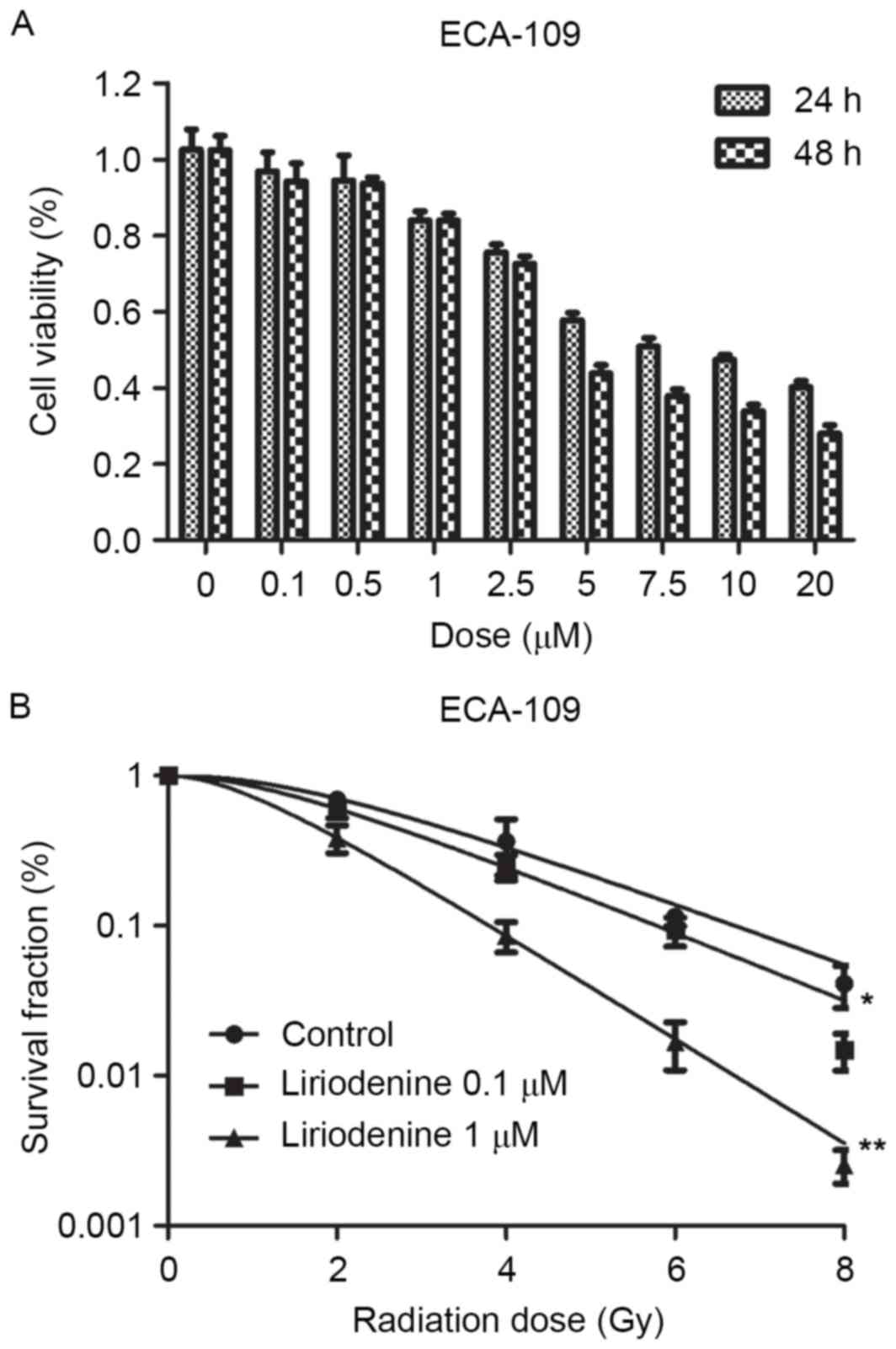
Liriodenine decreased proliferation of ESCC ECA-109
cells in a dose-dependent manner (Fig.
1A; P<0.01). Liriodenine at a low cytotoxic concentration
(0.1 and 1 µM) was selected to assess the sensitizing effects
towards radiotherapy. Liriodenine plus radiotherapy exhibited a
marked reduction in the colony formation of ESCC cells, as shown in
Fig. 1B (P<0.01). The
sensitization enhancement ratios of liriodenine concentrations at
0.1 and 1 µM were 1.11 and 1.69, and survival fraction (Gy) were
0.6 and 0.38 in ECA-109 cells according to methods in a clonogenic
assay, respectively (Table I). The
ability to form colonies was used as an indication of resistance to
IR. The combination treatment of liriodenine and radiation
significantly promoted the radiosensitivity of ECA-109 cells.
 | Table I.Radiosensitization effects of
liriodenine on esophageal squamous cell carcinoma ECA-109
cells. |
Table I.
Radiosensitization effects of
liriodenine on esophageal squamous cell carcinoma ECA-109
cells.
| ECA-109
treatment | D0 | Dq | SF2 | SER |
|---|
| Control | 2.11 | 3.13 | 0.69 |
|
| Liriodenine 0.1
µM | 1.91 | 2.17 | 0.60 | 1.11 |
| Liriodenine 1 µM | 1.25 | 1.44 | 0.38 | 1.69 |
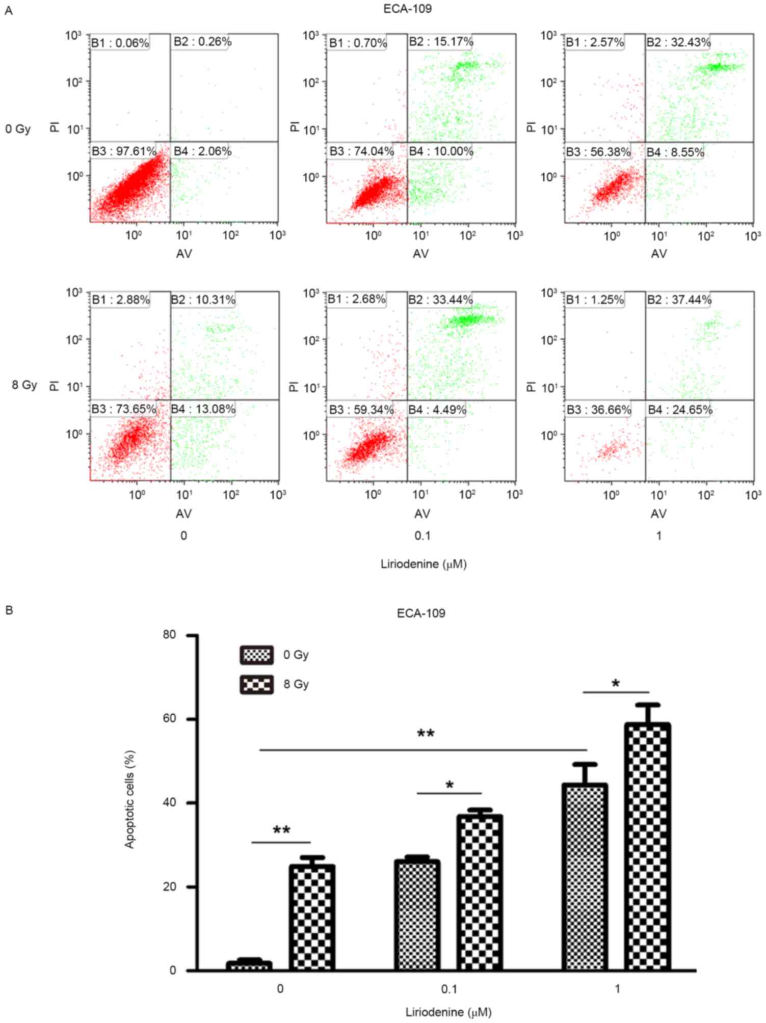
Treatment with radiation and
liriodenine together increase apoptosis and modify cell cycle
distribution
Whether liriodenine induced radiosensitization due
to increased apoptosis and changes to the cell cycle was
investigated. ECA-109 cells treated with liriodenine and IR
together exhibited significantly increased apoptosis rates compared
with either treatment alone (Fig. 2;
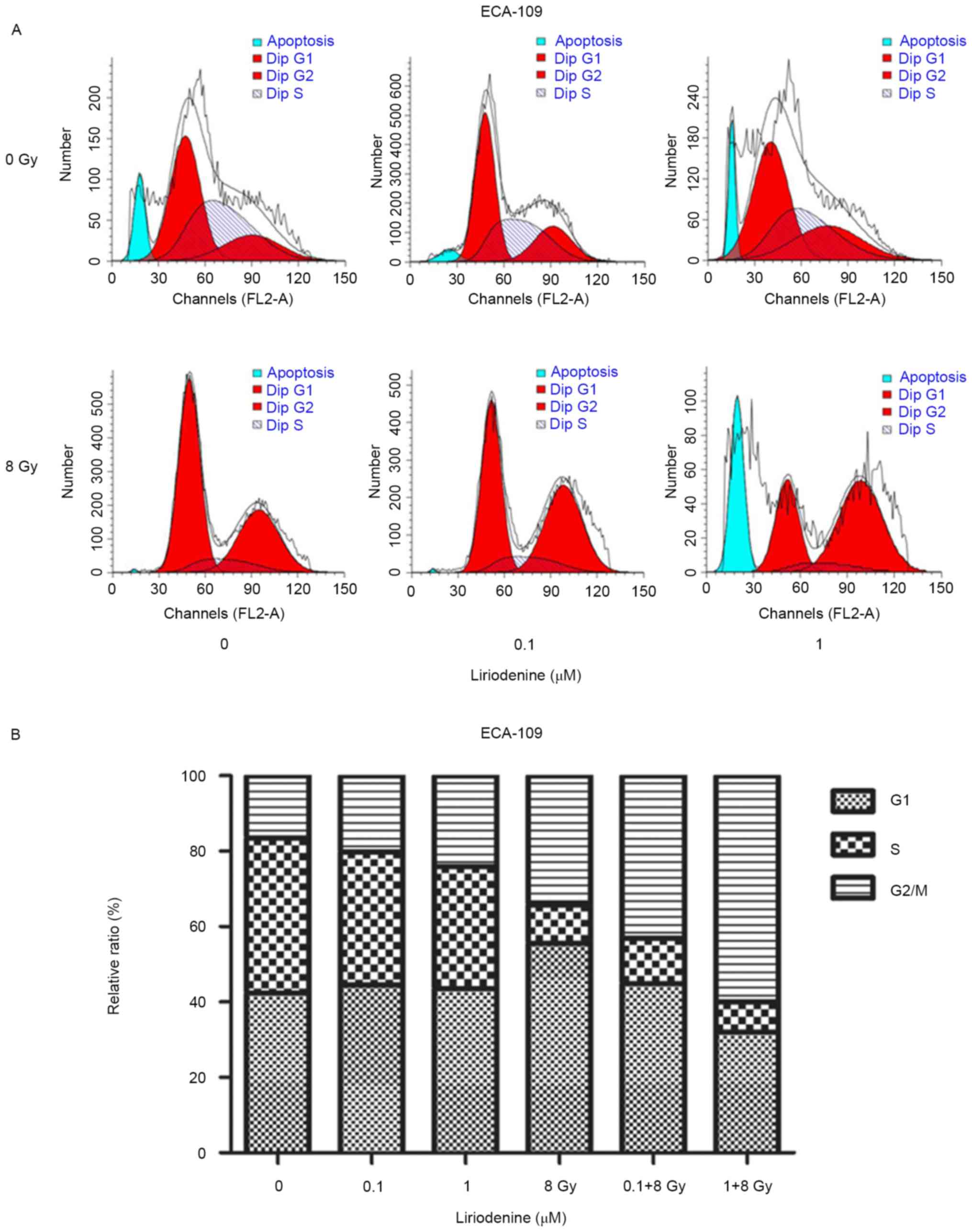
P<0.01). To investigate the antitumor effects of liriodenine,
changes in the cell cycle distribution were evaluated. The
variation of the percentage of cells in each phase of the cell
cycle is depicted in Fig. 3. Flow
cytometry analysis indicated that treatment with liriodenine and IR
resulted in a significant proportion of cells arrested in
G2/M phase (60.02% for ECA-109) (P<0.01) as compared
with single agent treatments. Exposure of the cells to liriodenine
or IR alone caused a small degree of accumulation of the cells in
G2/M phase, and a slight decrease in the proportion of
cells in G0/G1 phase. Therefore, liriodenine
appears to induce the arrest of cells in G2/M phase.
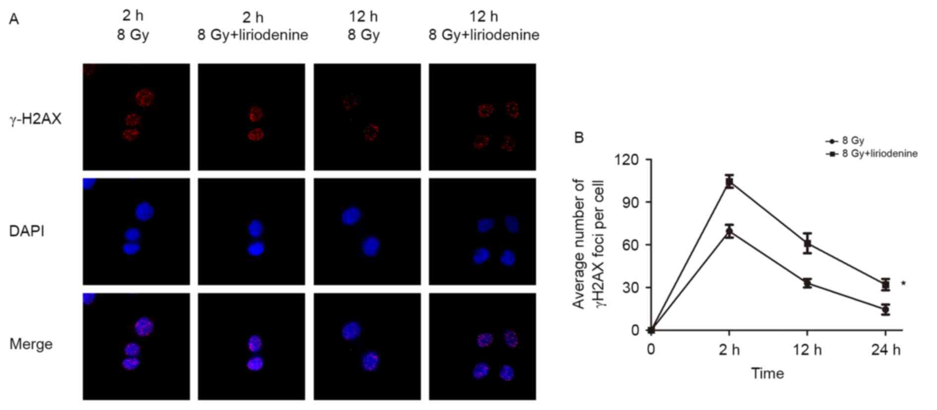
Liriodenine significantly decreases
double-strand break (DSB) repair in ECA-109 cells
ECA-109 cells with or without pretreatment with
liriodenine were harvested at 2, 12 and 24 h post-irradiation. The
mean numbers of γ-H2AX foci were calculated to examine the amount
of DSBs in cells. DNA repair following double-stranded DNA breaks
requires a variant histone protein called H2A.X. Radiation induces
phosphorylation of H2A.X at Ser139 following DNA breaks (15). Liriodenine primarily enhanced the
radiosensitivity of cells by delaying the repair of DNA damage. As
shown in Fig. 4, treatment with
liriodenine plus irradiation significantly delayed DNA DSB repair
compared with irradiation alone.
Liriodenine radiosensitizes ESCC cells
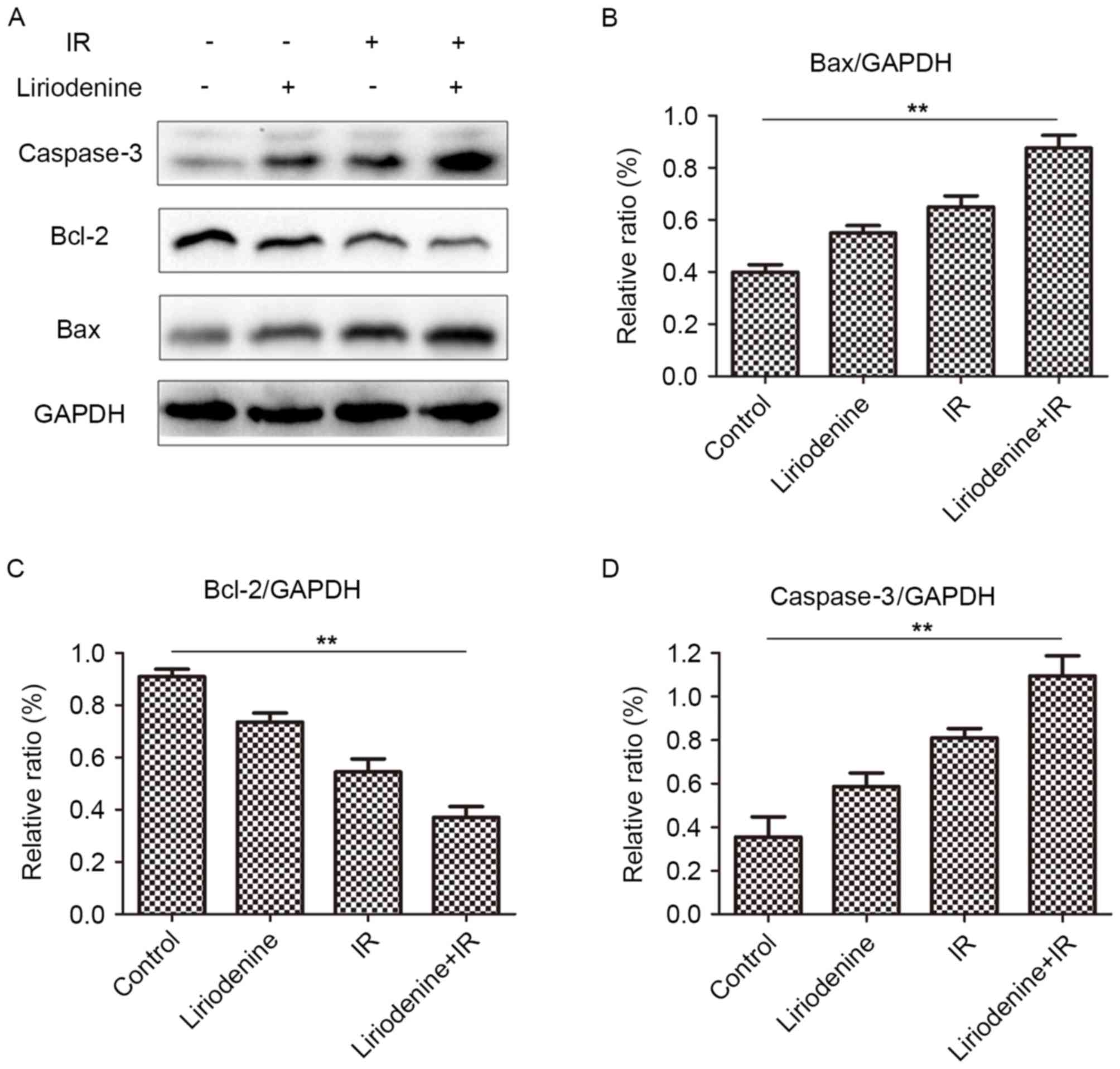
by regulating protein expression of Bax, Bcl-2 and Caspase-3
ESCC was radiosensitized by liriodenine through the
regulation of Bax, Bcl-2 and caspase-3 expression. Following
combinatorial treatment with liriodenine and radiation, changes in
the expression of Bax, Bcl-2 and caspase-3 were observed (Fig. 5). The expression of Bcl-2 protein
decreased, whereas that of Bax and caspase-3 increased in cells
that received combined treatment, compared with the cells treated
with liriodenine or radiation alone. These results indicated that
liriodenine induced changes in cell proliferation and
apoptosis.
Discussion
The aim of the present study was to evaluate the
radiosensitizing effect of liriodenine, an aporphine alkaloid from
Enicosanthellum pulchrum, Fissistigma glaucescens, Annona
glabra, Liriodendron tulipifera, and to investigate the
underlying molecular mechanisms through which this agent functions.
Liriodenine was first reported to exhibit tumor suppressive
properties in 1969 (6). In previous
studies, liriodenine was shown to inhibit cell proliferation in
hepatoma, ovarian, laryngocarcinoma and lung cancer (12–14).
Similarly, liriodenine significantly inhibited the proliferation of
melanoma cells in a cell viability assay (16). Results demonstrated that liriodenine,
particularly when combined with radiation therapy, induced a
significant decrease in the viability and proliferation of ESCC
cells in a dose-dependent manner in vitro.
It has previously been reported that liriodenine
affects cell cycle distribution. Li et al (16) revealed that liriodenine and cisplatin
significantly induced cell cycle arrest at the G2/M
phase and the S phase in human hepatoma BEL-7404 cells, while cell
cycle arrest at G2/M was also observed in human lung
adenocarcinoma A549 cells treated with liriodenine (13). In addition, combination treatment with
liriodenine and glaucine induced a moderate accumulation of cells
in G2/M phase in the human melanoma A375.S2 cells
(17). In the present study, it was
revealed that IR and nanomolar concentration of liriodenine
significantly induced cell cycle arrest at the G2/M
phase. The present results are thus in agreement with those of
previous studies.
Radiotherapy represents the primary modality of
treatment for patients with unresectable ESCC. However, despite
this treatment, 5-year survival rates remain poor, which is
attributed to radiation resistance (18). Previous studies have reported that
liriodenine may induce apoptosis in human lung, ovarian and
laryngocarcinoma cancer cells (12–14). In
the present study, compared with liriodenine or IR alone,
liriodenine and IR together strongly induced apoptosis in ESCC
cells. The radiosensitizing effect of liriodenine may be mediated
by numerous molecular pathways. The results of the present study
indicated that cell cycle arrest and apoptosis represent the
primary effects of liriodenine treatment.
High Bcl-2 expression levels have been shown to lead
to radioresistance in cancer patients, and radiation-induced
activity of Bcl-2 family members are known to cause radioresistance
in human prostate cancer cell lines (19). Similarly, high expression of Bax may
sensitize patients with head and neck cancer to radiotherapy
(20). As such, several direct Bax
activators have been use to overcome resistance to chemotherapy and
radiotherapy (21). Caspases are
central for the induction of apoptosis (22). Hypofractionated RT induces cell death
only in caspase-3-proficient breast cancer cells (23). In the present study, increased
expression of Bax and caspase-3 was observed following treatment
with liriodenine and IR, whereas that of Bcl-2 was significantly
decreased. As such, these results indicated that liriodenine may
induce apoptosis of radioresistant ESCC cells by regulating the
level of the key apoptotic-associated proteins.
In summary, the present study demonstrated that
liriodenine affects ESCC cell viability and proliferation, which
was associated with changes in cell cycle distribution. Liriodenine
may also induce apoptosis of radioresistant ESCC cell lines by
increasing expression of Bax and caspase-3, and decreasing that of
Bcl-2, indicating that liriodenine may be a promising anticancer
agent in ESCC radiation therapy.
Acknowledgements
The authors would like to thank Professor Xinchen
Sun (The First Affiliated Hospital of Nanjing Medical University,
Nanjing, China) for his technical support.
Funding
The present study was supported by Key Academic
Discipline of Jiangsu Province ‘Medical Aspects of Specific
Environments’, A Project Funded by the Priority Academic Program
Development of Jiangsu Higher Education Institutions (grant no.
JX10231801), A Phase III Clinical Trial Funded by Wuxi Hospital
Management Center (grant no. YGZXM14033).
Availability of data and materials
All data generated or analyzed during this study are
included in this published article.
Authors' contributions
GW and GC performed the experiments, analyzed the
data and wrote the manuscript. JZ participated in flow cytometry
analysis. JZ and JC helped with western blot analysis and data
collection. HZ and FZ conceived the idea, designed the study and
helped draft the manuscript. All authors read and approved the
final manuscript.
Ethics approval and consent to publish
Not applicable.
Patient consent for publication
Not applicable.
Competing interests
The authors declare that they have no competing
interests.
References
|
1
|
Ferlay J, Soerjomataram I, Dikshit R, Eser
S, Mathers C, Rebelo M, Parkin DM, Forman D and Bray F: Cancer
incidence and mortality worldwide: Sources, methods and major
patterns in GLOBOCAN 2012. Int J Cancer. 136:E359–E386. 2015.
View Article : Google Scholar : PubMed/NCBI
|
|
2
|
Kamangar F, Dores GM and Anderson WF:
Patterns of cancer incidence, mortality and prevalence across five
continents: Defining priorities to reduce cancer disparities in
different geographic regions of the world. J Clin Oncol.
24:2137–2150. 2006. View Article : Google Scholar : PubMed/NCBI
|
|
3
|
Pennathur A, Gibson MK, Jobe BA and
Luketich JD: Oesophageal carcinoma. Lancet. 381:400–412. 2013.
View Article : Google Scholar : PubMed/NCBI
|
|
4
|
Enzinger PC and Mayer RJ: Esophageal
cancer. N Engl J Med. 349:2241–2252. 2003. View Article : Google Scholar : PubMed/NCBI
|
|
5
|
Pennathur A, Farkas A, Krasinskas AM,
Ferson PF, Gooding WE, Gibson MK, Schuchert MJ, Landreneau RJ and
Luketich JD: Esophagectomy for T1 esophageal cancer: Outcomes in
100 patients and implications for endoscopic therapy. Ann Thorac
Surg. 87:1054–1055. 2009. View Article : Google Scholar
|
|
6
|
Warthen D, Gooden EL and Jacobson M: Tumor
inhibitors: Liriodenine, a cytotoxic alkaloid from Annona glabra. J
Pharm Sci. 58:637–638. 1969. View Article : Google Scholar : PubMed/NCBI
|
|
7
|
Bentley KW: Beta-phenylethylamines and the
isoquinoline alkaloids. Nat Prod Rep. 23:444–463. 2006. View Article : Google Scholar : PubMed/NCBI
|
|
8
|
Hsieh TJ, Liu TZ, Chern CL, Tsao DA, Lu
FJ, Syu YH, Hsieh PY, Hu HS, Chang TT and Chen CH: Liriodenine
inhibits the proliferation of human hepatoma cell lines by blocking
cell cycle progression and nitric oxide mediated activation of p53
expression. Food Chem Toxicol. 43:1117–1126. 2005. View Article : Google Scholar : PubMed/NCBI
|
|
9
|
Chang GJ, Wu MH, Wu YC and Su MJ:
Electrophysiological mechanisms for antiarrhythmic efficacy and
positive inotropy of liriodenine, a natural aporphine alkaloid from
Fissistigma glaucescens. Br J Pharmacol. 118:1571–1583. 1996.
View Article : Google Scholar : PubMed/NCBI
|
|
10
|
Clark AM, Watson ES, Ashfaq MK and Hufford
CD: In vivo efficacy of antifungal oxoaporphine alkaloids in
experimental disseminated candidiasis. Pharm Res. 4:495–498. 1987.
View Article : Google Scholar : PubMed/NCBI
|
|
11
|
Hufford CD, Funderburk MJ, Morgan JM and
Robertson LW: Two antimicrobial alkaloids from heartwood of
Liriodendron tulipifera L. J Pharm Sci. 64:789–792. 1975.
View Article : Google Scholar : PubMed/NCBI
|
|
12
|
Li L, Xu Y and Wang B: Liriodenine induces
the apoptosis of human laryngocarcinoma cells via the upregulation
of p53 expression. Oncol Lett. 9:1121–1127. 2015. View Article : Google Scholar : PubMed/NCBI
|
|
13
|
Chang HC, Chang FR, Wu YC and Lai YH:
Anti-cancer effect of liriodenine on human lung cancer cells.
Kaohsiung J Med Sci. 20:365–371. 2004. View Article : Google Scholar : PubMed/NCBI
|
|
14
|
Hsieh TJ, Liu TZ, Chern CL, Tsao DA, Lu
FJ, Syu YH, Hsieh PY, Hu HS, Chang TT and Chen CH: Liriodenine
inhibits the proliferation of human hepatoma cell lines by blocking
cell cycle progression and nitric oxide-mediated activation of p53
expression. Food Chem Toxicol. 43:1117–1126. 2005. View Article : Google Scholar : PubMed/NCBI
|
|
15
|
Almeida R, Fernández-Justel JM,
Santa-María C, Cadoret JC, Cano-Aroca L, Lombraña R, Herranz G,
Agresti A and Gómez M: Chromatin conformation regulates the
coordination between DNA replication and transcription. Nat Commun.
9:15902018. View Article : Google Scholar : PubMed/NCBI
|
|
16
|
Li YL, Qin QP, Liu YC, Chen ZF and Liang
H: A platinum (II) complex of liriodenine from traditional Chinese
medicine (TCM): Cell cycle arrest, cell apoptosis induction and
telomerase inhibition activity via G-quadruplex DNA stabilization.
J Inorg Biochem. 137:12–21. 2014. View Article : Google Scholar : PubMed/NCBI
|
|
17
|
Chiu CC, Chou HL, Wu PF, Chen HL, Wang HM
and Chen CY: Bio-functional constituents from the stems of
liriodendron tulipifera. Molecules. 17:4357–4372. 2012. View Article : Google Scholar : PubMed/NCBI
|
|
18
|
Efimova EV, Liang H, Pitroda SP, Labay E,
Darga TE, Levina V, Lokshin A, Roizman B, Weichselbaum RR and
Khodarev NN: Radioresistance of Stat1 overexpressingtumour cellsis
associated with suppressed apoptotic responseto cytotoxic agents
and increased IL6-IL8 signalling. Int J Radiat Biol. 85:421–431.
2009. View Article : Google Scholar : PubMed/NCBI
|
|
19
|
Ezekwudo D, Shashidharamurthy R, Devineni
D, Bozeman E, Palaniappan R and Selvaraj P: Inhibition ofexpression
of anti-apoptotic protein Bcl-2 and induction of cell death in
radioresistant human prostate adenocarcinoma cell line (PC-3)
bymethyl jasmonate. Cancer Lett. 270:277–285. 2008. View Article : Google Scholar : PubMed/NCBI
|
|
20
|
Csuka O, Remenár E, Koronczay K,
Doleschall Z and Németh G: Predictive value of p53, Bcl2 and bax in
the radiotherapy of head and neck cancer. Pathol Oncol Res.
3:204–210. 1997. View Article : Google Scholar : PubMed/NCBI
|
|
21
|
Liu Z, Ding Y, Ye N, Wild C, Chen H and
Zhou J: Direct activation of bax protein for cancer therapy. Med
Res Rev. 36:313–341. 2016. View Article : Google Scholar : PubMed/NCBI
|
|
22
|
Modjtahedi N, Giordanetto F, Madeo F and
Kroemer G: Apoptosis-inducing factor: Vital and lethal. Trends Cell
Biol. 16:264–272. 2006. View Article : Google Scholar : PubMed/NCBI
|
|
23
|
Kötter B, Frey B, Winderl M, Rubner Y,
Scheithauer H, Sieber R, Fietkau R and Gaipl US: The in vitro
immunogenic potential of caspase-3 proficient breast cancer cells
with basal low immunogenicity is increased by hypofractionated
irradiation. Radiat Oncol. 10:1972015. View Article : Google Scholar : PubMed/NCBI
|