Introduction
Hepatocellular carcinoma (HCC) is a common cancer
worldwide, particularly in East Asian countries, including Japan
(1). HCC is the sixth most commonly
occurring cancer and the third most common cause of
cancer-associated mortality worldwide in 2012 (2). Multiple risk factors have been
associated with the occurrence of HCC, including chronic liver
injury due to hepatitis B virus (HBV) or hepatitis C virus (HCV)
infection, autoimmune liver disease, drug-induced liver injury,
alcohol and aflatoxin B exposure (2–5). HCC had
one of the worst prognoses among any cancer with a 5-year survival
rate of 15–25% in United States and East Asian countries from 2007
to 2010, partially due to the resistance to chemotherapy and a high
recurrence rate (6,7). One of the most prevalent reasons for a
poor prognosis is the difficulty in early detection, and as a
result, curative therapy is no longer feasible at the time of
detection, due to intrahepatic and extrahepatic metastases
(2).
To assist the diagnosis of HCC, imaging techniques
used in screening, including ultrasonography, computed tomography
(CT) and/or magnetic resonance imaging (MRI), are notably
beneficial (2). However, in the case
of early HCC, the diagnosis of small lesions is relatively
inaccurate (8), and repeated
examination is costly. Other common approaches used in screening
for HCC in high-risk patients are serum tumor markers, including
α-fetoprotein (AFP) and protein induced by vitamin K absence or
antagonists-II (PIVKA-II), which can be measured simultaneously in
blood samples obtained for other liver function tests (9). However, the sensitivity and specificity
of high serum AFP and PIVKA-II levels for HCC are reported to range
from 39–64 and 76–91%, and 41–77 and 72–98%, respectively (10). Therefore, additional biomarkers that
can be used complementarily are required, particularly those
associated with early HCC.
MicroRNAs (miRNAs) are small, non-coding RNAs 18–25
nucleotides in length that suppress the translation of the target
mRNAs by binding to their 3′ untranslated region (11,12).
miRNAs control a number of important biological processes,
including cell proliferation, differentiation and development
(13–15), and specific miRNAs function as
oncogenes or tumor suppressors (16). The expression profiles of human
miRNAs indicate that specific miRNAs such as miR-15 and miR-16,
let-7, miR-34 are deregulated in cancer, and are differentially
expressed in various carcinoma types, including gastrointestinal,
urological, gynecological and lung cancer (17). Additionally, with respect to HCC, it
has been reported that the expression levels of a number of miRNAs
differ between cancerous and noncancerous specimens from radical
resection of patients with HCC (18). In our previous investigation, we
reported that the miRNA profile is different between HCC and normal
liver cell lines (19), and it is
hypothesized to exert significance as biomarkers (20).
It has previously been identified that circulating
miRNAs can exist stably in numerous body fluids, including the
peripheral blood (21), which can be
used for the diagnosis, evaluation and prognosis of colorectal,
esophageal, gastric and pancreatic cancer (22). When released from cells and tissues,
miRNA exists in exosome-encapsulated form or bound to protein or
lipid in the serum (23). The
research demonstrated that miRNAs are stable and detectable in the
serum and are not degraded by RNase. Since serum can be obtained
noninvasively and the miRNAs exhibit specificity to the disordered
tissue, application of circulating miRNA in diagnosis is expected;
however, its biological significance is unknown. Therefore, the
present study investigated the expression profile of circulating
miRNAs using serum samples from patients with HCV-associated HCC,
and analyzed whether a specific circulating miRNA could help in the
detection of early HCC.
Materials and methods
Patients and samples
In order to identify biomarkers of HCV-associated
HCC from among the candidate miRNAs, the present study examined
miRNA changes between the pre- and post-treatment serum of patients
with early stage (stage I or II) HCC according to the
Tumor-Node-Metastasis classification based on the criteria of the
Liver Cancer Study Group of Japan (24). Paired samples were obtained from a
total of 12 patients with HCC, who underwent curative treatment,
such as radiofrequency ablation or hepatectomy, in Kagawa
University Hospital (Kagawa, Japan), from April 2013 to April 2015.
The characteristics of the patients are summarized in Table I. All patients had HCC with chronic
hepatitis (CH) or liver cirrhosis (LC) due to HCV infection without
any other liver diseases, such as HBV infection and alcoholic,
autoimmune or metabolic liver diseases.
 | Table I.Clinical characteristics of
participants in the microarray analysis. |
Table I.
Clinical characteristics of
participants in the microarray analysis.
| Case | Sex | Age (years) | Genotype | HCV-RNA (log10
IU/ml) | AST (IU/l) | ALT (IU/l) | Platelet
(×109/l) | AFP (ng/ml) | AFP-L3 (%) | PIVKA-II
(AU/ml) | TNM stage | Treatment |
|---|
| 1 | M | 56 | 2a | 5.2 | 49 | 38 | 6.6 | Negative | Negative | Negative | T1N0M0 | RFA |
| 2 | M | 61 | 1b | 4.9 | 60 | 60 | 6.2 | 78 | 17.8 | Negative | T2N0M0 | Hepatectomy |
| 3 | M | 82 | 2b | 4.8 | 29 | 14 | 14.8 | Negative | Negative | 113 | T2N0M0 | Hepatectomy |
| 4 | F | 77 | 1b | 6.9 | 45 | 29 | 15.6 | Negative | Negative | 253 | T2N0M0 | RFA |
| 5 | M | 68 | 1b | 5.2 | 44 | 20 | 7.6 | Negative | Negative | 371 | T1N0M0 | RFA |
| 6 | M | 57 | 1b | 5.0 | 21 | 13 | 18.6 | Negative | Negative | 223 | T2N0M0 | Hepatectomy |
| 7 | M | 76 | 2a | 4.6 | 23 | 14 | 20.3 | Negative | Negative | Negative | T1N0M0 | RFA |
| 8 | M | 83 | 1b | 6.2 | 17 | 15 | 20.0 | Negative | Negative | 358 | T2N0M0 | RFA |
| 9 | F | 76 | 1b | 5.0 | 80 | 54 | 6.5 | 28 | Negative | Negative | T2N0M0 | RFA |
| 10 | F | 79 | 1b | 5.2 | 74 | 71 | 21.2 | Negative | Negative | Negative | T1N0M0 | RFA |
| 11 | M | 69 | 2a | 5.9 | 91 | 74 | 10.1 | 20 | Negative | 41 | T1N0M0 | RFA |
| 12 | F | 70 | 1b | 6.0 | 89 | 80 | 6.0 | 82 | 21 | Negative | T2N0M0 | RFA |
The present study examined candidate biomarkers
using paired serum samples from 12 patients with HCC pre- and
post-curative treatment. The pre-treatment samples were collected
prior to the first curative treatment, and the post-treatment
samples were collected following confirmation that there was no
long-term recurrence for at least 1 year following treatment. Tumor
marker measurement was performed every three months in all cases to
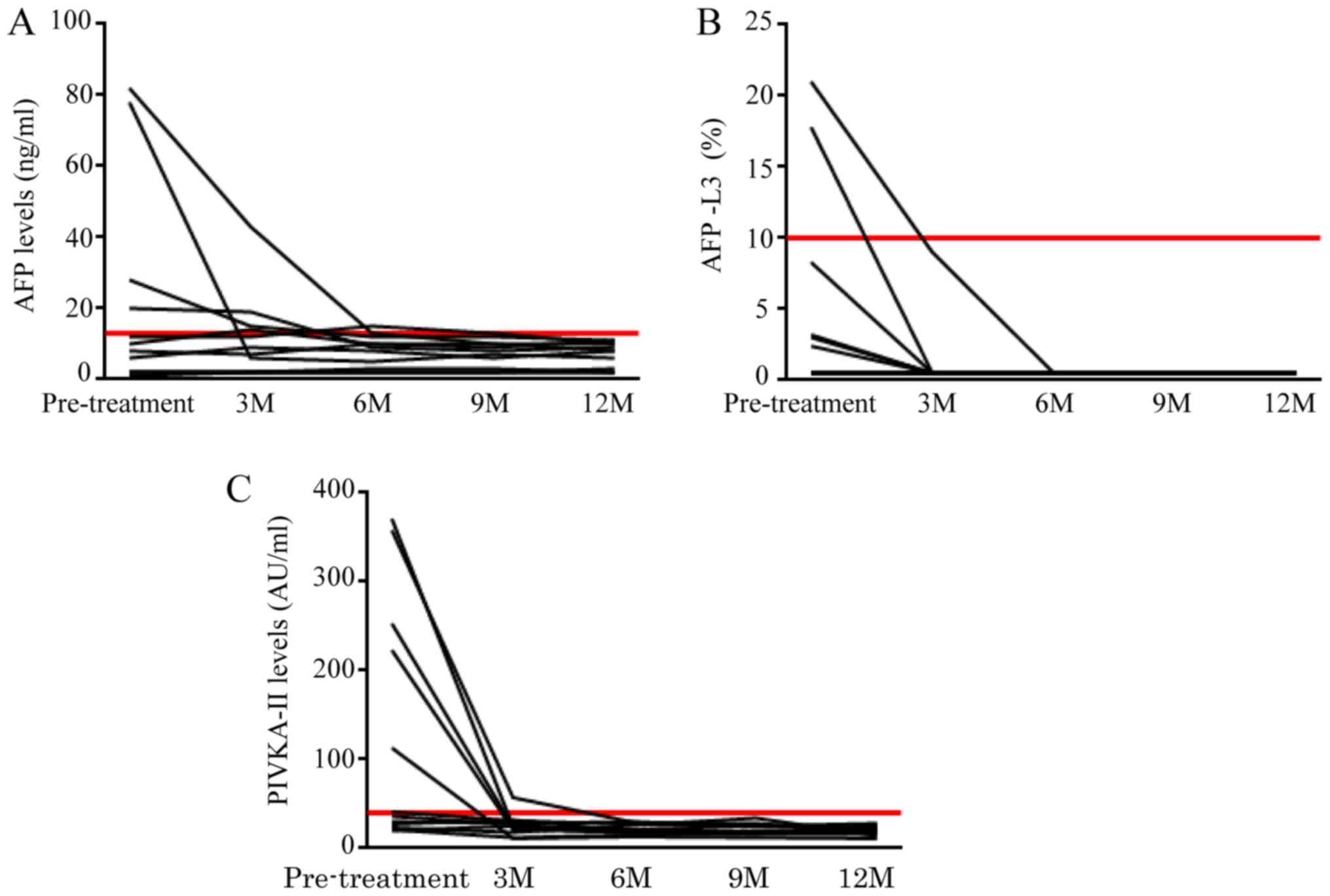
ensure there was no recurrence. Prior to treatment, 9 patients were
positive for at least one tumor maker (AFP, AFP-L3 and/or
PIVKA-II), yet were negative for all markers for at least 6 months
following treatment (Fig. 1A-C).
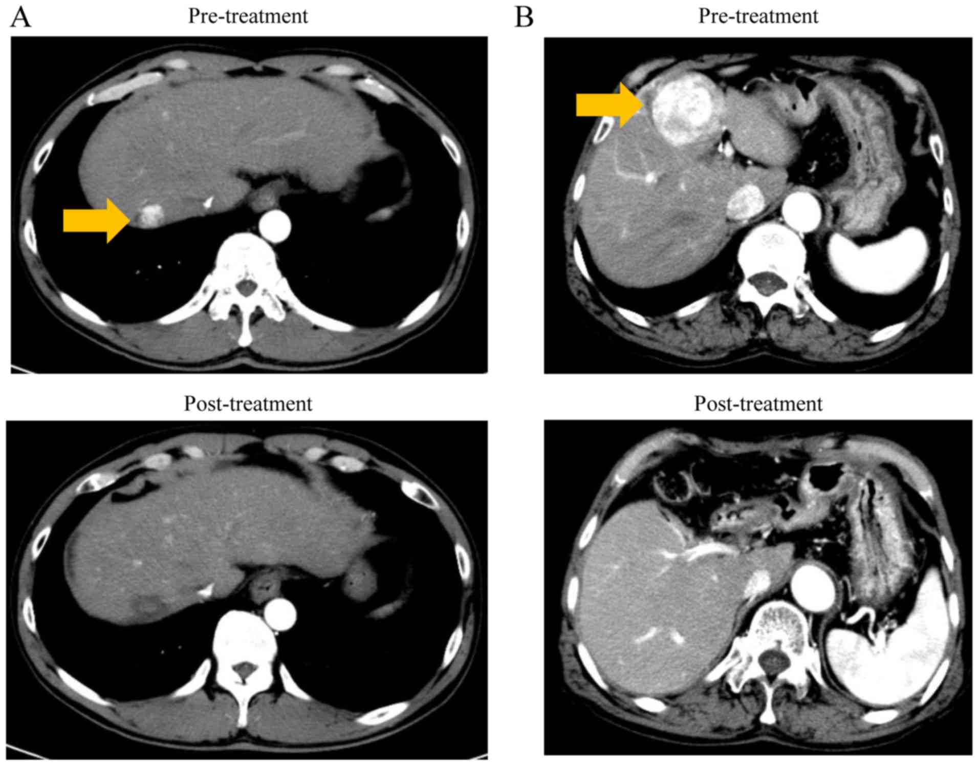
Additionally, imaging tests, including ultrasonography, dynamic CT
and/or MRI examination, were performed every three months and it
was confirmed that there was no recurrence. Dynamic CT images prior
to and 1 year following treatment in cases 1 and 2 are presented in
Fig. 2.
In the second experiment, the expression of the
specific miRNAs was examined using multiple serum samples from
individual patients with various liver diseases. A total of 40
individuals were enrolled including 10 age and sex matched patients
with CH, 10 patients with LC, 10 patients with early stage (stage I
or II) HCC and 10 patients with advanced stage (stage IV) HCC.
Characteristics of the patients are summarized in Table II. All subjects were patients with
liver disease associated with HCV infection, and patients with
other liver diseases were excluded. Serum samples were collected
from patients with HCC from the time of first diagnosis with HCC,
and patients with CH or LC prior to receiving antiviral therapy for
HCV.
| Table II.Clinical characteristics of
participants in the reverse transcription-quantitative polymerase
chain reaction analysis. |
Table II.
Clinical characteristics of
participants in the reverse transcription-quantitative polymerase
chain reaction analysis.
|
Characteristics | CH-C | LC-C | HCC-C stage I or
II | HCC-C stage IV |
|---|
| Individuals
(n) | 10 | 10 | 10 | 10 |
| Male/female
(n) | 7/3 | 7/3 | 7/3 | 7/3 |
| Mean age
(years) | 67.4±4.4 | 67.2±5.1 | 68.0±4.8 | 67.1±5.1 |
| Laboratory data
(median) |
|
|
|
|
| AST
(IU/l) | 40 (20–75) | 45 (13–77) | 50 (29–106) | 52 (40–485) |
| ALT
(IU/l) | 32 (13–99) | 43 (16–75) | 45 (17–104) | 34 (20–180) |
| Alb
(g/dl) | 4.1 (2.9–4.7) | 4.0 (3.4–4.7) | 3.9 (3.4–5.2) | 3.6 (2.6–3.8) |
| T.Bil
(mg/dl) | 0.7 (0.3–1.2) | 1.0 (0.3–2.1) | 1.1 (0.6–2.0) | 0.8 (0.4–2.8) |
| PT
(%) | 87 (59–130) | 78 (51–98) | 99 (72–111) | 79 (46–110) |
| Plt
(×104/mm3) | 18.0
(13.0–21.0) | 8.7 (5.9–9.8) | 11.3
(7.2–14.0) | 15.9
(8.4–22.9) |
| Tumor marker
(n) |
|
|
|
|
| AFP
positive | 1 | 2 | 2 | 7 |
| AFP-L3
positive | 0 | 0 | 1 | 8 |
|
PIVKA-II positive | 0 | 0 | 4 | 10 |
|
Negative | 9 | 8 | 5 | 0 |
| Child-Pugh score
(n) |
|
|
|
|
| A | 10 | 9 | 10 | 3 |
| B | 0 | 1 | 0 | 6 |
| C | 0 | 0 | 0 | 1 |
| FIB-4 index
(median) | 2.87
(1.17–3.78) | 5.12
(3.36–10.68) | 5.09
(2.69–9.21) | 4.03
(2.61–7.97) |
Written informed consent was obtained from all
participants, and the present study was approved by the Ethics
Committee of Kagawa University Hospital (Kagawa, Japan) (Ethics
approval Heisei 22–063).
Plasma preparation
Whole blood samples (5 ml) were collected from each
individual directly into RNase free tubes, followed by
centrifugation at 1,500 × g for 15 min at 4°C. The samples with
signs of hemolysis or chyle were excluded from the present study.
Each serum sample was immediately transferred to a RNase free tube
and stored at −80°C until subsequent analysis.
Total RNA extraction
RNA from total serum was extracted with a miRNeasy
Serum/Plasma kit (Qiagen GmbH, Hilden, Germany), according to the
manufacturer's protocol. To ensure RNA quality, only RNA sample
that exhibited A260/280 ratios between 1.9–2.1 were
selected. The A260/280 ratios were evaluated using the
Agilent 2100 Bioanalyzer (Agilent Technologies, Inc., Santa Clara,
CA, USA). RNA concentrations were measured using a NanoDrop 2000
spectrofluorometer (Thermo Fisher Scientific, Inc., Waltham, MA,
USA) and each sample was diluted with RNase free water.
miRNA microarray analysis
The RNA quantity was measured using a RNA 6000 Nano
kit (Agilent Technologies, Inc.), and the samples were labeled
using a miRCURY Hy3 Power Labeling kit (Exiqon; Qiagen GmbH) and
hybridized to the human miRNA Oligo Chip (v.21; Toray Industries,
Tokyo, Japan), which can analyze 2,555 miRNAs. Scanning was
performed using the 3D-Gene Scanner 3000 (Toray Industries, Inc.,
Tokyo, Japan). The 3D-Gene extraction version 1.2 software (Toray
Industries, Inc.) was used to calculate the raw signal intensity of
the images. The raw data were analyzed using the GeneSpring GX 10.0
software (Agilent Technologies, Inc.) to assess miRNA expression.
Quantile normalization was performed on raw data that were greater
than the background level.
Reverse transcription-quantitative
polymerase chain reaction (RT-qPCR) for miRNA validation
Due to the possibility of false positive results
obtained from the miRNA array analysis, the present study performed
qPCR using the same samples prior to and following treatment.
Furthermore, RT-qPCR was performed for the analysis of the
expression levels of specific miRNAs using 40 serum samples from
patients with HCV-associated liver diseases, including CH, LC and
HCC.
Initially, Caenorhabditis elegans miRNA,
cel-miR-39 (miRNeasy Serum/Plasma Spike-in control; Qiagen GmbH)
was added as an exogenous control during the process of total RNA
extraction. TaqMan microRNA assays (Applied Biosystems; Thermo
Fisher Scientific, Inc.) were adopted to determine the expression
levels of four miRNAs (assay ID: 002198 and target sequence:
5′-UCCCUGAGACCCUUUAACCUGUGA-3′ for hsa-miR-125a-5p; assay ID:
002340 and target sequence: 5′-UGAGGGGCAGAGAGCGAGACUUU-3′ for
hsa-miR-423-5p; assay ID: 46440 and target sequence:
5′-AGCCGCGGGGAUCGCCGAGGG-3′ for hsa-miR-3648; and assay ID: 000200
and target sequence: 5′-UCACCGGGUGUAAAUCAGCUUG-3′ for cel-miR-39).
To examine another two miRNAs, TaqMan Advanced miRNA Assays were
used (assay ID: 479553_mir and target sequence:
5′-CCCCGGGAACGUCGAGACUGGAGC-3′ for hsa-miR-1247-3p; assay ID:
479574_mir and target sequence: 5′-UCUCACUGUAGCCUCGAACCCC-3′ for
hsa-miR-1304-3p; and assay ID: 478293_mir and target sequence:
5′-UCACCGGGUGUAAAUCAGCUUG-3′ for cel-miR-39). miRNAs were reverse
transcribed using a TaqMan microRNA Reverse Transcription kit
(Applied Biosystems; Thermo Fisher Scientific, Inc.) and a TaqMan
Advanced miRNA cDNA Synthesis kit (Applied Biosystems; Thermo
Fisher Scientific, Inc.). qPCRs were performed using a MicroAmp
Fast Optical 96-Well Reaction Plate (Applied Biosystems; Thermo
Fisher Scientific, Inc.), and each well contained cDNA, 20X qPCR
assay, nuclease-free water and TaqMan Fast Advanced Master mix
(Applied Biosystems; Thermo Fisher Scientific, Inc.), according to
manufacturer's protocol. Using the ViiA7 Real-Time PCR System
(Applied Biosystems; Thermo Fisher Scientific, Inc.), samples were
denatured by incubation at 95°C for 20 sec. This was followed by 40
cycles of 1 sec at 95°C and 20 sec at 60°C.
The raw expression level was determined by the cycle
number at which the reaction crossed a predetermined quantification
cycle (Cq) identified for the miRNA probe. For relative expression
of each miRNA in each sample is determined using 2−ΔΔCq
method (25). For the validation of
miRNA changes between the pre- and post-treatment serum, the values
were calculated according to the following formula:
ΔCq=Cqtarget miRNA-Cqcel-miR-39, and
ΔΔCq=ΔCqpost-treatment sample-ΔCqpre-treatment
sample. For the analysis of the expression levels of specific
miRNAs from individual patients with various liver diseases, the
values were calculated according to the following formula;
ΔCq=Cqtarget miRNA-Cqcel-miR-39, and
ΔΔCq=ΔCq-meanΔCq of control group patients.
Furthermore, the expression profile of each
differentially-expressed miRNA was used to create receiver operator
characteristic (ROC) curves. This method displays the
discriminatory accuracy of the marker for distinguishing between
the non-HCC (patients with CH and LC) and HCC (patients with early
and advanced stage HCC) groups. Additionally, by using the ROC
curve, the area under the curve (AUC) value and the optimal cutoff
value were calculated.
Statistical analysis
All statistical analyses were performed using Prism
software version 6.0 (Graph Pad Software, Inc., La Jolla, CA, USA).
Normally distributed data were expressed as mean ± standard
deviation. Skewed data were described by the median and range. The
difference between normally distributed numeric variables was
analyzed by the Student's t-test, while non-normally distributed
variables were analyzed by Mann-Whitney U test. When comparing
multiple groups, one-way analysis of variance was conducted,
followed by Dunnett post-hoc test. All P-values were two-sided, and
P<0.05 was considered to indicate a statistically significant
difference.
Results
miRNA analysis pre- and post-curative
treatment
To determine miRNA changes between pre- and
post-curative treatment serums from the patients with early stage
HCC, the present study exhaustively analyzed 2,555 miRNA molecules
using a microarray. A total of 5 miRNAs were identified to be the
most significantly changed molecules (P<0.05), including
miR-125a-5p, miR-423-5p, miR-1247-3p, miR-1304-3p and miR-3648, all
of which were downregulated (Table
III).
| Table III.Serum miRNA levels were significantly
different between pre- and post-treatment. |
Table III.
Serum miRNA levels were significantly
different between pre- and post-treatment.
| miRNAs | Fold change
post-/pre-treatment | SD | P-value | Chromosome
location |
|---|
| miR-125a-5p | 0.74 | 0.23 | 0.00989 | 19 |
| miR-423-5p | 0.61 | 0.20 | 0.00151 | 17 |
| miR-1247-3p | 0.73 | 0.19 | 0.0066 | 14 |
| miR-1304-3p | 0.69 | 0.26 | 0.00952 | 11 |
| miR-3648 | 0.63 | 0.34 | 0.00384 | 21 |
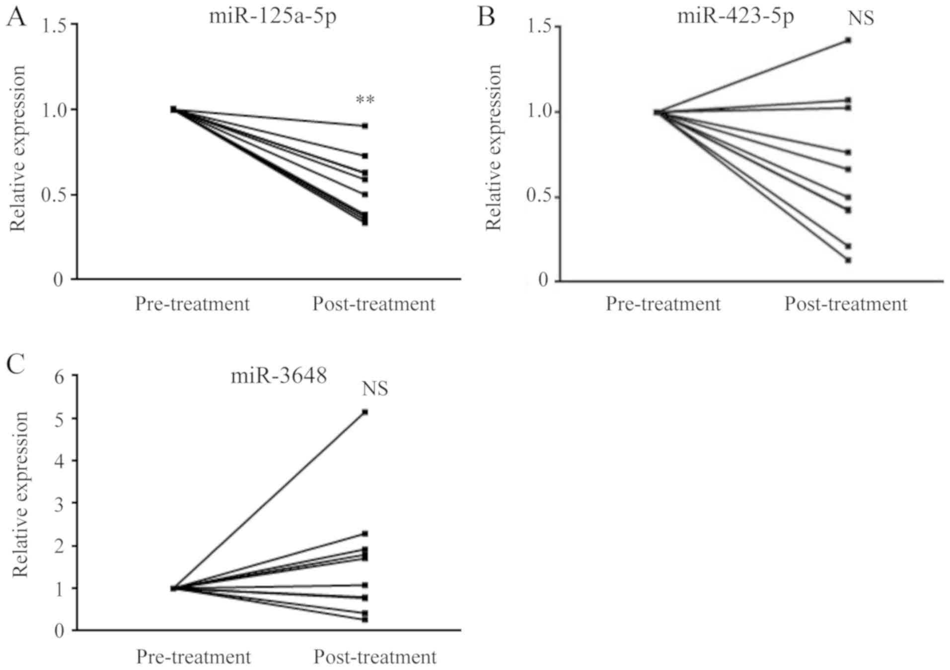
The 5 miRNAs selected by exhaustive analysis were
quantified by RT-qPCR. miR-125a-5p was downregulated post-treatment
in all 12 cases (Fig. 3A) and the
relative quantity (RQ) value was 0.57±0.27 (P<0.01). miR-423-5p
exhibited the opposite trend in expression in a few cases; however,
no significant change was observed by RT-qPCR analysis (Fig. 3B), and the RQ value was 0.71±0.56
(not significant). For miR-3648, the measured values varied from
case to case, and a certain trend could not be identified (Fig. 3C). miR-1247-3p and hsa-miR-1304-3p
did not yield stable results in the method used and could not be
detected in more than half of the cases.
Upregulation of miR-125a-5p in the
serum of patients with HCC
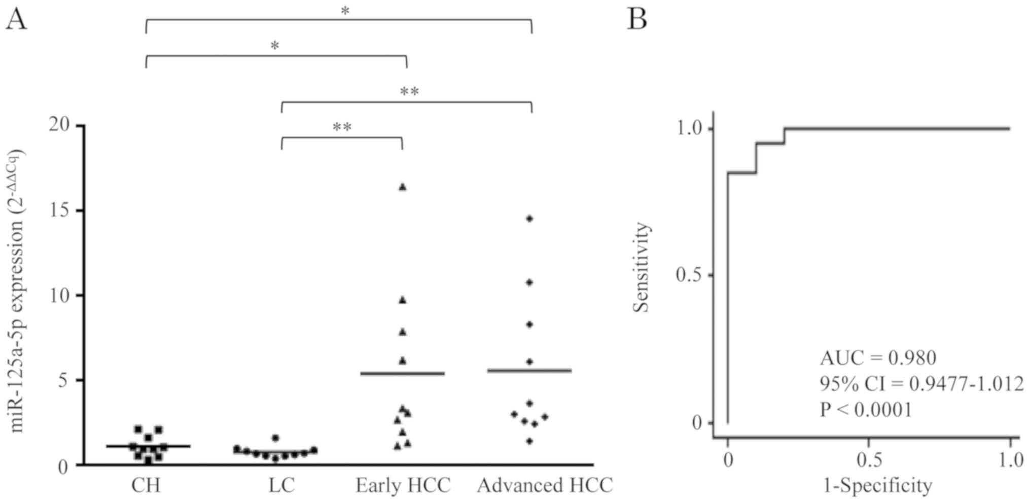
In order to determine whether miR-125a-5p was
differentially expressed in HCV-associated liver diseases, the
serum levels of miR-125a-5p in patients with HCV-associated CH, LC
and HCC were measured (Fig. 4A). The
results demonstrated that the miR-125a-5p expression was
significantly upregulated in patients with advanced stage HCC,
compared with patients with CH, with a RQ value of 5.60±4.34
(P<0.05). Additionally, miR-125a-5p was also significantly
upregulated in patients with early stage HCC when, compared with
patients with CH, with a RQ value of 5.43±4.84 (P<0.05). There
was no difference in miR-125a-5p expression between patients with
LC and patients with CH, with a RQ value of 0.82±0.34 (not
significant). Similar results were obtained in comparison with
patients with LC, as the levels of miR-125a-5p expression were
significantly upregulated in patients with advanced stage HCC,
compared with patients with LC, with a RQ value of 7.36±5.69
(P<0.01). Additionally, miR-125a-5p was significantly
upregulated in patients with early stage HCC, compared with
patients with LC, with a RQ value of 7.11±6.35 (P<0.01).
Diagnostic value of miR-125a-5p in
serum
In order to evaluate the diagnostic value of serum
miRNA-125a-5p in discriminating the HCC group (patients with early
and advanced stage HCC) from the non-HCC group (patients with CH
and LC), the optimal cutoff value for miR-125a-5p was the ROC curve
based on the RT-qPCR data. The AUC was 0.980 and the optimal cutoff
value was 2.476, which demonstrated a sensitivity of 0.8 and a
specificity of 1.0 (Fig. 4B).
Discussion
In the present study, serum samples obtained from
pre- and post-treatment patients with HCC, miRNAs underwent a
comprehensive examination and a number of miRNAs were selected as
biomarker candidates for HCC. The present study demonstrated that
serum miR-125a-5p levels are significantly reduced in
post-treatment samples, and that the levels in patients in the
early and advanced stages of HCC were significantly increased,
compared with patients with HCV-associated chronic liver disease.
These results indicated that miR-125a-5p has potential as a
biomarker for early detection of HCV-associated HCC and evaluation
following treatment. Additionally, among patients with early stage
HCC, the elevation of miR-125a-5p level was observed in 4/5 cases,
which were negative for tumor markers. These results indicated that
serum miR-125a-5p is a valuable biomarker that could be in
conjunction with conventional HCC tumor markers, including AFP and
PIVKA-II. These events may reflect a longer increase in serum
miR-125a-5p at cancer initiation rather than during
progression.
Among the characteristic miRNAs contained in the
serum of patients with cancer, circulating miR-21 plays an
important role and has been reported to be associated with various
carcinoma types, including colorectal (26), pancreatic (27), ovarian (28) and pharyngeal cancer (29). It was also reported that miR-21 is
upregulated in the serum of patients with HCC (20,30).
Furthermore, miR-718 has also been reported as a characteristic
miRNA in the serum of patients with HCC (31); however, in the present study,
comprehensive analysis revealed no significant change in miR-21 and
miR-718. The discrepancy between the present data and previous
reports may be explained by the difference in the methods used, as
in the present study, 2,555 miRNAs were comprehensively analyzed,
which was a notably larger number of molecules, compared with the
previous reports.
miR-125a is located at 19q13, and has been reported
that miR-125a targeted genes that suppress and control cancer,
including tumor protein P53 (32),
cyclin dependent kinase inhibitor 1A (33), Erb-B2 receptor tyrosine kinase 2
(ERBB2) and ERBB3 (34). In HCC cell
lines, studies also reported that miR-125a inhibits the migration
and invasion via suppression of phosphoinositide
3-kinase/AKT/mechanistic target of rapamycin kinase signaling
pathway (35), and miR-125a-5p
inhibits cell proliferation by downregulation of ERBB3 (36). The results demonstrated that
miR-125a-5p may serve a tumor-suppressive role in HCC
carcinogenesis.
Previous studies also demonstrated that the
expression of miR-125a-5p is downregulated in a number of human
cancer types, including breast (37), ovarian (38), lung (39) and gastric cancer (40) tissues. miR-125a-5p is also
downregulated in HCC tissues and may function as a tumor suppressor
(41,42). However, the clinical significance of
miR-125a-5p in serum of patients with HCC has yet to be completely
elucidated. Additionally, it was previously unclear whether miRNA
from cancer tissue are up- or downregulated in the serum of
patients with HCC; however, the present study demonstrated that
miR-125a-5p is upregulated in the serum of patients with HCC. Our
hypothesis is that investigating the biological role of miR-125a-5p
may be beneficial in understanding the pathology of HCC.
However, it should be considered that the present
study examined the serum miR-125a-5p level in patients with chronic
liver disease and only HCC caused by HCV infection. However, it has
previously been reported that miR-125a-5p can also target a viral
sequence and interfere with the expression of HBV surface antigen
(43). Another independent study
also reported that miR-125a-5p levels are correlated with HBV DNA
concentrations in the liver and plasma, and that miR-125a-5p is
upregulated in the patients with high viral load (44). As the level of miR-125a-5p may change
due to the viral load and the degree of HBV-induced CH, the present
study excluded the cases with persistent HBV infection. Therefore,
the present study selected only the samples with HCV-associated
HCC. Another limitation of the present study is that miR-125a-5p
levels in healthy controls without liver diseases were not
examined. Therefore, future investigations should examine whether
the miR-125a-5p measurement is different in HBV-associated liver
diseases, compared with healthy controls. However, in countries
such as Japan, where the majority of HCC is caused by HCV
infection, miR-125a-5p may be beneficial for diagnosis and
follow-up following treatment.
In conclusion, miRNA expression profile in the serum
of patients with HCV-associated HCC, and in particular, the serum
miR-125a-5p levels changed pre- and post-treatment in patients with
HCV-associated HCC. Irrespective of the clinical stage, the
miR-125a-5p level was identified to be elevated in the serum of
patients with HCC. Therefore, serum miR-125a-5p may serve as a
noninvasive biomarker for the diagnosis of early carcinogenesis in
HCV-associated chronic liver diseases.
Acknowledgements
The authors would like to thank Ms. Kayo Hirose, Ms.
Keiko Fujikawa, Ms. Miwako Watanabe, Ms. Megumi Okamura and Ms.
Fuyuko Kokado (Department of Gastroenterology and Neurology, Kagawa
University, Japan) for their skillful technical assistance.
Funding
No funding was received.
Availability of data and materials
The datasets used and/or analyzed during the current
study are available from corresponding author on reasonable
request.
Authors' contributions
KOu designed the study and wrote the manuscript.
KOu, KF and AM carried out the major experiments. HI, MN, TT and TS
analyzed and interpreted the data. TN, HY, SM, JT, HK, KOk, YS and
TM designed the study and conducted the experiments. All authors
read and approved the final version of the manuscript.
Ethics approval and consent to
participate
The present study was approved by the Ethics
Committee of Kagawa University Hospital (Kagawa, Japan) (Ethics
approval: Heisei 22-063). Written informed consent was obtained
from all participants.
Patient consent for publication
The patients provided written informed consent for
the publication of any data.
Competing interests
The authors declare that they have no competing
interests.
Glossary
Abbreviations
Abbreviations:
|
HCC
|
hepatocellular carcinoma
|
|
miRNA
|
microRNA
|
|
HCV
|
hepatitis C virus
|
|
CH
|
chronic hepatitis
|
|
LC
|
liver cirrhosis
|
|
HBV
|
hepatitis B virus
|
|
CT
|
computed tomography
|
|
MRI
|
magnetic resonance imaging
|
|
AFP
|
α-fetoprotein
|
|
PIVKA-II
|
protein induced by vitamin K absence
or antagonists-II
|
|
qPCR
|
quantitative polymerase chain
reaction
|
References
|
1
|
McGlynn KA and London WT: The global
epidemiology of hepatocellular carcinoma: Present and future. Clin
Liver Dis. 15:223–243. 2011. View Article : Google Scholar : PubMed/NCBI
|
|
2
|
Forner A, Llovet JM and Bruix J:
Hepatocellular carcinoma. Lancet. 379:1245–1255. 2012. View Article : Google Scholar : PubMed/NCBI
|
|
3
|
El-Seraq HB: Epidemiology of viral
hepatitis and hepatocellular carcinoma. Gastroenterology.
142:1264–1273.e1. 2012. View Article : Google Scholar : PubMed/NCBI
|
|
4
|
Yoeman AD, Al-Chalabi T, Karani JB,
Quaglia A, Devlin J, Mieli-Vergani G, Bomford A, O'Grady JG,
Harrison PM and Heneghan MA: Evaluation of risk factors in the
development of hepatocellular carcinoma in autoimmune hepatitis:
Implications for follow-up and screening. Hepatology. 48:863–870.
2008. View Article : Google Scholar : PubMed/NCBI
|
|
5
|
Zhang W, He H, Zang M, Wu Q, Zhao H, Lu
LL, Ma P, Zheng H, Wang N, Zhang Y, et al: Genetic features of
aflatoxin-associated hepatocellular carcinoma. Gastroenterology.
153:249–262.e2. 2017. View Article : Google Scholar : PubMed/NCBI
|
|
6
|
Altekruse SF, Henley SJ, Cucinelli JE and
McGlynn KA: Changing hepatocellular carcinoma incidence and liver
cancer mortality rates in the United States. Am J Gastroenterol.
109:542–553. 2014. View Article : Google Scholar : PubMed/NCBI
|
|
7
|
Kim SR, Kudo M, Hino O, Han KH, Chung YH
and Lee HS; Organizing Committee of Japan-Korea Liver Symposium, :
Epidemiology of hepatocellular carcinoma in Japan and Korea. A
review. Oncology. 75 (Suppl 1):S13–S16. 2008. View Article : Google Scholar
|
|
8
|
Murakami T, Kim T, Oi H, Nakamura H,
Igurashi H, Matsushita M, Okamura J and Kozuka T: Detectability of
hypervascular hepatocellular carcinoma by arterial phase images of
MR and spiral CT. Acta Radiol. 36:372–376. 1995. View Article : Google Scholar : PubMed/NCBI
|
|
9
|
Park SJ, Jang JY, Jeong SW, Cho YK, Lee
SH, Kim SG, Cha SW, Kim YS, Cho YD, Kim HS, et al: Usefulness of
AFP, AFP-L3, and PIVKA-II, and their combinations in diagnosing
hepatocellular carcinoma. Medicine (Baltimore). 96:e58112017.
View Article : Google Scholar : PubMed/NCBI
|
|
10
|
Tomimaru Y, Eguchi H, Nagano H, Wada H,
Kobayashi S, Marubashi S, Tanemura M, Tomokuni A, Takemasa I,
Umeshita K, et al: Circulating microRNA-21 as a novel biomarker for
hepatocellular carcinoma. J Hepatol. 56:167–175. 2012. View Article : Google Scholar : PubMed/NCBI
|
|
11
|
Esquela-Kerscher A and Slack FJ:
Oncomirs-microRNAs with a role in cancer. Nat Rev Cancer.
6:259–269. 2006. View
Article : Google Scholar : PubMed/NCBI
|
|
12
|
Bartel DP: MicroRNAs: Genomics,
biogenesis, mechanism, and function. Cell. 116:281–297. 2004.
View Article : Google Scholar : PubMed/NCBI
|
|
13
|
Carleton M, Cleary MA and Linsley PS:
MicroRNAs and cell cycle regulation. Cell Cycle. 6:2127–2132. 2007.
View Article : Google Scholar : PubMed/NCBI
|
|
14
|
Harfe BD: MicroRNAs in vertebrate
development. Curr Opin Genet Dev. 15:410–415. 2005. View Article : Google Scholar : PubMed/NCBI
|
|
15
|
Garzon R, Calin GA and Croce CM: MicroRNA
in cancer. Annu Rev Med. 60:167–179. 2009. View Article : Google Scholar : PubMed/NCBI
|
|
16
|
Melo SA and Esteller M: Dysregulation of
microRNAs in cancer: Playing with fire. FEBS Lett. 585:2087–2099.
2011. View Article : Google Scholar : PubMed/NCBI
|
|
17
|
Osaki M, Takeshita F and Ochiya T:
MicroRNAs as biomarkers and therapeutic drugs in human cancer.
Biomarkers. 13:658–670. 2008. View Article : Google Scholar : PubMed/NCBI
|
|
18
|
Budhu A, Jia HL, Forgues M, Liu CG,
Goldstein D, Lam A, Zanetti KA, Ye QH, Qin LX, Croce CM, et al:
Identification of metastasis-related microRNAs in hepatocellular
carcinoma. Hepatology. 47:897–907. 2008. View Article : Google Scholar : PubMed/NCBI
|
|
19
|
Morishita A, Iwama H, Fujihara S, Sakamoto
T, Fujita K, Tani J, Miyoshi H, Yoneyama H, Himoto T and Masaki T:
MicroRNA profiles in various hepatocellular carcinoma cell lines.
Oncol Lett. 12:1687–1692. 2016. View Article : Google Scholar : PubMed/NCBI
|
|
20
|
Morishita A and Masaki T: MicroRNAs as
possible biomarkers for hepatocellular carcinoma. Hepatol Res.
48:499–501. 2018. View Article : Google Scholar : PubMed/NCBI
|
|
21
|
Gibbings DJ, Ciaudo C, Erhardt M and
Voinnet O: Multivesicular bodies associate with components of miRNA
effector complexes and modulate miRNA activity. Nat Cell Biol.
11:1143–1149. 2009. View
Article : Google Scholar : PubMed/NCBI
|
|
22
|
Yin C, Zhou X, Yan J and Zhang G:
Potential role of circulating miR-21 in the diagnosis and prognosis
of digestive system cancer: A systematic review and meta-analysis.
Medicine (Baltimore). 94:e21232015. View Article : Google Scholar : PubMed/NCBI
|
|
23
|
Mitchell PS, Parkin RK, Kroh EM, Fritz BR,
Wyman SK, Pogosova-Agadjanyan EL, Peterson A, Noteboom J, O'Briant
KC, Allen A, et al: Circulating microRNAs as stable blood-based
markers for cancer detection. Proc Natl Acad Sci USA.
105:10513–10518. 2008. View Article : Google Scholar : PubMed/NCBI
|
|
24
|
Liver Cancer Study Group of Japan. In, .
General rules for the clinical and pathological study of primary
liver cancer. 3rd. Tokyo, Kanehara: 2010
|
|
25
|
Livak KJ and Schmittgen TD: Analysis of
relative gene expression data using real-time quantitative PCR and
the 2(-Delta Delta C(T)) method. Method. 25:402–408. 2001.
View Article : Google Scholar
|
|
26
|
Ogata-Kagata H, Izumiya M, Kurioka D,
Honma Y, Yamada Y, Furuta K, Gunji T, Ohta H, Okamoto H, Soneda H,
et al: Circulating exosomal microRNAs as biomarkers of colon
cancer. PLoS One. 9:e929212014. View Article : Google Scholar : PubMed/NCBI
|
|
27
|
Que R, Ding G, Chen J and Cao L: Analysis
of serum exosomal microRNAs and clinicopathologic features of
patients with pancreatic adenocarcinoma. World J Surg Oncol.
11:2192013. View Article : Google Scholar : PubMed/NCBI
|
|
28
|
Taylor DD and Gercel-Talor C: MicroRNA
signatures of tumor-derived exosomes as diagnostic biomarkers of
ovarian cancer. Gynecol Oncol. 110:13–21. 2008. View Article : Google Scholar : PubMed/NCBI
|
|
29
|
Wang J, Zhou Y, Lu J, Sun Y, Xiao H, Liu M
and Taian L: Combined detection of serum exosomal miR-21 and HOTAIR
as diagnostic and prognostic biomarkers for laryngeal squamous cell
carcinoma. Med Oncol. 31:1482014. View Article : Google Scholar : PubMed/NCBI
|
|
30
|
Wang H, Hou L, Li A, Duan Y, Gao H and
Song X: Expression of serum exosomal microRNA-21 in human
hepatocellular carcinoma. Biomed Res Int.
2014:8648942014.PubMed/NCBI
|
|
31
|
Sugimachi K, Matsumura T, Hirata H, Uchi
R, Ueda M, Ueo H, Shinden Y, Iguchi H, Eguchi H, Shirabe K, et al:
Identification of a bona fide microRNA biomarker in serum exosomes
that predicts hepatocellular carcinoma recurrence after liver
transplantation. Br J Cancer. 112:532–538. 2015. View Article : Google Scholar : PubMed/NCBI
|
|
32
|
Zhang Y, Gao JS, Tang X, Tucker LD,
Quesenberry P, Rigoutsos I and Ramratnam B: MicroRNA 125a and its
regulation of the p53 tumor suppressor gene. FEBS Lett.
583:3725–3730. 2009. View Article : Google Scholar : PubMed/NCBI
|
|
33
|
Wu S, Huang S, Ding J, Zhao Y, Liang L,
Liu T, Zhan R and He X: Multiple microRNAs modulate p21Cip1/Waf1
expression by directly targeting its 3′ untranslated region.
Oncogene. 29:2302–2308. 2010. View Article : Google Scholar : PubMed/NCBI
|
|
34
|
Scott GK, Goga A, Bhaumik D, Berger CE,
Sullivan CS and Benz CC: Coordinate suppression of ERBB2 and ERBB3
by enforced expression of micro-RNA miR-125a or miR-125b. J Biol
Chem. 282:1479–1486. 2007. View Article : Google Scholar : PubMed/NCBI
|
|
35
|
Tang H, Li RP, Liang P, Zhou YL and Wang
GW: miR-125a inhibits the migration and invasion of liver cancer
cells via suppression of the PI3K/AKT/mTOR signaling pathway. Oncol
Lett. 10:681–686. 2015. View Article : Google Scholar : PubMed/NCBI
|
|
36
|
Li G, Zhang W, Gong L and Huang X:
MicroRNA-125a-5p inhibits cell proliferation and induces apoptosis
in hepatitis B virus-related hepatocellular carcinoma by
downregulation of ErbB3. Oncol Res. 27:449–458. 2019. View Article : Google Scholar : PubMed/NCBI
|
|
37
|
O'Day E and Lal A: MicroRNAs and their
target gene in breast cancer. Breast Cancer Res. 12:2012010.
View Article : Google Scholar : PubMed/NCBI
|
|
38
|
Nam EJ, Yoon H, Kim SW, Kim H, Kim YT, Kim
JH, Kim JW and Kim S: MicroRNA expression profiles in serous
ovarian carcinoma. Clin Cancer Res. 14:2690–2695. 2008. View Article : Google Scholar : PubMed/NCBI
|
|
39
|
Wang G, Mao W, Zheng S and Ye J: Epidermal
growth factor receptor-regulated miR-125a-5p-a metastatic inhibitor
of lung cancer. FEBS J. 276:5571–5578. 2009. View Article : Google Scholar : PubMed/NCBI
|
|
40
|
Nishida N, Mimori K, Fabbri M, Yokobori T,
Sudo T, Tanaka F, Shibata K, Ishii H, Doki Y and Mori M:
MicroRNA-125a-5p is an independent prognostic factor in gastric
cancer and inhibits the proliferation of human gastric cancer cells
in combination with Trastuzumab. Clin Cancer Res. 17:2725–2733.
2011. View Article : Google Scholar : PubMed/NCBI
|
|
41
|
Li W, Xie L, He X, Li J, Tu K, Wei L, Wu
J, Guo Y, Ma X, Zhang P, et al: Diagnostic and prognostic
implications of microRNAs in human hepatocellular carcinoma. Int J
Cancer. 123:1616–1622. 2008. View Article : Google Scholar : PubMed/NCBI
|
|
42
|
Bi Q, Tang S, Xia L, Du R, Fan R, Gao L,
Jin J, Liang S, Chen Z, Xu G, et al: Ectopic expression of miR-125a
inhibits the proliferation and metastasis of hepatocellular
carcinoma by targeting MMP11 and VEGF. PLoS One. 7:e401692012.
View Article : Google Scholar : PubMed/NCBI
|
|
43
|
Potenza N, Papa U, Mosca N, Zerbini F,
Nobile V and Rosso A: Human microRNA hsa-miR-125a-5p interferes
with expression of hepatitis B virus surface antigen. Nucleic Acid
Res. 39:5157–5163. 2011. View Article : Google Scholar : PubMed/NCBI
|
|
44
|
Coppola N, Potenza N, Pisaturo M, Mosca N,
Tonziello G, Signoriello G, Messina V, Sagnelli C, Russo A and
Sagnelli E: Liver microRNA hsa-miR-125a-5p in HBV chronic
infection: Correlation with HBV replication and disease
progression. PLoS One. 8:e653362013. View Article : Google Scholar : PubMed/NCBI
|