Introduction
Lung squamous cell carcinoma (SqCC) is a subtype of
non-small cell lung cancer (NSCLC) and represents 25–30% of all
lung cancer cases (1). Compared with
patients that develop other subtypes, such as adenocarcinoma or
large cell carcinoma, patients that develop SqCC are commonly
tobacco users or older adults (2).
Although the incidence of SqCC has steadily decreased in recent
decades, SqCC remains a common malignancy, accounting for
>400,000 new cases worldwide each year (3).
Despite being the second most common histological
type of NSCLC, the treatment options for lung SqCC remain limited.
Surgery is regarded as the primary treatment modality; however,
only ~25% of tumors are suitable for potentially curative resection
(4). Chemotherapy or a combination
of chemotherapy and radiotherapy is often used to improve patient
survival (5). However, the treatment
results for lung SqCC have remained stagnant over the last decade
The 5-year survival rate for lung SqCC is as low as 20%, primarily
due to chemotherapy or radiation resistance (6). In addition, treatment-associated side
effects often occur, even at usual therapeutic doses (7). Therefore, there is an urgent need to
identify novel target therapies, especially those using
less-harmful natural materials, for the treatment of lung SqCC.
Pterostilbene
(trans-3,5-dimethoxy-4′-hydroxy-stilbene) is a natural dimethyl
analog of resveratrol primarily found in grapes and blueberries
(8). Pterostilbene has been recently
examined as it has been revealed to exert several pharmacological
effects similar to those of resveratrol, such as antioxidant,
anticancer and anti-inflammatory activities (9). Compared with resveratrol, pterostilbene
has greater potential in clinical applications due to its higher
bioavailability, including increased oral absorption, better
metabolic stability and a higher potential for cellular uptake
(10). Previous studies have
reported that pterostilbene can inhibit the growth of tumors in
vitro and in vivo in different types of cancer (10,11). In
lung cancer, pterostilbene has been reported as a potent anticancer
compound; however, there is limited data presently available
regarding the role of pterostilbene in lung SqCC or validation
in vivo (11). Therefore, the
present study aimed to determine the in vitro and in
vivo antitumor activities of pterostilbene in a human lung SqCC
cell line. In addition, the possible molecular mechanisms
responsible for the anticancer activity of pterostilbene were
investigated.
Materials and methods
Cell culture and materials
Although epidermal growth factor receptor (EGFR)
mutations serve an important therapeutic role in patients with
NSCLC, EGFR mutations are rarely (0–14.6%) identified in lung SqCC
(12). Therefore, the present study
used EGFR-negative SqCC cell lines (13,14).
NCI-H520 and NCI-H226 cells, which are human lung SqCC cell lines,
which were purchased from the Food Industry Research and
Development Institute (15,16). These cells were maintained in
RPMI-1640 supplemented with 10% FBS and an antibiotic-antimycotic
agent containing amphotericin B, penicillin and streptomycin. All
cell culture reagents were purchased from Invitrogen (Thermo Fisher
Scientific, Inc.). The cells were cultured at 37°C in a humidified
incubator with 5% CO2. The stock dose of pterostilbene
(Sigma-Aldrich; Merck KGaA) was 50 mM and was dissolved in a DMSO
solution (Sigma-Aldrich; Merck KGaA).
Cell viability assay
H520 cells (3×10⁴) were seeded into a
24-well plate (Corning, Inc,), and allowed to adhere overnight. The
cells were then treated with 1.56, 3.13, 6.25, 12.5 25 and 50 µM
pterostilbene for 24 and 48 h. Following 24 and 48 h treatment,
cells were incubated with 200 µl 0.5 mg/ml MTT (Sigma-Aldrich;
Merck KGaA) for 4 h. Cells treated with 0.1% DMSO were used as the
control. The formazan was then dissolved in DMSO, and the optical
density (OD) value at 570 nm was measured using an ELISA reader
(Tecan Group, Ltd.). The IC50 was calculated by polynomial
regression analysis using Microsoft Excel software version 2016
(Microsoft Corporation), and the mean (OD) ± SD for each group of
triplicates was calculated.
Propidium iodide (PI) staining
H520 cells (3×10⁵) were seeded into a
6-well plate (Corning, Inc.), and allowed to adhere overnight in
the aforementioned culture conditions. The next day, the cells were
treated with 12.5, 25 and 50 µM pterostilbene for 48 h. Following
48 h treatment, cells were collected into a flow tube containing
trypsin-EDTA (Gibco; Thermo Fisher Scientific, Inc.) and
centrifuged at 1,000 × g for 5 min at 4°C. Then, the cells were
fixed for 16 h in 75% ethanol at −20°C. The cells were washed using
PBS and incubated with 500 µl PBS with 0.1% (v/v) Triton X-100, 100
µg/ml RNase A and 50 µg/ml PI (Sigma-Aldrich; Merck KGaA) for 30
min at room temperature. The sub-G1 population containing apoptotic
cells was detected using an Accuri™ C5 flow cytometer (BD
Biosciences). Data were further analyzed with the C6 Accuri system
software 1.0.264.21 (BD Biosciences).
Annexin-FITC apoptotic assay
H520 cells (3×10⁵) were seeded into a
6-well plate, and allowed to adhere for 24 h in the aforementioned
culture conditions. The cells were then treated with 12.5–50 µM
pterostilbene for 48 h. At these time points, the cells were
collected into a flow tube containing trypsin-EDTA (Gibco; Thermo
Fisher Scientific, Inc.) and centrifuged at 1,000 × g for 5 min at
4°C. Then, the cells were stained according to the protocol of an
Annexin V-FITC apoptosis detection kit (cat. no. 556547; BD
Biosciences) for 15 min at room temperature. Finally, the green and
red fluorescence of Annexin V/PI were detected with an Accuri™ C5
cytometer in the FL-1 and FL-2 graphs. Data were further analyzed
with the C6 Accuri system software 1.0.264.21.
JC-1 staining
H520 cells (3×10⁵) were seeded into a
6-well plate. The next day, the cells were treated with 12.5–50 M
of pterostilbene for 48 h. Following 48 h treatment, the cells were
collected into a flow tube containing trypsin-EDTA (Gibco; Thermo
Fisher Scientific, Inc.) and centrifuged at 1,000 × g for 5 min at
4°C. The cells were then stained with 2 µM JC-1 (10 µg/ml;
Sigma-Aldrich; Merck KGaA) for 10 min at room temperature. The red
and green fluorescence were detected with an Accuri™ C5 cytometer
in the FL-1 and FL-2 graphs. Data were further analyzed with the C6
Accuri system software 1.0.264.21.
Western blot analysis
H520 cells (5×10⁵) were seeded onto a
6-cm plate. The next day, the cells were treated with 12.5–50 µM
pterostilbene for 48 h. Following 48 h treatment, the cells were
collected into a flow tube containing trypsin-EDTA (Gibco; Thermo
Fisher Scientific, Inc.) and centrifuged at 1,000 × g for 5 min at
4°C. The cells were lysed in RIPA buffer containing 1% protease
inhibitor cocktail and 2% PMSF (all from Sigma-Aldrich; Merck KGaA)
on ice for 30 min, and the protein concentration in the cell
lysates was quantified using a bicinchoninic acid (BCA) protein
assay kit (Thermo Fisher Scientific, Inc.). Cytosolic fractions
were isolated using the Mitochondria/Cytosol Fraction kit
(BioVision, Inc.) according to the manufacturer's instructions and
the protein concentration in the cytosolic fraction was quantified
using the BCA protein assay kit. The samples were separated using
12% SDS-PAGE and transferred onto a PVDF membrane (EMD Millipore).
The membrane was blocked with a blocking buffer containing 5%
non-fat milk for 1 h at room temperature and incubated with primary
antibodies at 4°C overnight. The primary antibodies used were as
follows: Anti-cyclin A (1:500; cat. no. sc-239, clone BF683; Santa
Cruz Biotechnology, Inc.), anti-cyclin E (1:500; cat. no. sc-247,
clone HE12; Santa Cruz Biotechnology, Inc.), anti-CDK2 (1:500; cat.
no. sc-6248, clone D12; Santa Cruz Biotechnology, Inc.), anti-p21
(1:1,000; cat. no. 2947, clone 12D1; Cell Signaling Technology,
Inc.), anti-p27 (1:1,000; cat. no. 3686, clone D69C12; Cell
Signaling Technology, Inc.), anti-cytochrome c (1:500; cat.
no. 4280, clone 136F3; Cell Signaling Technology, Inc.), anti-Bax
(1:1,000; cat. no. 5023, clone D2E11; Cell Signaling Technology,
Inc.), anti-Bcl-2 (1:1,000 dilution; cat. no. 4223, clone D55G8;
Cell Signaling Technology, Inc.) and anti-GAPDH (1:2,000 dilution;
cat. no. sc-32233, clone 6C5; Santa Cruz Biotechnologies, Inc.).
The next day, membranes were incubated with horseradish peroxidase
AffiniPure goat anti-rabbit immunoglobulin G (H+L) (1:2,000; cat.
no. 111-035-144; Jackson ImmunoResearch Laboratories, Inc.) or
horseradish peroxidase AffiniPure Goat Anti-mouse IgG (H+L)
(1:2,000; cat. no. 111-035-003; Jackson ImmunoResearch
Laboratories, Inc.) at 4°C overnight. Finally, a Western
Lightning® Plus-ECL kit reagent (PerkinElmer, Inc.) was
added to the membranes so the immunofluorescence signals could be
detected using Hansor Luminescence Image system (Hansor). All bands
in the blots were normalized to the level of GAPDH for each lane.
The intensity of the bands was quantified using ImageJ 1.47
software for Windows (National Institutes of Health).
Caspase activity assay
H520 cells (3×10⁵) were seeded into a
6-well plate. The next day, the cells were treated with 12.5–50 M
pterostilbene for 48 h. Following 48 h treatment, the cells were
collected into a flow tube containing trypsin-EDTA and centrifuged
at 1,000 × g for 5 min at 4°C to remove the supernatant. The cells
were stained according to the manufacturer's protocol for the
CaspGLOW™ fluorescein active caspase-3/-8/-9 staining kit
(BioVision, Inc.).
Animal experimentation
Female BALB/c athymic nude mice (18–22 g,
6-week-old) were purchased from the National Laboratory Animal
Center (Taipei, Taiwan). A total of 12 animals were categorized
into two groups, each comprising 6 mice (n=6). During the whole
period of experimental study, animals were housed under standard
environmental conditions of temperature and humidity (22±2°C and
60±10%, respectively) and with 12-h light/dark cycle. All animals
had free access to regular rat chow and water ad libitum. All
animal experiments were approved by the Institutional Animal Care
and Use Committee of National Chung Hsing University.
Tumor xenograft model
H520 cells (1×10⁷ in 100 µl) was mixed
with 0.2 ml of extracellular matrix gel (BD Biosciences) that was
inoculated under the skin of the nude mice. When the tumor volume
had reached ~10 mm3, the nude mice (n=6 per cage) were
intraperitoneally administered 50 mg/kg pterostilbene or the
vehicle control (10% DMSO + 90% glyceryl trioctanoate;
Sigma-Aldrich; Merck KGaA) every other day continuously until day
37. On day 38, the nude mice were sacrificed using 30 psi
CO2 for 15–20 sec until cardiac arrest. Subsequently,
the tumors were excised from the nude mice, images of the tumors
were obtained, the tumors were weighed and the tumor volumes were
measured three times a week using an electronic caliper (Mitutoyo
Inc.). Tumor volume was calculated according to the following
formula: (a × b2 ×0.5), where a and b indicate the
tumor's long and short diameters, respectively. The dosage and
route of pterostilbene injection were based on previous research
(17). To evaluate the toxicity of
the pterostilbene treatment on the mice, the weights of the body,
heart, lung, liver, kidney and spleen were recorded.
Statistical analysis
For comparison between two groups, an unpaired
two-tailed t-test (Student's t-test) was conducted. One-way or
two-way ANOVA followed by Tukey's HSD post hoc test was used to
compare multiple groups. Data are presented as the mean ± standard
deviation. All statistical analyses were performed using GraphPad
Prism version 5.0 (GraphPad Software, Inc.). P<0.05 was
considered to indicate a statistically significant difference.
Results
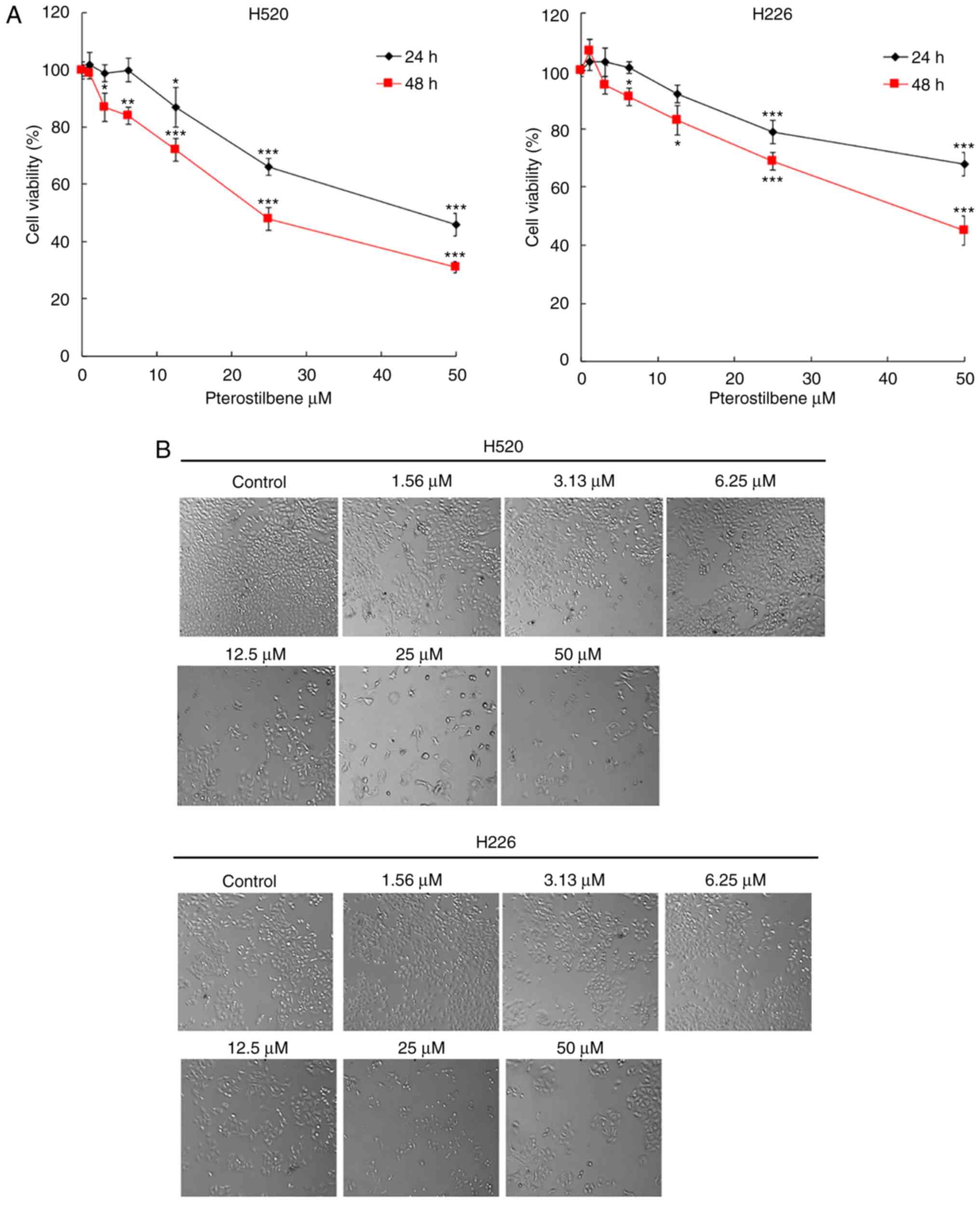
Cytotoxic effect of pterostilbene on
the viability of H520 tumor cells
The cytotoxic effect of pterostilbene on the
viability of SqCC cancer cell lines was determined using an MTT
assay. In the present study, H520 and H226 human SqCC cancer cell
lines were treated with different concentrations of pterostilbene
for 24 and 48 h. As illustrated in Fig.
1A, pterostilbene induced cytotoxicity in human SqCC cancer
cell lines in a dose-dependent manner. Notably, these two SqCC cell
lines exhibited different sensitivities to pterostilbene; the IC50
values of pterostilbene for H520 cells were 47.7±5.3 and 31.4±4.6
µM at 24 and 48 h, respectively, while the IC50 values for H226
cells were >50 and 44.3±3.7 µM at 24 and 48 h, respectively. In
addition, the cell morphology and shape were assessed using an
inverted microscope; this assessment indicated apoptotic
morphological changes, including cell shrinkage and cytoplasmic
blebbing, in the treated cells (Fig.
1B). The results demonstrated that the H520 cell line was
highly sensitive to pterostilbene treatment; therefore H520 was
selected for the subsequent analysis and evaluation of the
cytotoxic potency of pterostilbene.
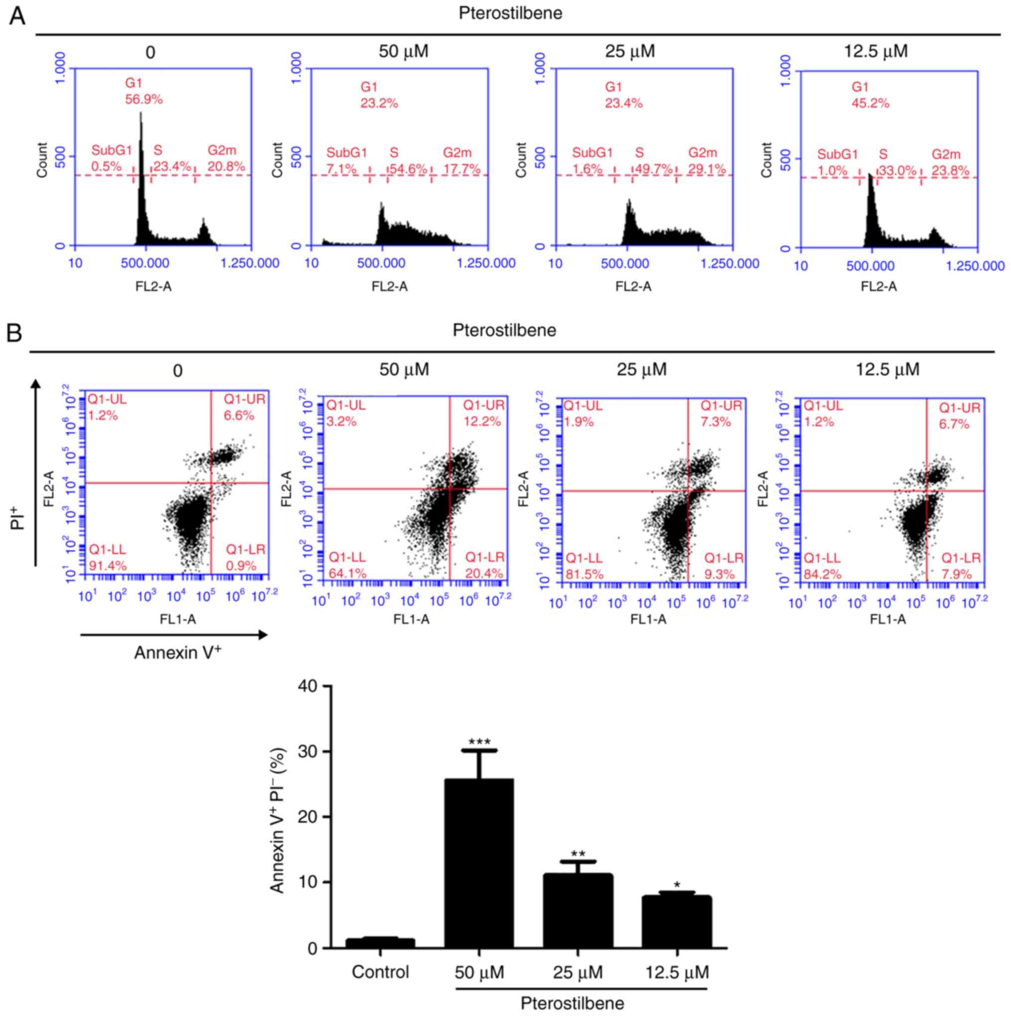
Pterostilbene induced S phase arrest
and apoptosis in H520 cells
The present study subsequently investigated the
impact of pterostilbene on different cell cycle phases in H520
cells. The H520 cells were exposed to different concentrations of
pterostilbene for 48 h and were subjected to flow cytometric
analysis following staining with PI. As Fig. 2A illustrates, the percentage of cells
in S phase was markedly increased following exposure to 12.5–50 µM
pterostilbene for 48 h. Additionally, the sub-G1 cell population,
which is indicative of cell mortality, was increased in the
presence of 50 µM pterostilbene. To confirm whether apoptotic
mechanisms may have been involved in the cell death associated with
treatment with pterostilbene, H520 cells were treated with
pterostilbene for 48 h and subjected to flow cytometric analysis
following Annexin V-FITC and PI staining. A quantitative flow
cytometric analysis revealed that the percentages of early
apoptotic H520 cells (Annexin V+/PI−, lower
right quadrant) were increased by pterostilbene in a dose-dependent
manner (Fig. 2B). Taken together,
these results indicated that pterostilbene induced S phase arrest
and early apoptosis in H520 SqCC cells.
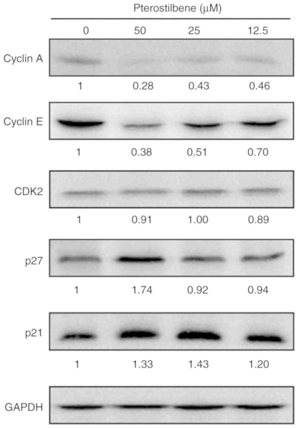
Pterostilbene decreased S phase
regulatory CDK and cyclin levels in H520 cells
To investigate the molecular mechanisms of
pterostilbene in the induction of S phase arrest, the present study
examined the effect of pterostilbene on the expression of key cell
cycle regulators of S phase progression through western blot
analysis. The Cip/Kip family members, p21 (Cip1) and p27 (Kip1),
can inhibit the activity of cyclin E-CDK2 and cyclin A-CDK2
complexes. The results indicated that treatment with pterostilbene
reduced the protein expression of cyclin A and cyclin E, but did
not affect the expression of CDK2 (Fig.
3), as indicated by the fold change in the band intensity
compared with that of the vehicle-treated cells. In addition, p21
and p27 protein expression was increased by treatment with
pterostilbene (Fig. 3). These
findings suggested that pterostilbene induced S phase arrest by
regulating the expression of S phase cell cycle regulatory proteins
in the H520 SqCC cell lines.
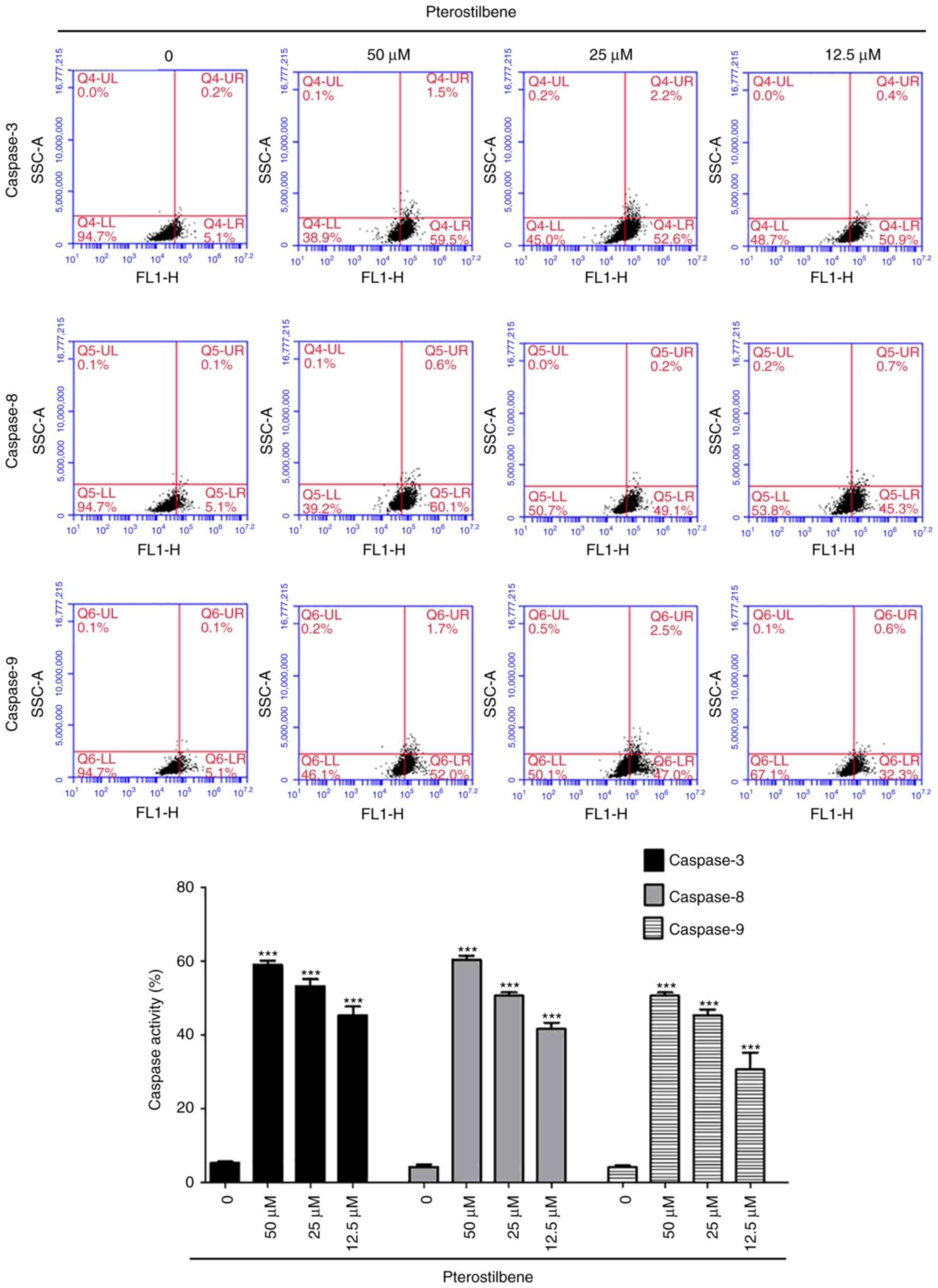
Pterostilbene stimulated the activity
of caspase-3, −8 and −9 in H520 cells
In order to determine whether pterostilbene induced
apoptotic cell death in H520 cells through the activation of
caspase-3, −8 and −9, H520 cells were treated with pterostilbene
for 48 h, harvested and examined using flow cytometry.
Pterostilbene increased the activity of caspase-3, −8 and −9
(Fig. 4) in a dose-dependent manner
and induced cell death through the activation of caspase-3, −8 and
−9 in H520 cells.
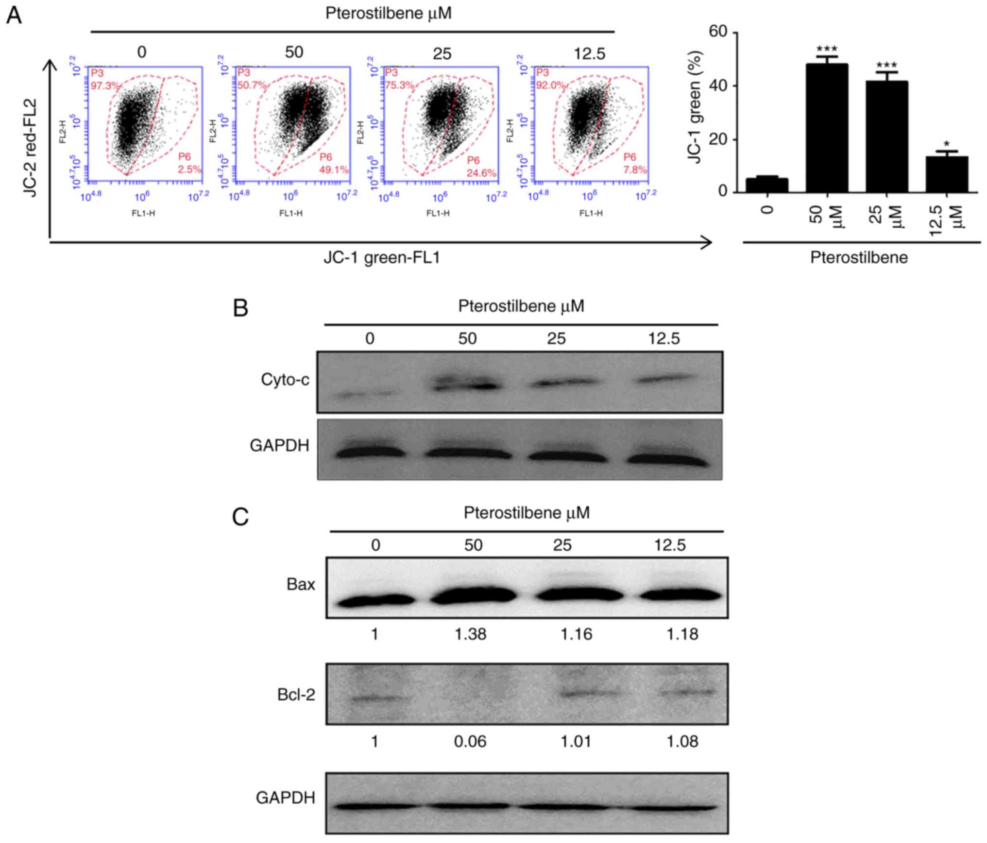
Pterostilbene depolarized the
mitochondrial membrane potential and increased cytochrome c
release
The loss of mitochondrial membrane has a major role
in the induction of apoptosis. Therefore, the membrane-permeable
JC-1 dye was used to examine the mitochondrial membrane in H520
cells that has been treated with pterostilbene. As presented in
sFig. 5A, following treatment with pterostilbene, a dose-dependent
increase in the green fluorescence was observed in H520 cells,
suggesting that pterostilbene produced significant mitochondrial
depolarization. Cytochrome c release from the mitochondria
to the cytosol is also a key initial step in apoptosis. Therefore,
in order to determine whether cytochrome c was released
during pterostilbene-induced apoptosis, western blot analysis was
performed to analyze the cytochrome c level in the cytosolic
fractions of H520 cells that had been exposed to pterostilbene. As
presented in Fig. 5B, an increase in
cytochrome c at 48 h was observed in the cytosolic fraction
of the pterostilbene-treated cells, compared with the cytochrome
c level in the control cells. In terms of the expression of
Bcl-2 family members, such as the anti-apoptotic protein Bcl-2 and
the pro-apoptotic protein Bax, have been identified during critical
events in the mitochondrial apoptotic pathway; therefore, the
present study further investigated the effects of pterostilbene on
the expression of Bax and Bcl-2 in H520 cells by western blot
analysis, and there was an increase in Bax protein expression and a
decrease in Bcl-2 expression in H520 cells (Fig. 5C). These data suggested that
pterostilbene induced the caspase-dependent mitochondrial apoptotic
pathways in H520 cells.
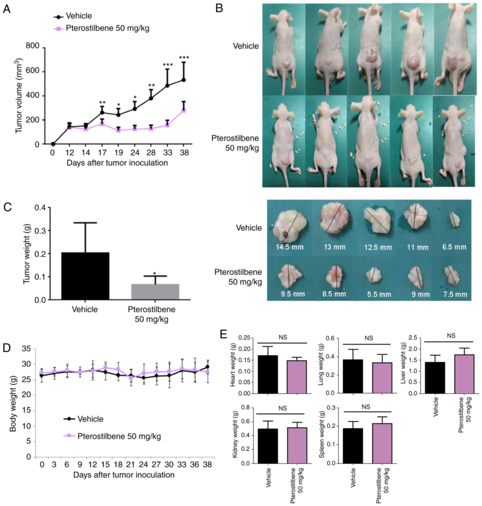
Pterostilbene activity against H520
×enografts in nude mice
The present study then investigated whether
pterostilbene had an effect against H520 ×enografts in nude mice.
As illustrated in Fig. 6A-C,
compared with the effect of the vehicle control, pterostilbene (50
mg/kg) was able to reduce the tumor volume (Fig. 6A and B) and weight (Fig. 6C) (day 38), indicating that
pterostilbene also inhibited the growth of SqCC cells in
vivo. The toxic effect of treatment with pterostilbene was
investigated in H520 ×enograft models. As illustrated in Fig. 6D and E, there was no significant
difference in body weight (Fig. 6D),
or heart, lung, liver, kidney, and spleen weights (Fig. 6E) between the pterostilbene-treated
group and the vehicle-treated group, which suggested that treatment
with pterostilbene did not obviously exhibit adverse side
effects.
Discussion
The present investigated the inhibitory effect of
pterostilbene on tumor growth in the EGFR-negative lung SqCC cell
line H520 and the potential signaling pathways through which
pterostilbene operates. Consistent with a previous study,
pterostilbene inhibited lung SqCC cell viability in a
concentration- and time-dependent manner (18). The data obtained by the present study
also indicated that pterostilbene promoted mortality of cancer
cells through S phase cell cycle arrest and the induction of
intrinsic and extrinsic apoptosis. Additionally, to the best of our
knowledge, the BALB/c athymic nude mouse xenograft model experiment
is the first to report the in vivo antitumor effect of
pterostilbene in lung SqCC. Therefore, pterostilbene may be a
potent therapeutic agent either alone or combined with another
chemotherapeutic agent in the treatment of lung SqCC.
Lung cancer remains the leading cause of
malignancy-associated mortality worldwide (1). Despite early diagnosis and advances in
therapeutic strategies, outcomes have remained poor in recent
decades (19). The primary cause of
treatment failure is resistance to standard therapy, especially
apoptosis-inducing drugs (18).
Further, chemotherapy-associated side effects are major concerns.
Thus, it is desirable to identify novel drugs that contain less
harmful natural materials to complement and improve the efficacy of
traditional chemotherapeutics. Previous studies have demonstrated
that naturally occurring compounds in foods such as fruits or
vegetables may exert a chemopreventive and therapeutic effect
against cancers (20,21). As a natural phytoalexin present in
grapes and berries, resveratrol is considered to be a potential
anticancer drug due to its antioxidant, anti-inflammatory, and
antiproliferative activities (22).
However, its poor absorption and rapid metabolism limit the
potential clinical applications of resveratrol (18). Pterostilbene, the natural
dimethylated analog of resveratrol, was shown to have similar
structure and function to resveratrol, but a higher
bioavailability, longer half-life and lower toxicity, thus
demonstrating greater potential for clinical applications (9). Although numerous studies have reported
that pterostilbene exhibits the hallmarks of a valuable and
well-tolerated anticancer agent, few studies have investigated its
anticancer mechanisms in lung SqCC (11,23).
In agreement with recent research reporting the
antitumor effect of pterostilbene on various malignant cancers
(24–26), the results of the present study
demonstrated that pterostilbene inhibited lung SqCC tumor growth.
Although the pterostilbene mechanisms of action have not yet been
completely elucidated, pterostilbene may influence multiple
signaling pathways critical to cancer development. It has been
reported that pterostilbene could induce cell cycle arrest and
apoptosis in numerous types of cancer cells, including gastric,
lung, and pancreatic cancer cells (11). The results of the present study
indicated that pterostilbene induced cell cycle arrest at the S
phase through apoptosis-related caspase and Bcl-2 family proteins.
Western blot analysis revealed that pterostilbene increased the
expression of p21 and p27, and decreased the expression of cyclin A
and E. The cyclin A- and E-CDK2 complexes are the most important
proteins in S phase that regulate progression to the G2/M phase. If
the cyclin A or E complex is inhibited, the cell cycle is arrested
in S phase, leading to the inhibition of cell proliferation and the
promotion of apoptosis (27). The
results of the present study also indicated that the expression
levels of p21 and p27, which are CDK inhibitors that can inhibit
the majority of cyclin-CDK complexes, were increased by
pterostilbene (27).
Although cells can die due to non-apoptotic
mechanisms, apoptosis is the principal mechanism of cancer cell
death for numerous chemotherapeutic agents (28). Therefore, the induction of apoptosis
is considered to be a potent therapeutic strategy for the
eradication of tumors. Previous medical literature has reported
that pterostilbene exerts proapoptotic activities (29,30). In
order to examine this hypothesis, the present study analyzed the
activity of caspase-3, −8 and −9. These caspases are the enzymes
involved in the effector phase of apoptosis. Caspase-3 is the most
critical component in the apoptotic pathway and can be activated by
caspase-8 or caspase-9, which are the two key proteins in the
extrinsic and intrinsic apoptotic pathways, respectively (31). Previously, Schneider et al
(29) revealed that pterostilbene
upregulates caspase-3/7 activity in SqCC (SK-MES-1) cell lines,
suggesting a pterostilbene-induced mitochondrial mechanism of
apoptosis. The results of the present study indicated that
pterostilbene increased the expression of caspase-3, −8, and −9,
suggesting that pterostilbene induced the apoptosis of lung SqCC
cells in a caspase-dependent manner, potentially through the
activation of the extrinsic and intrinsic apoptotic pathways. These
results are similar to those obtained by Xie et al (10) who observed an increase in
caspase-dependent apoptosis in multiple myeloma cells. In addition,
Pan et al (32) also reported
that pterostilbene promoted apoptosis and induced cell cycle arrest
in gastric cancer AGS cells, by activating the caspase cascade and
inducing changes in several cell cycle-regulating proteins, which
is in agreement with the results of the current study.
To summarize, the present study demonstrated the
antitumor effect of pterostilbene in lung SqCC, and revealed that
pterostilbene could cause cell cycle arrest, induce apoptosis in
lung SqCC cell lines in vitro, and inhibit tumor growth
in vivo in a mouse xenograft model. Furthermore, recent
in vivo safety analyses have revealed that pterostilbene
does not harm mice or humans (23,33). In
this regard, as it is a natural compound that exhibits low
toxicity, pterostilbene may exhibit potential to treat lung SqCC in
a clinical setting. Further study is required to evaluate the
efficacy of pterostilbene if it is combined with different
chemotherapeutic regimens or with radiation.
Acknowledgements
Not applicable.
Funding
No funding was received.
Availability of data and materials
The analyzed datasets generated during the study are
available from the corresponding author upon reasonable
request.
Authors' contributions
KTT, SHL and CCY conceived and designed the study.
KTT, PWC, SL and TMK performed the experiments. KTT, SHL, and CCY
wrote the paper. KTT, SHL, SL and CCY reviewed and edited the
manuscript. All authors read and approved the manuscript and agree
to be accountable for all aspects of the research in ensuring that
questions about the accuracy or integrity of any part of the work
are appropriately investigated and resolved.
Ethics approval and consent to
participate
All animal experimental procedures were carried out
following the Guide for the Care and Use of Laboratory Animals of
the Institutional Animal Care and Use Committee of National Chung
Hsing University (Taichung, Taiwan); the procedures were approved
by the Research Ethics Committee of National Chung Hsing
University.
Patient consent for publication
Not applicable.
Competing interests
The authors declare that they have no competing
interests.
References
|
1
|
Siegel R, Ma J, Zou Z and Jemal A: Cancer
statistics, 2014. CA Cancer J Clin. 64:9–29. 2014. View Article : Google Scholar : PubMed/NCBI
|
|
2
|
Okamoto T, Suzuki Y, Fujishita T, Kitahara
H, Shimamatsu S, Kohno M, Morodomi Y, Kawano D and Maehara Y: The
prognostic impact of the amount of tobacco smoking in non-small
cell lung cancer-differences between adenocarcinoma and squamous
cell carcinoma. Lung Cancer. 85:125–130. 2014. View Article : Google Scholar : PubMed/NCBI
|
|
3
|
Gandara DR, Hammerman PS, Sos ML, Lara PN
Jr and Hirsch FR: Squamous cell lung cancer: From tumor genomics to
cancer therapeutics. Clin Cancer Res. 21:2236–2243. 2015.
View Article : Google Scholar : PubMed/NCBI
|
|
4
|
Yang CC, Fong Y, Lin LC, Que J, Ting WC,
Chang CL, Wu HM, Ho CH, Wang JJ and Huang CI: The age-adjusted
Charlson comorbidity index is a better predictor of survival in
operated lung cancer patients than the Charlson and Elixhauser
comorbidity indices. Eur J Cardiothorac Surg. 53:235–240. 2018.
View Article : Google Scholar : PubMed/NCBI
|
|
5
|
Ettinger DS, Wood DE, Aisner DL, Akerley
W, Bauman J, Chirieac LR, D'Amico TA, DeCamp MM, Dilling TJ,
Dobelbower M, et al: Non-small cell lung cancer, version 5.2017,
NCCN clinical practice guidelines in oncology. J Natl Compr Canc
Netw. 15:504–535. 2017. View Article : Google Scholar : PubMed/NCBI
|
|
6
|
Ettinger DS, Akerley W, Borghaei H, Chang
AC, Cheney RT, Chirieac LR, D'Amico TA, Demmy TL, Govindan R,
Grannis FW Jr, et al: Non-small cell lung cancer, version 2.2013. J
Natl Compr Canc Netw. 11:645–653. 2013. View Article : Google Scholar : PubMed/NCBI
|
|
7
|
Shi A, Zhu G, Wu H, Yu R, Li F and Xu B:
Analysis of clinical and dosimetric factors associated with severe
acute radiation pneumonitis in patients with locally advanced
non-small cell lung cancer treated with concurrent chemotherapy and
intensity-modulated radiotherapy. Radiat Oncol. 5:352010.
View Article : Google Scholar : PubMed/NCBI
|
|
8
|
Rimando AM, Kalt W, Magee JB, Dewey J and
Ballington JR: Resveratrol, pterostilbene, and piceatannol in
vaccinium berries. J Agric Food Chem. 52:4713–4719. 2004.
View Article : Google Scholar : PubMed/NCBI
|
|
9
|
Estrela JM, Ortega A, Mena S, Rodriguez ML
and Asensi M: Pterostilbene: Biomedical applications. Crit Rev Clin
Lab Sci. 50:65–78. 2013. View Article : Google Scholar : PubMed/NCBI
|
|
10
|
Xie B, Xu Z, Hu L, Chen G, Wei R, Yang G,
Li B, Chang G, Sun X, Wu H, et al: Pterostilbene inhibits human
multiple myeloma cells via ERK1/2 and JNK pathway in vitro and in
vivo. Int J Mol Sci. 17(pii): E19272016. View Article : Google Scholar : PubMed/NCBI
|
|
11
|
McCormack D and McFadden D: Pterostilbene
and cancer: Current review. J Surg Res. 173:e53–e61. 2012.
View Article : Google Scholar : PubMed/NCBI
|
|
12
|
Prabhakar CN: Epidermal growth factor
receptor in non-small cell lung cancer. Transl Lung Cancer Res.
4:110–118. 2015.PubMed/NCBI
|
|
13
|
Tang Z, Du R, Jiang S, Wu C, Barkauskas
DS, Richey J, Molter J, Lam M, Flask C, Gerson S, et al: Dual
MET-EGFR combinatorial inhibition against T790M-EGFR-mediated
erlotinib-resistant lung cancer. Br J Cancer. 99:911–922. 2008.
View Article : Google Scholar : PubMed/NCBI
|
|
14
|
Zhang H, Zhan C, Ke J, Xue Z, Zhang A, Xu
K, Shen Z, Yu L and Chen L: EGFR kinase domain mutation positive
lung cancers are sensitive to intrapleural perfusion with
hyperthermic chemotherapy (IPHC) complete treatment. Oncotarget.
7:3367–3378. 2016.PubMed/NCBI
|
|
15
|
Zhang H, Li N, Chen Y, Huang LY, Wang YC,
Fang G, He DC and Xiao XY: Protein profile of human lung squamous
carcinoma cell line NCI-H226. Biomed Environ Sci. 20:24–32.
2007.PubMed/NCBI
|
|
16
|
Zang JP and Wei R: Effects of Cx43 gene
modification on the proliferation and migration of the human lung
squamous carcinoma cell line NCI-H226. Genet Mol Res.
14:13110–13119. 2015. View Article : Google Scholar : PubMed/NCBI
|
|
17
|
Li K, Dias SJ, Rimando AM, Dhar S, Mizuno
CS, Penman AD, Lewin JR and Levenson AS: Pterostilbene acts through
metastasis-associated protein 1 to inhibit tumor growth,
progression and metastasis in prostate cancer. PLoS One.
8:e575422013. View Article : Google Scholar : PubMed/NCBI
|
|
18
|
Mannal PW, Alosi JA, Schneider JG,
McDonald DE and McFadden DW: Pterostilbene inhibits pancreatic
cancer in vitro. J Gastrointest Surg. 14:873–879. 2010. View Article : Google Scholar : PubMed/NCBI
|
|
19
|
Wang BY, Huang JY, Cheng CY, Lin CH, Ko J
and Liaw YP: Lung cancer and prognosis in taiwan: A
population-based cancer registry. J Thorac Oncol. 8:1128–1135.
2013. View Article : Google Scholar : PubMed/NCBI
|
|
20
|
Siddiqui IA, Adhami VM, Ahmad N and
Mukhtar H: Nanochemoprevention: Sustained release of bioactive food
components for cancer prevention. Nutr Cancer. 62:883–890. 2010.
View Article : Google Scholar : PubMed/NCBI
|
|
21
|
Riboli E, Slimani N and Kaaks R:
Identifiability of food components for cancer chemoprevention. IARC
Sci Publ. 23–31. 1996.PubMed/NCBI
|
|
22
|
Suh N, Paul S, Hao X, Simi B, Xiao H,
Rimando AM and Reddy BS: Pterostilbene, an active constituent of
blueberries, suppresses aberrant crypt foci formation in the
azoxymethane-induced colon carcinogenesis model in rats. Clin
Cancer Res. 13:350–355. 2007. View Article : Google Scholar : PubMed/NCBI
|
|
23
|
Riche DM, McEwen CL, Riche KD, Sherman JJ,
Wofford MR, Deschamp D and Griswold M: Analysis of safety from a
human clinical trial with pterostilbene. J Toxicol.
2013:4635952013. View Article : Google Scholar : PubMed/NCBI
|
|
24
|
Pei HL, Mu DM and Zhang B: Anticancer
activity of pterostilbene in human ovarian cancer cell lines. Med
Sci Monit. 23:3192–3199. 2017. View Article : Google Scholar : PubMed/NCBI
|
|
25
|
Kong Y, Chen G, Xu Z, Yang G, Li B, Wu X,
Xiao W, Xie B, Hu L, Sun X, et al: Pterostilbene induces apoptosis
and cell cycle arrest in diffuse large B-cell lymphoma cells. Sci
Rep. 6:374172016. View Article : Google Scholar : PubMed/NCBI
|
|
26
|
Wawszczyk J, Kapral M, Hollek A and
Weglarz L: In vitro evaluation of antiproliferative and cytotoxic
properties of pterostilbene against human colon cancer cells. Acta
Pol Pharm. 71:1051–1055. 2014.PubMed/NCBI
|
|
27
|
Mann MB and Kaldis P: Cell cycle
transitions and Cdk inhibition in melanoma therapy: Cyclin' through
the options. Cell Cycle. 10:13492011. View Article : Google Scholar : PubMed/NCBI
|
|
28
|
Aleo E, Henderson CJ, Fontanini A, Solazzo
B and Brancolini C: Identification of new compounds that trigger
apoptosome-independent caspase activation and apoptosis. Cancer
Res. 66:9235–9244. 2006. View Article : Google Scholar : PubMed/NCBI
|
|
29
|
Schneider JG, Alosi JA, McDonald DE and
McFadden DW: Pterostilbene inhibits lung cancer through induction
of apoptosis. J Surg Res. 161:18–22. 2010. View Article : Google Scholar : PubMed/NCBI
|
|
30
|
Chen RJ, Ho CT and Wang YJ: Pterostilbene
induces autophagy and apoptosis in sensitive and chemoresistant
human bladder cancer cells. Mol Nutr Food Res. 54:1819–1832. 2010.
View Article : Google Scholar : PubMed/NCBI
|
|
31
|
Aral K, Aral CA and Kapila Y: The role of
caspase-8, caspase-9, and apoptosis inducing factor in periodontal
disease. J Periodontol. 90:288–294. 2019. View Article : Google Scholar : PubMed/NCBI
|
|
32
|
Pan MH, Chang YH, Badmaev V, Nagabhushanam
K and Ho CT: Pterostilbene induces apoptosis and cell cycle arrest
in human gastric carcinoma cells. J Agric Food Chem. 55:7777–7785.
2007. View Article : Google Scholar : PubMed/NCBI
|
|
33
|
Ruiz MJ, Fernandez M, Pico Y, Mañes J,
Asensi M, Carda C, Asensio G and Estrela JM: Dietary administration
of high doses of pterostilbene and quercetin to mice is not toxic.
J Agric Food Chem. 57:3180–3186. 2009. View Article : Google Scholar : PubMed/NCBI
|