Introduction
Undifferentiated embryonal sarcoma of the liver
(UESL), also known as malignant mesenchymoma or embryonic sarcoma,
is a rare disease (1) that rarely
occurs in adults; 90% of UESL cases occur in children aged 6–10
years (2). There is no evident
gender discrepancy in the disease. UESL is often misdiagnosed as
other types of hepatic malignant tumors. The diagnosis of UESL
relies on postoperative pathology and immunostaining analysis
(3). Due to its high malignancy,
UESL often metastasizes to the lung, peritoneum and pleura,
resulting in a poor prognosis (4).
Currently, therapy for UESL mainly comprises surgery with
postoperative radio-chemotherapy (5). In the current study, we present a case
of UESL. The patient’s family provided consent for the study.
Case report
A 7-year-old female child was admitted to the
Department of Hepatobiliary Surgery of the Shandong Cancer Hospital
(Jinan, China) on December 29 2009, with an augmented swelling in
the upper abdomen and a palpable massive irregular tumor that had
been identified >10 days previously. A magnetic resonance
imaging (MRI) scan revealed a massive focal lesion in the right
lobe of the liver. The lesion was diagnosed as a malignant tumor
with a high possibility of hepatoblastoma. The patient did not
present with fever, bellyache, abdominal distention, diarrhea,
jaundice, nausea or vomiting.
Physical examination revealed that scleral or
systematic mucocutaneous icterus and swollen lymph nodes were not
present. The patient had ectasia of the upper abdomen, particularly
in the right superior belly. The liver under the rib edge was
palpable for 7 cm, and the surface tuberculum crossing the left of
the ventral median line was not palpable for 1 cm. No percussion
pain was observed in the hepatic renal region.
The laboratory examination results were as follows:
α-fetoprotein (AFP), 0.853 ng/ml; carbohydrate-associated antigen
(CA) 19-9, 23.39 U/ml; carcinoembryonic antigen (CEA), 0.788 ng/ml;
alanine transaminase (ALT), 68.3 μmol/l; aspartate
transaminase (AST), 63.8 μmol/l; albumin, 32.01 g/l; albumin
and globulin ratio, 1.25; total bilirubin (Tbil), 19.7
μmol/l; direct bilirubin (Dbil), 2.5 μmol/l; indirect
bilirubin (Ibil), 17.2 μmol/l; alkaline phosphatase (ALP),
162 μmol/l; γ-glutamyl transpeptidase (GGT), 80
μmol/l.
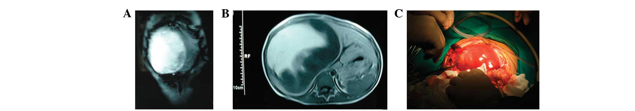
The MRI examination revealed a massive tumor
(15×13×10 cm) in the right lobe of the liver (Fig. 1A and B), which demonstrated a high
central and low periphery mixed signal shadow on the T1-weighted
image (T1W1) and a mixed high signal on T2W1. Certain partitions
were observed to be present in the interior of the tumor and the
boundaries remained clear. The patient’s right kidney was pushed
down by the pressure, which enhanced the scanning of the peripheral
solid parts with a strap type of persistent potentiation; the
boundaries were clear and no marked abnormalities were observed in
the left hepatic lobe. Following admission, the patient underwent
partial excision of the right hepatic lobe and the left inner lobe
on December 2, 2009. The perioperative results revealed that a
small level of clear and transparent pale yellow ascites at a
volume of 80 ml was present in the abdominal cavity (Fig. 1C). A massive tumor with a smooth and
glossy surface was observed in the right hepatic lobe, and the
Glisson’s capsule remained intact. Moreover, in the patient’s left
hepatic lobe, the first and second porta hepatis had been removed
by the pressure, and the inferior vena cava was distorted.
The following clinical pathology results were
observed by the naked eye. The size of the tumor in the hepatic
right lobe was ∼17.0 cm, and the incisal surface was occupied by a
gray-yellow/red/white swelling. Additionally, the texture of the
swelling was impalpable and soft. Necrosis was observed and the
swollen tissue demonstrated the partial stick-slip phenomenon.
Further clinicopathological results were obtained using a light
microscope. Under the light microscope (Figs. 1 and 2), the tumor cells demonstrated embryonic
mesenchymal differentiation and lacked epithelial characteristics.
Moreover, the tumor cells exhibited fusiform and star shapes with
undetermined boundaries and clear heteromorphism. In addition,
changes in the interstitial mucus and the periodic acid-Schiff
(PAS)-positive eosinophilic body, both inside and outside the tumor
cells, were identified. Furthermore, the remnant hepatic cells and
the hyperplastic bile duct were observed in the boundaries of the
tumor, and there were no changes in hepatic cirrhosis. The
postoperative pathological diagnosis was undifferentiated sarcoma
in the right hepatic lobe without tumor tissue in the gall
bladder.
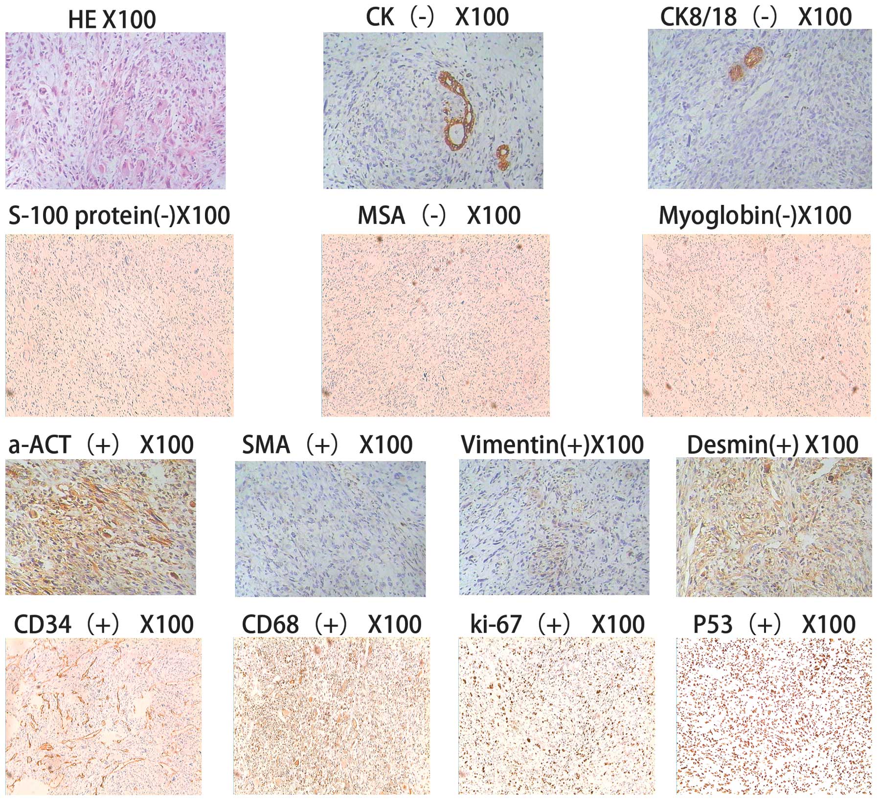 | Figure 2Light microscopy. The tumor cells
exhibited embryonic mesenchymal differentiation and lacked
epithelial characteristics. Moreover, the tumor cells demonstrated
fusiform and star shapes, which have undetermined boundaries and
clear heteromorphism. Changes in the interstitial mucus and the
periodic acid-Schiff (PAS)-positive eosinophilic body, both inside
and outside the tumor cells, were evident. The remnant hepatic
cells and hyperplastic bile duct are visible in the boundaries of
the tumor, and there were no changes in the hepatic cirrhosis. The
immunohistochemistry results are as follows: Broad spectrum
creatine kinase (CK)(−), cytokeratin 8/18(CK8/18)(−), S-100(−),
MSA(−), myoglobin(−), SMA foci(+), a-ACT foci(+), desmin foci(+),
vimentin foci(+), CD34(+), CD68(+), Ki-67(+) and P53(+). |
The immunohistochemistry results were as follows:
Broad spectrum creatine kinase (CK)(−), cytokeratin
8/18(CK8/18)(−), S-100(−), MSA(−), myoglobin(−), SMA foci(+), a-ACT
foci(+), desmin foci(+), vimentin foci(+), CD34(+), CD68(+),
Ki-67(+) and P53(+).
The karyotype analysis revealed that the chromosomal
aberrations accounted for the remaining 5% of all chromosomes,
which involved chromosomes 6, 11, 12 and 14, as well as the X
chromosome. The aberrations included fourcentric chromosomes,
deletion, disruption and marker chromosomes.
Discussion
UESL is a rare hepatic mesenchymal tumor that was
first reported and classified by Stocker et al in 1978
(1). The incidence of the disease
has no significant gender difference. In addition, 90% of patients
are children aged 6–10 years, and the diesease accounts for 5–8% of
hepatic tumors in children. The tumor is mainly localized or found
in the hepatic right lobe (59%), while it rarely develops in the
hepatic left lobe (22%) or the bilateral lobe (20%). UESL typically
has a diameter of 10–25 cm with a solitary clear boundary (6). Hemorrhaging, necrosis and cystic
degeneration are frequently observed, while clinical manifestations
include abdominal mass, pain, fever and, rarely, jaundice.
An ultrasonography scan reveals a mixed echoic mass
containing an irregular anechoic region or multiple small capsular
spaces of different sizes. The solid region exhibits a mixture of
high- and low-level echos. By contrast, a computed tomography (CT)
scan reveals cystic lesions with low density that is reflected as a
fluid. Therefore, the difference between CT and ultrasonography
results is a critical characteristic of this disease. However, both
scans reveal cystic lesions when hemorrhage, necrosis and
liquefaction account for the major part of the tumor. An enhanced
CT scan of the tumor solid part, septation and pseudo-capsule shows
different degrees of reinforcement. However, in the present study,
MRI revealed that the T1W1 scan mainly detected a low or equal
signal and high signal foci, which were a reflection of a
hemorrhage in the tumor. Additionally, the T2W1 scan frequently
exhibited a mixed high signal. Positron emission tomography
(PET)-CT scans are also used in the diagnosis of UESL, and they
have a critical diagnostic value for UESL patients, particularly
those with metastasis of an extra-hepatic organ.
The immunohistochemistry results reveal the positive
expression of SMA, a-ACT, desmin, vimentin and actin in UESL
patients, and a minority of cases are positive for PCNA, CK8/18 and
p53; while AFP, S-100, CEA, CA 19-9 and cytokeratin have negative
expression. The indices of the patient were as follows: SMA
foci(+), a-ACT foci(+), desmin foci(+), vimentin foci(+), broad
spectrum CK(−), CK8/18(−), AFP(−), CEA(−) and CA19-9(−).
Generally, a definite diagnosis of UESL is not able
to be determined preoperatively; the diagnosis relies on
postoperative pathological analysis and immunohistochemical
results. UESL ought to be differentiated from hepatoblastoma,
embryonal rhabdomyosarcoma, hepatic mesenchymal hamartoma and
hepatic echinococcosis. Hepaoblastoma, which mainly occurs in
infants aged <3 years, is composed of primary hepatic
parenchymal cells with small cellular heteromorphism, little
karyokinesis, increased blood sinus and positive expression of
endosomal membrane protein, vimentin and AFP revealed by
immunohistochemistry. By contrast, embryonal rhabdomyosarcoma
mainly occurs in infants aged <6 years, and the tumor is mainly
composed of striated muscle maternal cells in different phases and
primary mesenchymal cells. The myosin often exhibits strong
positive expression by immunohistochemistry, and transverse
striation may be observed by preservative associated transient
hyperfluorescence (PATH) staining. With hepatic mesenchymal
hamartoma, which mainly occurs in infants aged <1 year, the
tumor is composed of a mucus matrix and a large number of star and
fusiform primary mesenchymal cells, whereas the tumor cells
themselves do not exhibit heteromorphism. By contrast, hepatic
echinococcosis is a local parasitic disease that mainly occurs in
adults aged 20–40 years. Casoni and complement fixation tests are
used to diagnose this disease, and the positive rate is 90–95%,
which has a critical diagnostic value. A case history combined with
imaging analysis has been demonstrated to be beneficial for the
discrimination of UESL. A previous study has revealed that UESL is
often misdiagnosed as echinococcosis of the liver (2). Previous studies have demostrated that
cases of UESL have exhibited an amplification and deletion in
chromosomes 1q, 5p, 8p and 12q, and a translocation of 19q13.4
(7), as well as mutation of the p53
gene (8). Therefore, the detection
of molecular genetics is beneficial for the differential diagnosis
of UESL. In this case, chromosomal aberration accounted for the
remaining 5% of all chromosomes, which invovled chromosomes 6, 11,
12 and 14, as well as the X chromsome. However, the patient’s
parents’ and sisters’ chromosomes were normal, which indicated that
genetic mutations may induce tumorigenesis, and thus further
studies ought to be performed.
UESL is generally considered to be a highly invasive
malignant tumor in hepatic primary mesenchymal tissue with distant
metastasis. Lee et al described a case of UESL in a child
with distant metastases in the lung and adrenal glands (4). The prognosis of UESL was not observed
to correlate with the size and degree of differentiation of the
tumor; however, it was found to correlate with invasion, diffusion
and metastasis (9). In recent
years, survival rates have significantly improved due to
improvements in therapy, and long-term survival cases have been
reported (9). The key management of
the disease is total resection followed by postoperative combined
therapeutic measures, including chemotherapy, radiotherapy and
interventional therapy, which are able to significantly improve
survival rates. Yu et al suggested that the best method for
improving survival is total resection, regardless of whether the
tumor is ruptured (10). Uchiyama
et al proposed that surgical resection combined with
chemotherapy is the most effective therapy for UESL patients with
tumor rupture. Additionally, the authors suggested that
chemotherapy containing 3–4 types of drugs, such as adriamycin,
cisplatin and others, demonstrated beneficial therapeutic effects
and significantly improved survival rates (5). Li et al demonstrated that a
UESL patient who underwent interventional therapy and surgical
resection exhibited a prolonged survival compared with patients who
underwent surgical resection only (3). McCarthy et al described a case
of a pregnant UESL patient who underwent trans-catheter ablation to
control the development of the tumor (11). Certain studies have demonstrated
that hepatic transplantation is an effective therapeutic method for
UESL patients whose tumor is not able to be resected or who have
postoperative recurrence of the tumor. This method may potentially
significantly improve survival rates and times (12).
In the present case, blockage of the blood flow in
half the liver was performed while the patient was under total
anesthesia on December 29, 2009. A massive tumor with a smooth and
glossy surface was observed in the right hepatic lobe and the
Glisson’s capsule remained intact. Moreover, in the patient’s left
hepatic lobe, the first and second porta hepatis had been removed
by the pressure, and the inferior vena cava had been distorted. The
surgery ran smoothly. The hepatic right lobe and partial left
internal lobe were resected. The patient recovered successfully,
and a chemotherapy regimen of epirubicin at 20 mg for days 1–2 and
cisplatin at 15 mg for days 1–3 was administered at 14-day
intervals following surgery. Subsequently, epirubicin at 20 mg for
days 1–2 and cisplatin at 15 mg for days 1–3 were administered in
the second and third cycle of chemotherapy on July 27, 2010.
Currently, the patient’s physical status is normal, and no local
recurrence or metastasis has been observed. We conclude that
preoperative and/or postoperative interventional therapy, combined
with radiotherapy and chemotherapy, may improve survival rates and
times in certain cases. The precise timing of the total surgical
resection is crucial to prevent invasive growth of the tumor, and a
liver transplantation is the most effective therapy for a patient
whose tumor cannot be surgically resected.
Acknowledgements
This study was financially supported
by The 973 Project of China (Grant No. 2011CB504302) and Projects
of the Natural Science Foundation of Shandong (Grant No.
ZR2010HL018 and No. Y2008C30). The authors would like to thank the
other members of their laboratories for their valuable
comments.
References
|
1
|
Stocker JT and Ishak KG: Undifferentiated
(embryonal) sarcoma of the liver: report of 31 cases. Cancer.
42:336–348. 1978. View Article : Google Scholar : PubMed/NCBI
|
|
2
|
Faraj W, Mukherji D, El Majzoub N,
Shamseddine A, Shamseddine A and Khalife M: Primary
undifferentiated embryonal sarcoma of the liver mistaken for
hydatid disease. World J Surg Oncol. 8:582010. View Article : Google Scholar : PubMed/NCBI
|
|
3
|
Li XW, Gong SJ, Song WH, Zhu JJ, Pan CH,
Wu MC and Xu AM: Undifferentiated liver embryonal sarcoma in
adults: a report of four cases and literature review. World J
Gastroenterol. 16:4725–4732. 2010. View Article : Google Scholar : PubMed/NCBI
|
|
4
|
Lee MK, Kwon CG, Hwang KH, Choe W, Kim JE,
Tchah H and Jeon IS: F-18 FDG PET/CT findings in a case of
undifferentiated embryonal sarcoma of the liver with lung and
adrenal gland metastasis in a child. Clin Nucl Med. 34:107–108.
2009. View Article : Google Scholar : PubMed/NCBI
|
|
5
|
Uchiyama M, Iwafuchi M, Yagi M, Iinuma Y,
Kanada S, Yamazaki S, Ohtaki M and Shirai Y: Treatment of ruptured
undifferentiated sarcoma of the liver in children: a report of two
cases and review of the literature. J Hepatobiliary Pancreat Surg.
8:87–91. 2001. View Article : Google Scholar : PubMed/NCBI
|
|
6
|
Pachera S, Nishio H, Takahashi Y, Yokoyama
Y, Oda K, Ebata T, Igami T and Nagino M: Undifferentiated embryonal
sarcoma of the liver: case report and literature survey. J
Hepatobiliary Pancreat Surg. 15:536–544. 2008. View Article : Google Scholar : PubMed/NCBI
|
|
7
|
Webber EM, Morrison KB, Pritchard SL and
Sorensen PH: Undifferentiated embryonal sarcoma of the liver:
results of clinical management in one center. J Pediatr Surg.
34:1641–1644. 1999. View Article : Google Scholar : PubMed/NCBI
|
|
8
|
Sangkhathat S, Kusafuka T, Nara K, Yoneda
A and Fukuzawa M: Non-random p53 mutations in pediatric
undifferentiated (embryonal) sarcoma of the liver. Hepatol Res.
35:229–234. 2006. View Article : Google Scholar : PubMed/NCBI
|
|
9
|
Bisogno G, Pilz T, Perilongo G, Ferrari A,
Harms D, Ninfo V, Treuner J and Carli M: Undifferentiated sarcoma
of the liver in childhood: a curable disease. Cancer. 94:252–257.
2002. View Article : Google Scholar : PubMed/NCBI
|
|
10
|
Yu DC, Tandon R, Bohlke AK, Steiner RB,
Haque S and Florman SS: Resection of a large, ruptured,
undifferentiated (embryonal) sarcoma of the liver in a child: a
case report and review of the literature. J La State Med Soc.
161:41–44. 2009.PubMed/NCBI
|
|
11
|
McCarthy FP, Harris M and Kornman L:
Management of undifferentiated embryonal sarcoma of the liver in
pregnancy. Obstet Gynecol. 109:558–560. 2007. View Article : Google Scholar : PubMed/NCBI
|
|
12
|
Kelly MJ, Martin L, Alonso M and Altura
RA: Liver transplant for relapsed undifferentiated embryonal
sarcoma in a young child. J Pediatr Surg. 44:e1–e3. 2009.
View Article : Google Scholar : PubMed/NCBI
|