Application of POLARIC™ fluorophores in an in vivo tumor model

  • Authors:
    • Nako Maishi
    • Taisuke Kawamoto
    • Noritaka Ohga
    • Koji Yamada
    • Kosuke Akiyama
    • Kazuyuki Yamamoto
    • Takahiro Osawa
    • Yasuhiro Hida
    • Kyoko Hida
  • View Affiliations

  • Published online on: July 18, 2013     https://doi.org/10.3892/or.2013.2620
  • Pages: 1695-1700
Metrics: Total Views: 0 (Spandidos Publications: | PMC Statistics: )
Total PDF Downloads: 0 (Spandidos Publications: | PMC Statistics: )


Abstract

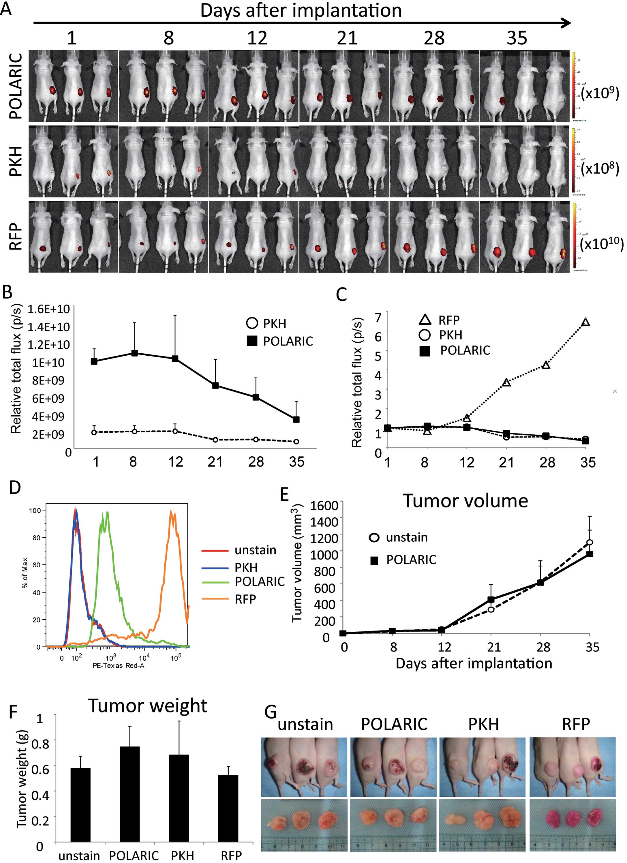
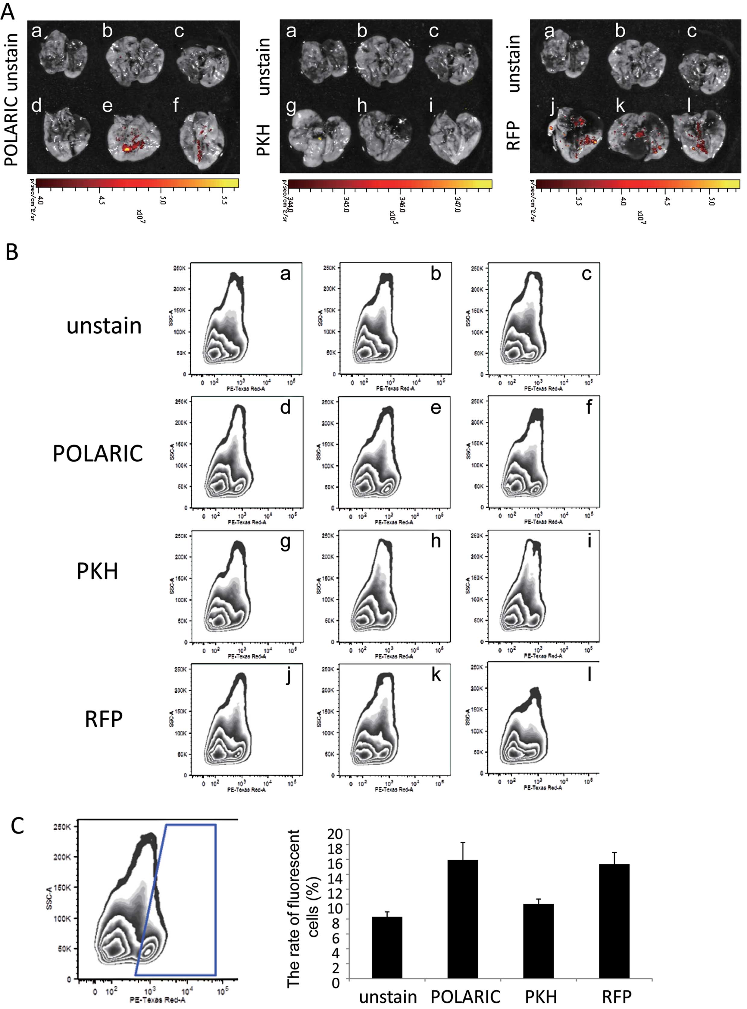
Fluorescent and luminescent tools are commonly used to study the dynamics of cancer progression and metastases in real‑time. Fluorophores have become essential tools to study biological events. However, few can sustain fluorescence long enough during long-term studies. In the present study, we focused on a series of new amphiphilic fluorophores known as POLARIC™, which emit strong fluorescence in lipid bilayers and can be readily modified using the Suzuki-Miyaura cross‑coupling reaction. Appropriate chemical modifications of substituent groups can improve target-site specificity, reduce cytotoxicity and prolong emission. Therefore, in contrast to conventional fluorescent probes, these fluorophores show promise for long-term monitoring of biological processes. In the present study, we conducted long-term observations of tumor growth and metastasis using a POLARIC derivative as a novel fluorescent probe. For this purpose, we studied the metastatic melanoma cell line A375-SM, which proliferates at a high rate. We compared the characteristics of the POLARIC probe with the commercially available fluorescent dye PKH26 and fluorescent protein mRFP1. A375-SM cells were labeled with these fluorescent probes and orthotopically implanted into nude mice. The fluorescence emitted by POLARIC was detected more than five weeks after implantation without causing detectable harmful effects on tumor growth. By contrast, fluorescence of cells labeled with PKH26 could not be detected at this same time. Furthermore, POLARIC-, but not PKH26-labeled cells, were also detected in lung metastases. These results indicate that labeling cells with POLARIC fluorophores can significantly extend the time course of in vivo studies on tumor cell growth.
View Figures
View References

Related Articles

Journal Cover

October 2013
Volume 30 Issue 4

Print ISSN: 1021-335X
Online ISSN:1791-2431

Sign up for eToc alerts

Recommend to Library

Copy and paste a formatted citation
x
Spandidos Publications style
Maishi N, Kawamoto T, Ohga N, Yamada K, Akiyama K, Yamamoto K, Osawa T, Hida Y and Hida K: Application of POLARIC™ fluorophores in an in vivo tumor model. Oncol Rep 30: 1695-1700, 2013
APA
Maishi, N., Kawamoto, T., Ohga, N., Yamada, K., Akiyama, K., Yamamoto, K. ... Hida, K. (2013). Application of POLARIC™ fluorophores in an in vivo tumor model. Oncology Reports, 30, 1695-1700. https://doi.org/10.3892/or.2013.2620
MLA
Maishi, N., Kawamoto, T., Ohga, N., Yamada, K., Akiyama, K., Yamamoto, K., Osawa, T., Hida, Y., Hida, K."Application of POLARIC™ fluorophores in an in vivo tumor model". Oncology Reports 30.4 (2013): 1695-1700.
Chicago
Maishi, N., Kawamoto, T., Ohga, N., Yamada, K., Akiyama, K., Yamamoto, K., Osawa, T., Hida, Y., Hida, K."Application of POLARIC™ fluorophores in an in vivo tumor model". Oncology Reports 30, no. 4 (2013): 1695-1700. https://doi.org/10.3892/or.2013.2620