Introduction
Immunotherapy has been found to be effective for
bladder cancer, especially in non-muscle invasive diseases.
Treatment of non-muscle invasive disease bladder cancers resulted
in a reduction in the risk of recurrence and progression (1,2).
Although the mechanisms for BCG in delaying recurrence and
prolonging survival remain poorly understood (2,3), the
modulation of the expression of a variety of cytokines and immune
cell maturation have been demonstrated with BCG treatment even
though their functional status and effects remain controversial
(1).
Programmed death ligand-1 (PD-L1) is a T-cell
regulatory molecule that may be expressed on the surface of tumor
and tumor-infiltrating immune cells. The PD-L1/PD-1 pathway has
been shown to be important in cancer progression (4). PD-L1 is frequently found to be
overexpressed in tumors. By binding to PD-1, it may inhibit the
activation of cytotoxic T lymphocytes in order to evade the host
immune response preventing tumors from cytotoxic T
lymphocyte-induced killing. Other than PD-1, PD-L1 also interacts
with B7.1 to further suppress the tumor antigen-induced activation
of cytotoxic T lymphocytes.
Tumor PD-L1 protein expression was found to be
associated with a higher grade of tumors and a poorer survival rate
in urothelial cancers (5). PD-L1
has been previously identified as an independent determinant for
stage progression (6). Among other
T-cell co-regulatory molecules, such as B7-H3 and PD-1,
investigated in urothelial cell carcinoma of the bladder, only
PD-L1 expression levels were found to be associated with an
increased pathological stage and an independent predictor for
all-cause mortality after cystectomy (7). Microarray technology was recently used
to examine the expression of PD-L1, PD-1 and B7-H3 in urothelial
carcinoma of the bladder (8). The
results of that study showed that the expression levels of the
three molecules were correlated with each other in the primary
tumors, while the expression of PD-L1 was associated with an
increased risk of overall mortality in the subgroup of patients
with organ-confined disease.
Efforts have begun to delineate the role of the
PD-L1 pathway in metastatic urothelial bladder cancer. Inhibition
of the PD-L1 pathway by the investigational molecule, MPDL3280A,
which blocks PD-1/PD-L1 and B7.1/PD-L1 interactions, thereby
completely inhibiting the PD-L1-mediated immunosuppressive signals,
has shown great clinical activity (9). Patients whose urothelial bladder
tumors or their associated tumor-infiltrating lymphocytes had PD-L1
expression of an immunohistochemical (IHC) score of 2+ or 3+ had an
objective response rate of 40 and 50%, respectively (9). Those results led to a breakthrough
therapy designation status for MPDL3280A in the treatment of
metastatic urothelial bladder cancer granted by FDA in the USA,
indicating that PD-L1 is an important target that should be
investigated.
Although IHC is a standard test for protein
overexpression, the resulting interpretation is often solely visual
and highly subjective. For instance, diagnosis of the HER2
overexpression levels in breast cancer often requires complementary
fluorescence in situ hybridization analysis for those
patients with an IHC staining score of 2+, due to the difficulty of
defining a true-positive result, when the staining itself is
moderate. Assessment of gene expression at the transcriptional
level by quantitative tests removes the subjectivity of
interpreting IHC results. In the present study, we aimed to analyze
the mRNA expression of related molecules of the PD-L1 pathway and
the prognostic significance of the players in the PD-L1 pathway in
early bladder cancer, by investigating the associations of the
expression of PD-L1, B7.1 and PD-1 and the clinicopathological
parameters in three bladder cancer patient cohorts using available
microarray and patient data.
Materials and methods
Extraction of clinical and microarray
gene expression data from bladder cancer patient datasets
Three bladder cancer patient datasets, GSE13507
(n=256) (10), GSE32894 (n=308)
(11) and GSE32548 (n=131)
(12) were identified from the Gene
Expression Omnibus database based on the following search criteria:
i) bladder or urothelial cancer, ii) available information on
clinicopathological parameters, iii) available information on the
PD-L1, B7.1 and PD-1 gene expression levels and iv) with a sample
size of ≥100. The GEO website has standardized URLs for its
individual datasets, such as the overall summary information
regarding the microarray dataset GSE13507 accessed at http://www.ncbi.nlm.gov/geo/query/acc.cgi?acc=GSE13507.
For each GEO data series, links are provided at the bottom of the
page to the Series Matrix File(s), which contain the expression
values for each gene (probe set) and each microarray. The URLs to
the Series Matrix File(s) are also standardized. For GSE13507, the
URL was ftp://ftp.ncbi.nlm.nih.gov/pub/geo/DATA/SeriesMatrix/GSE13507.
The files in gzip format were then unzipped to the tab-delimited
text format, which contained detailed information for statistical
analysis. R scripting was used to extract the expression values of
a small number of genes (probe sets) of interest and the clinical
data from the data matrices downloaded from GEO.
Correlations of gene expression levels
and clinical data
The statistical analyses were performed using
SPSS19.0. The associations between the expression levels of the
genes were analyzed by the Spearman’s rank test. The expression
levels were then divided into high and low levels using the higher
quartile expression level as the cut-off point for the Kaplan-Meier
survival analysis. The results were compared by the log-rank test.
The patients were divided into three groups based on the expression
levels of PD-L1 and B7.1. The PD-L1/B7.1 low group comprised
patients expressing the two genes at low levels, the PD-L1/B7.1
intermediate group comprising patients expressing one of the two
genes at high levels, and the PD-L1/B7.1 high group comprising
patients expressing the two genes at high levels. The survival time
of the patients stratified by this grouping method was analyzed by
the Kaplan-Meier survival analysis as described above. The
univariate Cox regression and multivariate analyses were performed
for the clinicopathological parameters, including gender, age,
T-stage, tumor type and grade and PD-L1 expression. The
multivariate Cox regression analysis with forward stepwise
selection and an entry limit of p<0.05 was performed to identify
independent predictors of survival in the patient cohort. Muscle
invasive bladder cancer specimens were examined using the
Chi-square test.
Identification of PD-L1 co-expressing
genes
The patients were stratified into two groups based
on the expression levels of PD-L1 as described above. The gene
expression patterns of patients in the PD-L1 low subgroup and those
in the PD-L1 high subgroup (whose survival was significantly
poorer) were compared. Probe sets that were differentially
expressed between the two subgroups were identified by the
two-sample Welch’s t-test. This test was used to avoid the type I
error due to unequal variances of the values of the probe sets
between the subgroups. Briefly, a Welch’s t-test was applied to
each probe set corresponding to a certain gene in the data matrix
using our own Java application MyStats. P-values and the
differential expression in fold-changes for all the probe sets were
generated as tab-delimited worksheets of Excel for subsequent
analysis.
Identification of therapeutic targets for
bladder cancer patients overexpressing PD-L1
Patients who had tumors expressing a high level of
PD-L1 were stratified into two groups based on their survival
status (alive or deceased). Differential expression of different
probe sets between patients in the PD-L1 high-alive subgroup and
those in the PD-L1 high-deceased subgroup were identified as
described above.
Results
Association between the expression levels
of PD-L1, B7.1 and PD-1 with clinicopathological parameters and
survival in bladder cancer
Three independent microarray datasets were
identified in the GEO database when we limited our search to
include only bladder cancer datasets with a sample size of >100
patients and available clinical data, including survival and tumor
grade. The three datasets were GSE13507 (n=256), GSE32894 (n=308)
and GSE32548 (n=131). The expression levels of PD-L1, B7.1 and PD-1
were extracted and correlated with available clinical data for the
three datasets.
In GSE13507 (n=256) (10), PD-L1 and B7.1 expression did not
vary significantly between normal bladder (n=10), surrounding
mucosa (n=58), bladder cancer (n=165) and recurrent tumor (n=23),
while the expression of PD-1 was significantly reduced in bladder
cancer compared to normal bladder or surrounding mucosa (Welch’s
t-test, p<0.001; post-hoc Games-Howell test, p=0.024 and
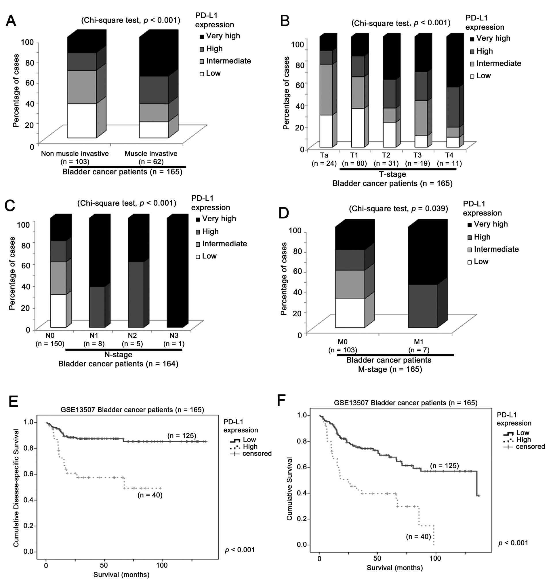
p=0.001, respectively). Muscle invasive bladder cancer specimens
expressed significantly more PD-L1 (Fig. 1A; Chi-square test, p<0.001) and
B7.1 (data not shown). The expression levels of PD-L1 were
significantly higher with a higher T stage (Fig. 1B; Chi-square test, p<0.001) and
were also significantly higher with a higher N stage of the tumors
(Fig. 1C; Chi-square test,
p<0.001). The expression levels of the three genes were
significantly higher in tumors of high grade than those of low
grade (data not shown). In addition, the mRNA expression of PD-L1
was significantly higher (Welch’s t-test, p=0.039) in primary
tumors with M1 (n=7) stage than those with M0 stage (n=158),
although the number of specimens with M1 stage was small (Fig. 1D). We also investigated whether
PD-L1 expression was associated with patient survival by
stratifying the cohort into two sub-cohorts based on their PD-L1
expression. One sub-cohort comprised the quartile of patients with
the highest PD-L1 expression, and the second sub-cohort that of the
remaining three quartiles. Notably, the high expression of PD-L1
mRNA (top 25%) in the primary tumors was significantly associated
with a reduced overall survival time (Fig. 1E; log-rank test, p<0.001) and a
shorter disease-specific survival time of patients (log-rank test,
p<0.001). No significant association was observed for the
expression of B7.1 or PD-1.
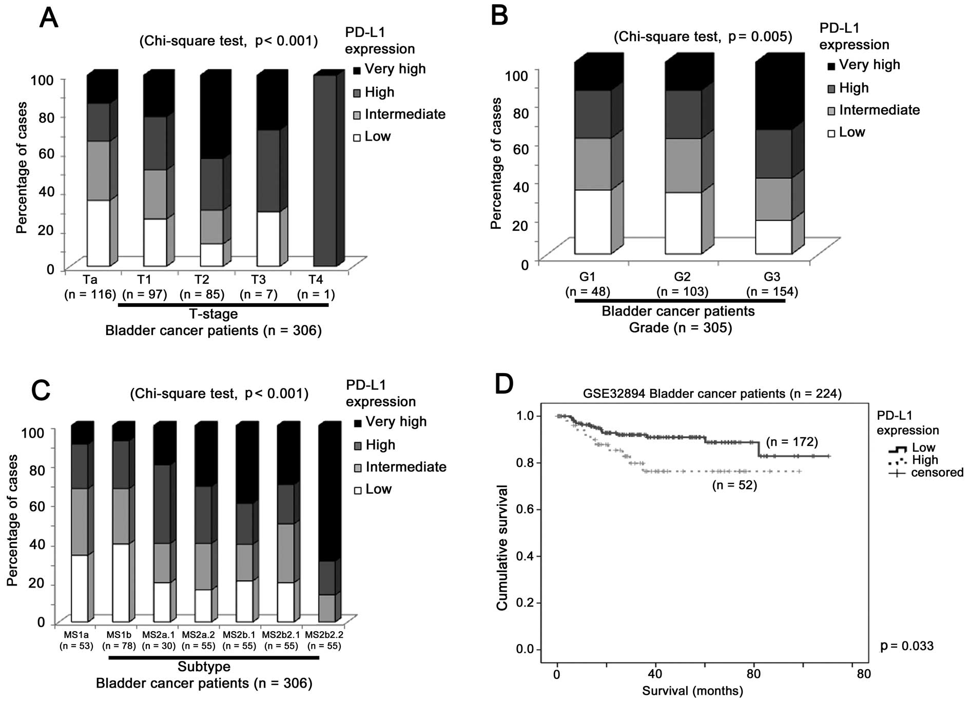
In GSE32894 (n=308) (11), we found that the expression level of
PD-L1 was significantly higher in tumors of higher stage (Fig. 2A, p<0.001) and grade (Fig. 2B, p=0.005). The expression levels of
B7.1 and PD-1 were also significantly higher in tumors of a higher
stage and grade (data not shown). The mRNA expression of PD-L1
(Fig. 2C, p<0.001) and B7.1
(data not shown), but not that of PD-1 (data not shown), was
significantly higher in subtype MS2b2.2, which is one of the two
subtypes with a poorer prognosis compared to the remaining five
subtypes (11). Similar to the
other datasets, a high level of PD-L1 (top 25%) expression was
significantly associated with a shorter overall survival time
(Fig. 2D; log-rank test, p=0.033).
Again, this association was not observed for B7.1 or PD-1
expression.
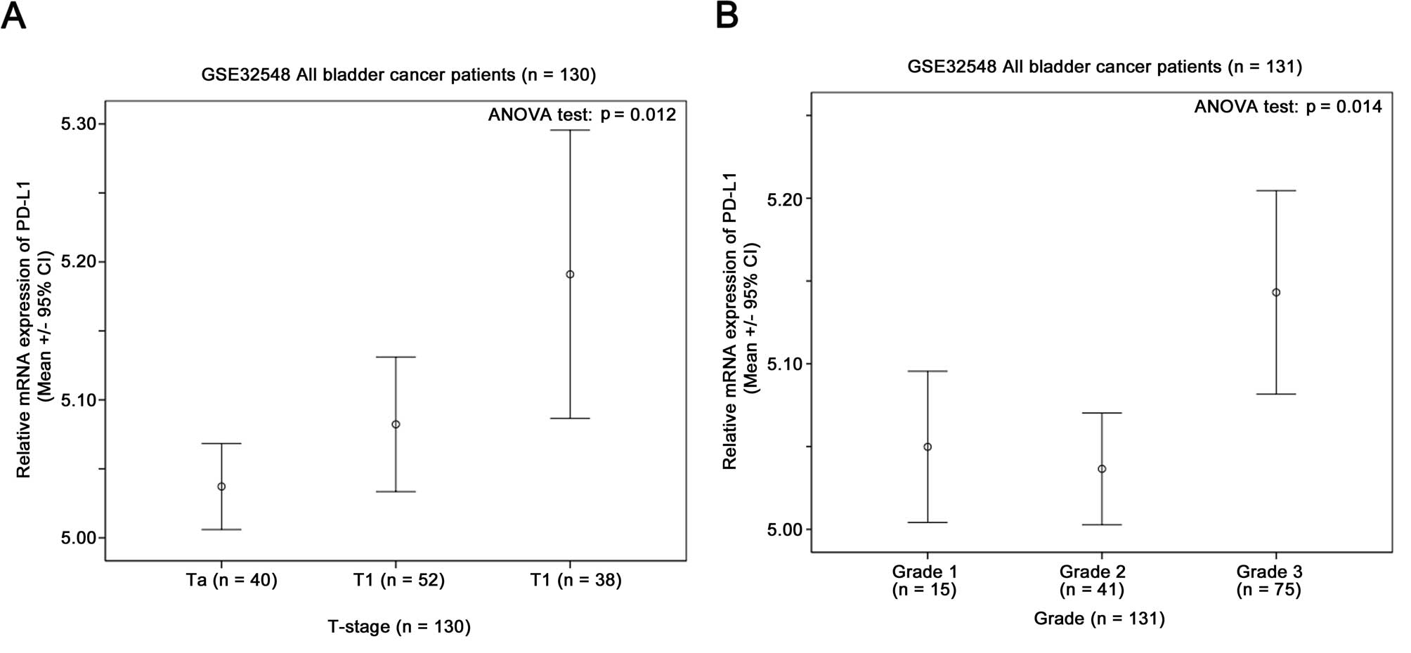
In GSE32548 (n=131) (12), the PD-L1 mRNA expression level was
significantly higher in patients with a higher T-stage (Fig. 3A, p=0.012) and tumor grade (Fig. 3B, p=0.014). The same was true for
B7.1 and PD-1, the expression of which was enhanced with the
increasing T-stage and tumor grade (data not shown).
The results suggested that a high level of the PD-L1
expression predicts a poor prognosis of bladder cancer patients in
terms of patient survival. While an association between the B7.1
expression and the survival of bladder cancer patients was not
demonstrated, its high level of expression was associated
significantly with tumors with more aggressive phenotypes.
Correlations between the expression of
PD-L1, B7.1 and PD-1
The correlations of the mRNA expression of the three
genes were then studied to determine whether they are
co-over-expressed in bladder cancer. The three genes were highly
correlated with each other in the radical cystectomy specimens in
the three datasets analyzed (Fig.
4). In GSE13507 (n=256), the expression of PD-L1 was
significantly positively correlated with that of B7.1 and PD-1
(Fig. 4A and D; Spearman’s rank
test, r=0.233, p<0.001; and r=0.194, p=0.002, respectively),
while the B7.1 expression also significantly positively correlated
with the PD-1 expression (data not shown). In GSE32894 (n=308), the
expression of PD-L1 was significantly positively correlated with
that of B7.1 and PD-1 (Fig. 4B and
E; Spearman’s rank test, r=0.317, p<0.001; and r=0.268,
p<0.001, respectively), while the B7.1 expression was also
significantly positively correlated with the PD-1 expression (data
not shown). In GSE32548 (n=131), the expression of PD-L1 was
significantly positively correlated with that of B7.1 and PD-1
(Fig. 4C and F; Spearman’s rank
test, r=0.456, p<0.001; and r=0.285, p=0.001, respectively),
while the B7.1 expression was also significantly positively
correlated with the PD-1 expression (data not shown). These results
suggested that the mRNA expression of PD-L1 is highly correlated
with that of B7.1 and PD-1.
Association of co-overexpression of PD-L1
and B7.1, and survival in bladder cancer
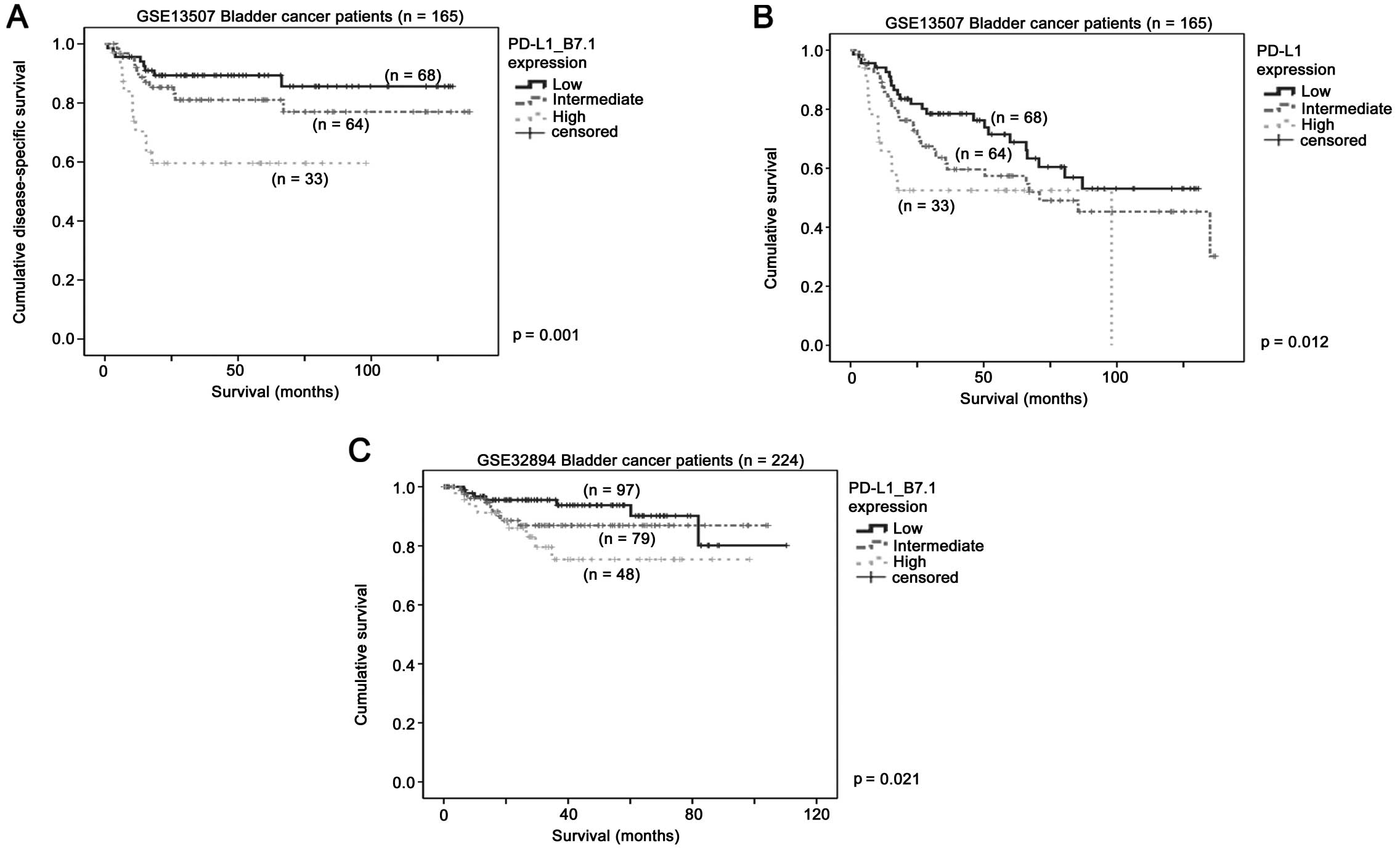
We investigated whether the correlations between
PD-L1 and B7.1, and between PD-L1 and PD-1 may be of prognostic
significance. Patients expressing high levels of PD-L1 and B7.1 had
a shorter survival time than those patients expressing low levels
of PD-L1 and B7.1 (Fig. 5). In
GSE13507, patients with bladder cancer expressing high levels of
PD-L1 and B7.1 had a mean overall survival of 62 months compared to
115 months for those with bladder cancer expressing low levels of
PD-L1 and B7.1 (Fig. 5A; log-rank
test, p=0.001). Similarly, patients with bladder cancer expressing
high levels of the two genes had a mean disease-free survival of
only 56 months, which was significantly shorter than 89 months for
those patients with bladder cancer expressing low levels of the two
genes (Fig. 5B; log-rank test,
p=0.012). In addition, similar results were obtained in GSE32894.
Patients with bladder cancer expressing high level of the two genes
had a mean survival of 79 months, which was significantly shorter
than 100 months for those patients with their bladder cancers
expressing low levels of the two genes (Fig. 5C; log-rank test, p=0.021). However,
no significant association was detected for the co-expression of
PD-L1 and PD-1 or that of B7.1 and PD-1. These results suggested
that the co-overexpression of PD-L1 and B7.1, but not that of the
other combinations, is important for bladder cancer
progression.
PD-L1 expression is an independent
predictor for survival in bladder cancer
Since the PD-L1 expression was the most significant
factor among PD-L1, PD-1 and B7.1, in predicting survival in
bladder cancer, we investigated whether the association between
PD-L1 expression and survival is independent of other predictive
clinicopathological parameters. Since the GSE13507 dataset had a
high sample number as well as a high event rate, this dataset was
optimal for identifying independent predictive factors. As shown in
Tables I and II, using multivariate Cox regression
analysis, PD-L1 expression, T stage, tumor type and age were
independent predictors for disease-specific survival, while for
PD-L1 expression, T stage and age were independent predictors for
overall survival, respectively. This analysis suggested that the
PD-L1 expression is an important prognostic factor in bladder
cancer independent of the T stage as well as whether it is muscle
invasive or not.
 | Table ICox regression analysis of
disease-specific survival in GSE13507 bladder cancer dataset. |
Table I
Cox regression analysis of
disease-specific survival in GSE13507 bladder cancer dataset.
| Clinicopathological
variables | Univariate analysis
| Multivariate analysis
|
|---|
| Hazard ratio (95%
CI) | P-value | Hazard ratio (95%
CI) | P-value |
|---|
| Gender |
| Female (n=30) | 1 | Reference | | |
| Male (n=135) | 0.477
(0.220–1.032) | 0.060 | | |
| Age (years)
(n=165) | 1.051
(1.016–1.088) | 0.004 | 1.061
(1.020–1.096) | 0.001 |
| T stage |
| Early (n=135) | 1 | Reference | 1 | Reference |
| Advanced (n=30) | 12.747
(6.129–26.509) | <0.001 | 3.261
(1.464–7.267) | 0.004 |
| Tumor type |
| Non-muscle invasive
(n=103) | 1 | Reference | 1 | Reference |
| Muscle invasive
(n=62) | 24.719
(7.501–81.457) | <0.001 | 13.342
(3.556–50.065) | <0.001 |
| Grade |
| Low (n=105) | 1 | Reference | | |
| High (n=60) | 5.980
(2.755–12.980) | <0.001 | | |
| PD-L1 expression |
| Low (n=125) | 1 | Reference | 1 | Reference |
| High (n=40) | 4.049
(2.020–8.117) | <0.001 | 2.972
(1.442–6.127) | 0.003 |
| Table IICox regression analysis of overall
survival in GSE13507 bladder cancer dataset. |
Table II
Cox regression analysis of overall
survival in GSE13507 bladder cancer dataset.
| Clinicopathological
variables | Univariate analysis
| Multivariate
analysis
|
|---|
| Hazard ratio (95%
CI) | P-value | Hazard ratio (95%
CI) | P-value |
|---|
| Gender |
| Female (n=30) | 1 | Reference | | |
| Male (n=135) | 0.641
(0.361–1.138) | 0.129 | | |
| Age (years)
(n=165) | 1.070
(1.044–1.096) |
<0.001 | 1.066
(1.039–1.093) |
<0.001 |
| T stage |
| Early (n=135) | 1 | Reference | 1 | Reference |
| Advanced
(n=30) | 4.400
(2.611–7.414) |
<0.001 | 4.576
(2.668–7.846) |
<0.001 |
| Tumor type |
| Non-muscle
invasive (n=103) | 1 | Reference | | |
| Muscle invasive
(n=62) | 2.897
(1.791–4.684) |
<0.001 | | |
| Grade |
| Low (n=105) | 1 | Reference | | |
| High (n=60) | 2.740
(1.694–4.433) |
<0.001 | | |
| PD-L1
expression |
| Low (n=125) | 1 | Reference | 1 | Reference |
| High (n=40) | 3.025
(1.850–4.946) |
<0.001 | 2.273
(1.370–3.771) | 0.001 |
Identification of PD-L1 co-regulated
genes in bladder cancer
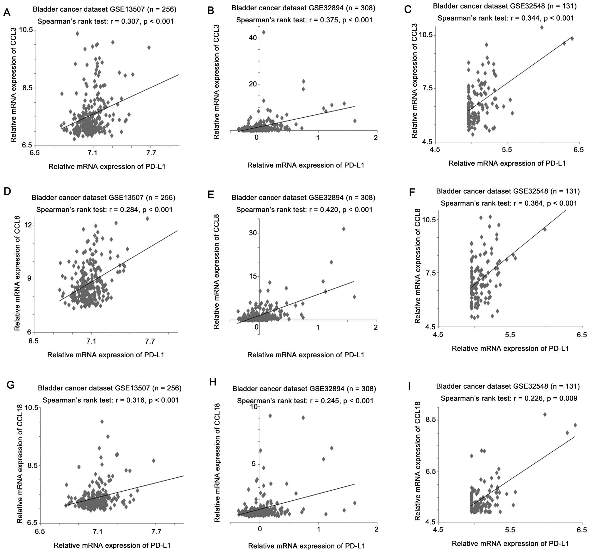
We investigated the differential gene expression
between bladder cancer expressing a high level of PD-L1 and those
expressing a low level of PD-L1. Of the genes that were correlated
with PD-L1 expression in bladder cancer, three chemokine (C-C
motif) ligand (CCL) genes, CCL3, CCL8 and CCL18, were among the top
30 differentially expressed genes with some of the lowest p-values
found during the comparison. Since CCLs have been suggested to play
an important role in malignant growth and metastasis (13), the correlations between PD-L1
expression and the CCL3, CCL8 and CCL18 expression levels were
investigated by the Spearman’s rank test. In GSE13507, PD-L1
expression was significantly positively correlated with the CCL3
(Fig. 6A; r=0.307, p<0.001),
CCL8 (Fig. 6D; r=0.284, p<0.001)
and CCL18 (Fig. 6G; r=0.316,
p<0.001) expression levels. In GSE32894, PD-L1 expression was
significantly positively correlated with CCL3 (Fig. 6B; r=0.375, p<0.001), CCL8
(Fig. 6E; r=0.420, p<0.001) and
CCL18 (Fig. 6H; r=0.245,
p<0.001) expression levels. In GSE32548, PD-L1 expression was
significantly positively correlated with CCL3 (Fig. 6C; r=0.344, p<0.001), CCL8
(Fig. 6F; r=0.364, p<0.001) and
CCL18 (Fig. 6I; r=0.226, p=0.009)
expression levels.
Identification of genes that predict
survival in patients with high level expression of PD-L1
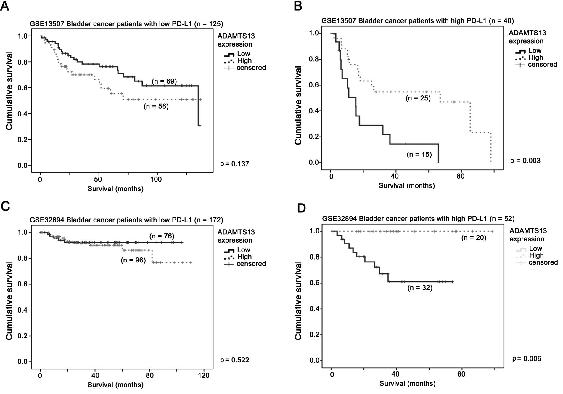
We investigated differentially expressed genes in
patients with a high level of PD-L1 between those who were still
alive and those who were deceased at the last follow-up. Patient
survival data were available in two of the datasets in the present
study. Only the ADAMTS13 expression was shown to confer further
prognostic value in those patients with bladder cancer already
expressing a high level of PD-L1, consistently, in the two datasets
with the survival time available. A lower median expression of the
ADAMTS13 gene was significantly associated with a shorter overall
survival time in the GSE13507 (Fig.
7B; log-rank test, p=0.003) and GSE32894 (Fig. 7D; log-rank test, p=0.006) datasets
only in patients whose bladder cancer expressed a high level of
PD-L1, but not in the other patients (Fig. 7B and D).
Discussion
The PD-L1 pathway is important in cancer
immunosurveillance (14), and is an
important target for cancer immunotherapy (15). MPDL3280A, a monoclonal antibody
targeting PD-L1, has shown promising response in the treatment of
bladder cancer in a phase I study (9). In the present study, we have shown
that the expression of PD-L1, B7.1 and PD-1 was associated with
more aggressive clinicopathological parameters of bladder cancer in
three independent cancer datasets, a highly consistent observation.
Additionally, a high level of expression of PD-L1, but not that of
B7.1 and PD-1, was associated with a shorter survival time in two
independent bladder cancer datasets, suggesting that PD-L1 is a
potential prognostic factor and a promising therapeutic target in
bladder cancer.
The expressions of PD-L1, B7.1 and PD-1 were
significantly correlated with each other. Thus, we investigated the
prognostic significance of their entwining relationships. Only the
PD-L1 and B7.1 co-overexpression was significantly associated with
a shorter survival time in bladder cancer patients. The latter
finding is important, as there is an important difference between
the antibody targeting PD-L1 and PD-1, i.e., while the anti-PD-L1
antibody may be able to block the PD-L1-PD-1 and PD-L1-B7.1
interactions, the anti-PD-1 antibody would be ineffective against
the PD-L1-B7.1 interaction (9,16),
thus rationalizing the use of an anti-PD-L1 antibody, MPDL3280A, in
clinical trials for the treatment of bladder cancer.
CCL3 has been shown to be produced by myeloid cells
from bladder cancer patients, while expansion of these myeloid
cells is associated with cancer-related inflammation and
tumor-induced immune suppression (17). The urinary concentration of CCL18
has been shown to be significantly elevated in bladder cancer
patients and a highly specific biomarker for the early detection of
bladder cancer (18,19). Since CCL18 expression is mainly
detected in inflammatory cells in bladder tumor specimens (20), it follows that a high level of
expression of CCLs in bladder cancer specimens may reflect the
amount of tumor infiltrating lymphocytes. Tumor-infiltrating
lymphocytes are an important phenomenon implicating activation of
the immune system for the killing of tumor cells, which has been
shown to correlate with patient survival (21). However, the overexpression of PD-L1
has been shown to be common in tumor cells and infiltrating
lymphocytes, and this suppresses the activation of these immune
cells and their ability to kill tumor cells (4,22). In
the present study, we have shown that the expression of PD-L1 in
bladder cancer specimens was significantly positively correlated
with the expression of CCLs, and we hypothesize that PD-L1
expression in tumor specimens may be associated with increased
tumor-infiltrating lymphocytes, although due to the high level of
expression of PD-L1, patients would still possess poorer
prognosis.
ADAMTS13 is a metalloprotease that is crucial in the
prevention of spontaneous microvascular thrombosis (23). ADAMTS13 has been found to play a
role in the regulation of leukocyte adhesion and extravasation
during inflammation (23,24). Tumor-associated inflammation may
induce the expression of various molecules in tumor sites and
create a microenvironment that promotes cancer development
(25). Since our data suggest that
expression of ADAMTS13 confers further prognostic value to PD-L1
high bladder cancer, we find it possible that ADAMTS13-modulated
inflammation may impair PD-L1-mediated cancer immunosurveillance.
Future studies may be useful to determine the interrelationship of
ADAMTS13 and PD-L1 in the progression of bladder cancer.
In conclusion, our data suggest that PD-L1 plays an
important role in bladder cancer progression, and that PD-L1 is a
promising therapeutic target for bladder cancer.
Acknowledgments
This study was supported by the National Science
Foundation for Young Scientists of China (grant no. 31301172), the
Natural Science Foundation of Fujian Province (grant no.
2014J01122), and the University of Macau Start-Up Research Grant
& Multi-Year Research Grant (grant no. SRG2014-00006-FHS &
MYRG2015-00065-FHS respectively). We would like to thank Omic
Science and Technology Limited for supporting the gene analysis in
the present study.
References
|
1
|
Liakou CI, Narayanan S, Ng Tang D,
Logothetis CJ and Sharma P: Focus on TILs: Prognostic significance
of tumor infiltrating lymphocytes in human bladder cancer. Cancer
Immun. 7:102007.PubMed/NCBI
|
|
2
|
Redelman-Sidi G, Glickman MS and Bochner
BH: The mechanism of action of BCG therapy for bladder cancer - a
current perspective. Nat Rev Urol. 11:153–162. 2014. View Article : Google Scholar : PubMed/NCBI
|
|
3
|
Alexandroff AB, Jackson AM, O’Donnell MA
and James K: BCG immunotherapy of bladder cancer: 20 years on.
Lancet. 353:1689–1694. 1999. View Article : Google Scholar : PubMed/NCBI
|
|
4
|
Chen DS, Irving BA and Hodi FS: Molecular
pathways: Next-generation immunotherapy - inhibiting programmed
death-ligand 1 and programmed death-1. Clin Cancer Res.
18:6580–6587. 2012. View Article : Google Scholar : PubMed/NCBI
|
|
5
|
Nakanishi J, Wada Y, Matsumoto K, Azuma M,
Kikuchi K and Ueda S: Overexpression of B7-H1 (PD-L1) significantly
associates with tumor grade and postoperative prognosis in human
urothelial cancers. Cancer Immunol Immunother. 56:1173–1182. 2007.
View Article : Google Scholar
|
|
6
|
Inman BA, Sebo TJ, Frigola X, Dong H,
Bergstralh EJ, Frank I, Fradet Y, Lacombe L and Kwon ED: PD-L1
(B7-H1) expression by urothelial carcinoma of the bladder and
BCG-induced granulomata: Associations with localized stage
progression. Cancer. 109:1499–1505. 2007. View Article : Google Scholar : PubMed/NCBI
|
|
7
|
Boorjian SA, Sheinin Y, Crispen PL, Farmer
SA, Lohse CM, Kuntz SM, Leibovich BC, Kwon ED and Frank I: T-cell
coregulatory molecule expression in urothelial cell carcinoma:
Clinicopathologic correlations and association with survival. Clin
Cancer Res. 14:4800–4808. 2008. View Article : Google Scholar : PubMed/NCBI
|
|
8
|
Xylinas E, Robinson BD, Kluth LA, Volkmer
BG, Hautmann R, Küfer R, Zerbib M, Kwon E, Thompson RH, Boorjian
SA, et al: Association of T-cell co-regulatory protein expression
with clinical outcomes following radical cystectomy for urothelial
carcinoma of the bladder. Eur J Surg Oncol. 40:121–127. 2014.
View Article : Google Scholar
|
|
9
|
Powles T, Vogelzang NJ, Fine GD, Eder JP,
Braiteh FS, Loriot Y, Zambrano CC, Bellmunt J, Burris HA, Teng SM,
et al: Inhibition of PD-L1 by MPDL3280A and clinical activity in
pts with metastatic urothelial bladder cancer (UBC). J Clin Oncol
(ASCO Meeting Abstracts). 32:50112014.
|
|
10
|
Lee JS, Leem SH, Lee SY, Kim SC, Park ES,
Kim SB, Kim SK, Kim YJ, Kim WJ and Chu IS: Expression signature of
E2F1 and its associated genes predict superficial to invasive
progression of bladder tumors. J Clin Oncol. 28:2660–2667. 2010.
View Article : Google Scholar : PubMed/NCBI
|
|
11
|
Sjödahl G, Lauss M, Lövgren K, Chebil G,
Gudjonsson S, Veerla S, Patschan O, Aine M, Fernö M, Ringnér M, et
al: A molecular taxonomy for urothelial carcinoma. Clin Cancer Res.
18:3377–3386. 2012. View Article : Google Scholar : PubMed/NCBI
|
|
12
|
Lindgren D, Sjödahl G, Lauss M, Staaf J,
Chebil G, Lövgren K, Gudjonsson S, Liedberg F, Patschan O, Månsson
W, et al: Integrated genomic and gene expression profiling
identifies two major genomic circuits in urothelial carcinoma. PLoS
One. 7:e388632012. View Article : Google Scholar : PubMed/NCBI
|
|
13
|
Kulbe H, Levinson NR, Balkwill F and
Wilson JL: The chemokine network in cancer - much more than
directing cell movement. Int J Dev Biol. 48:489–496. 2004.
View Article : Google Scholar
|
|
14
|
Corthay A: Does the immune system
naturally protect against cancer? Front Immunol. 5:1972014.
View Article : Google Scholar : PubMed/NCBI
|
|
15
|
Kyi C and Postow MA: Checkpoint blocking
antibodies in cancer immunotherapy. FEBS Lett. 588:368–376. 2014.
View Article : Google Scholar
|
|
16
|
Langer CJ: Emerging immunotherapies in the
treatment of non-small cell lung cancer (NSCLC): The role of immune
checkpoint inhibitors. Am J Clin Oncol. Mar 28–2014.Epub ahead of
print. View Article : Google Scholar : PubMed/NCBI
|
|
17
|
Eruslanov E, Neuberger M, Daurkin I,
Perrin GQ, Algood C, Dahm P, Rosser C, Vieweg J, Gilbert SM and
Kusmartsev S: Circulating and tumor-infiltrating myeloid cell
subsets in patients with bladder cancer. Int J Cancer.
130:1109–1119. 2012. View Article : Google Scholar
|
|
18
|
Urquidi V, Kim J, Chang M, Dai Y, Rosser
CJ and Goodison S: CCL18 in a multiplex urine-based assay for the
detection of bladder cancer. PLoS One. 7:e377972012. View Article : Google Scholar : PubMed/NCBI
|
|
19
|
Goodison S, Chang M, Dai Y, Urquidi V and
Rosser CJ: A multianalyte assay for the non-invasive detection of
bladder cancer. PLoS One. 7:e474692012. View Article : Google Scholar
|
|
20
|
Miyake M, Ross S, Lawton A, Chang M, Dai
Y, Mengual L, Alcaraz A, Giacoia EG, Goodison S and Rosser CJ:
Investigation of CCL18 and A1AT as potential urinary biomarkers for
bladder cancer detection. BMC Urol. 13:422013. View Article : Google Scholar : PubMed/NCBI
|
|
21
|
Zhang L, Conejo-Garcia JR, Katsaros D,
Gimotty PA, Massobrio M, Regnani G, Makrigiannakis A, Gray H,
Schlienger K, Liebman MN, et al: Intratumoral T cells, recurrence,
and survival in epithelial ovarian cancer. N Engl J Med.
348:203–213. 2003. View Article : Google Scholar : PubMed/NCBI
|
|
22
|
Chen DS and Mellman I: Oncology meets
immunology: The cancer-immunity cycle. Immunity. 39:1–10. 2013.
View Article : Google Scholar : PubMed/NCBI
|
|
23
|
Gandhi C, Khan MM, Lentz SR and Chauhan
AK: ADAMTS13 reduces vascular inflammation and the development of
early atherosclerosis in mice. Blood. 119:2385–2391. 2012.
View Article : Google Scholar :
|
|
24
|
Chauhan AK, Kisucka J, Brill A, Walsh MT,
Scheiflinger F and Wagner DD: ADAMTS13: A new link between
thrombosis and inflammation. J Exp Med. 205:2065–2074. 2008.
View Article : Google Scholar : PubMed/NCBI
|
|
25
|
Atsumi T, Singh R, Sabharwal L, Bando H,
Meng J, Arima Y, Yamada M, Harada M, Jiang JJ, Kamimura D, et al:
Inflammation amplifier, a new paradigm in cancer biology. Cancer
Res. 74:8–14. 2014. View Article : Google Scholar
|