Introduction
Consumption of soy-based foods has been associated
with a lower risk of breast cancer, the most frequently diagnosed
cancer in women worldwide (1,2). The
isoflavones genistein and daidzein are naturally occurring phenolic
compounds. The main sources of dietary isoflavones are soybeans,
although they are present in many herbs and foods of botanical
origin. Among the isoflavones, genistein is well-known to have a
hypolipidemic effect and prevent cardiovascular disease by
regulating lipid and carbohydrate metabolism (3,4).
Daidzein has been demonstrated to be a potent antioxidant and has
received much attention in relation to human health (5). In recent years, the anticancer
activity of daidzein has been given much attention. Magee et
al reported that daidzein suppressed MDA-MB-231 breast cancer
cell invasion by reducing matrix metalloproteinase (MMP)-2
activity, suggesting an important role of daidzein in breast
carcinogenesis (6). In another
study, the inhibitory effect of daidzein on AML-1 cells was weak on
day 3, yet it was evident on day 5 at concentrations of 20, 100,
200 and 400 µM (7). In order
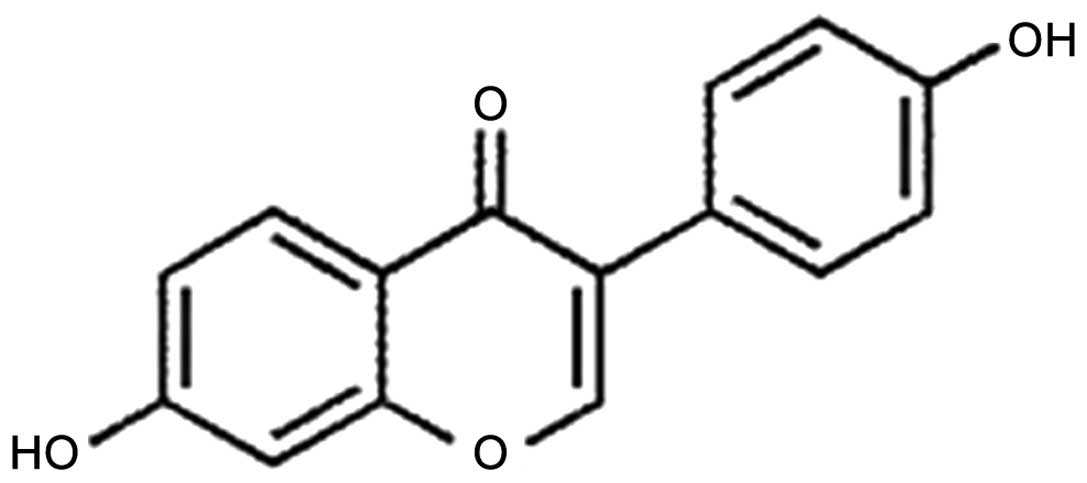
to obtain more insight into the anticancer activity of daiazein
(Fig. 1), in the present study, the
cytotoxic effect of daiazein on BEL-7402, A549, HeLa, HepG-2 and
MG-63 cell lines was investigated. Apoptosis, DNA damage, levels of
reactive oxygen species and mitochondrial membrane potential in
BEL-7402 cells was assessed by fluorescence microscopy. The
percentage of apoptotic cells and cell cycle arrest were
investigated by flow cytometry. The expression of Bcl-2 family
proteins was performed by western blot analysis. The results
demonstrated that daidzein induced BEL-7402 cell apoptosis through
an ROS-mediated mitochondrial dysfunction pathway.
Materials and methods
All reagents and solvents were purchased
commercially and used without further purification unless otherwise
noted. Ultrapure Milli-Q water was used in all experiments.
Daiazein was purchased from the Aladdin Industrial Corporation.
Dimethylsulfoxide (DMSO) and RPMI-1640 were purchased from Sigma.
HepG-2 (human hepatocellular carcinoma), A549 (human lung
carcinoma), BEL-7402 (hepatocellular), MG-63 (human osteosarcoma)
and HeLa (human cervical cancer) cell lines were purchased from the
American Type Culture Collection (ATCC; Manassas, VA, USA).
Cytotoxic activity in vitro
3-(4,5-Dimethylthiazol-2-yl)-2,5-diphenyltetrazolium
bromide (MTT) assay was used as previously described (8). Cells were placed in 96-well
micro-assay culture plates (8×10³ cells/well) and grown
overnight at 37°C in a 5% CO2 incubator. The compound
tested was then added to the wells to achieve final concentrations
ranging from 10−6 to 10−4 M. Control wells
were prepared by addition of culture medium (100 µl). The
plates were then incubated at 37°C in a 5% CO2 incubator
for 48 h. Upon completion of the incubation, stock MTT dye solution
(20 µl, 5 mg/ml) was added to each well. After 4 h, buffer
(100 µl) containing dimethylformamide (50%) and sodium
dodecyl sulfate (20%) was added to solubilize the MTT formazan. The
optical density of each well was measured with a microplate
spectrophotometer at a wavelength of 490 nm. The IC50
values were determined by plotting the percentage of cell viability
vs. concentration on a logarithmic graph and by reading the
concentration at which 50% of cells remained viable relative to the
control. Each experiment was repeated at least three times to
obtain the mean values. Five different tumor cell lines were used
in the present study: A549, BEL-7402, MG-63, HepG-2 and HeLa.
Apoptosis assay by AO/EB staining
method
BEL-7402 cells were seeded onto chamber slides in
6-well plates at a density of 2×10⁵ cells/well and
incubated for 24 h. The cells were cultured in RPMI-1640
supplemented with 10% of fetal bovine serum (FBS) and incubated at
37°C in 5% CO2. The medium was removed and replaced with
medium (final DMSO concentration, 0.05% v/v) containing daidzein
(30 µM) for 24 h. The medium was removed again, and the
cells were washed with ice-cold phosphate-buffer saline (PBS), and
fixed with formalin (4%, w/v). Cell nuclei were counterstained with
acridine orange (AO) and ethidium bromide (EB) (AO, 100
µg/ml; EB, 100 µg/ml) for 10 min. The cells were
observed and imaged with a fluorescence microscope (Nikon,
Yokohama, Japan) with excitation at 350 nm and emission at 460
nm.
Comet assay
DNA damage was investigated by means of comet assay.
BEL-7402 cells in culture medium were incubated with 30 and 60
µM of daidzein for 24 h at 37°C. The control cells were also
incubated for the same time periods. The cells were harvested by a
trypsinization process at 24 h. A total of 100 µl of 0.5%
normal agarose in PBS was dropped gently onto a fully frosted
microslide, covered immediately with a coverslip, and then placed
at 4°C for 10 min. The coverslip was removed after the gel had set.
The cell suspension (50 µl) (200 cells/µl) was mixed
with 50 µl of 1% low melting agarose preserved at 37°C. A
total of 100 µl of this mixture was applied quickly on top
of the gel, coated over the microslide, covered immediately with a
coverslip, and then placed at 4°C for 10 min. The coverslip was
again removed after the gel had set. A third coating of 50
µl of 0.5% low melting agarose was placed on the gel and
allowed to set at 4°C for 15 min. After solidification of the
agarose, the coverslips were removed, and the slides were immersed
in an ice-cold lysis solution (2.5 M NaCl, 100 mM EDTA, 10 mM Tris,
90 mM sodium sarcosinate, NaOH, pH 10, 1% Triton X-100 and 10%
DMSO) and placed in a refrigerator at 4°C for 2 h. All of the above
operations were performed under low lighting conditions to avoid
additional DNA damage. The slides, after removal from the lysis
solution, were placed horizontally in an electrophoresis chamber.
The reservoirs were filled with an electrophoresis buffer (300 mM
NaOH, 1.2 mM EDTA) until the slides were just immersed in it, and
the DNA was allowed to unwind for 30 min in electrophoresis
solution. Then the electrophoresis was carried out at 25 V and 300
mA for 20 min. After electrophoresis, the slides were removed,
washed thrice in a neutralization buffer (400 mM Tris, HCl, pH
7.5). Cells were stained with 20 µl of EB (20
µg/ml−) in the dark for 20 min. The slides were
washed in chilled distilled water for 10 min to neutralize the
excess alkali, air-dried and scored for comets by fluorescence
microscopy.
Reactive oxygen species (ROS)
detection
BEL-7402 cells were seeded into 6-well plates
(Costar, Corning Inc., Corning, New York, NY, USA) at a density of
2×10⁵ cells/well and incubated for 24 h. The cells were
cultured in RPMI-1640 medium supplemented with 10% of FBS and
incubated at 37°C in 5% CO2. The medium was removed and
replaced with medium (final DMSO concentration, 0.05% v/v)
containing daidzein (30 µM) for 24 h. The medium was removed
again. The fluorescent dye 2′,7′-dichlorodihydrofluorescein
diacetate (DCHF-DA; 10 µM) was added to the medium to cover
the cells. The treated cells were then washed with cold PBS-EDTA
twice, collected by trypsinization and centrifugation at 1,500 rpm
for 5 min. The cell pellets were then suspended in PBS-EDTA and
imaged with a fluorescence microscope. The fluorescent intensity
was determined by a microplate analyzer (Infinite M200; Tecan,
Switzerland) with excitation at 488 nm and emission at 525 nm.
Mitochondrial membrane potential
assay
BEL-7402 cells were treated for 24 h with daidzein
(30, 60 and 90 µM) in 12-well plates and were then washed
three times with cold PBS. The cells were detached with
trypsin-EDTA solution. Collected cells were incubated for 20 min
with 1 µg/ml of JC-1 in culture medium at 37°C in the dark.
Cells were immediately centrifuged to remove the supernatant. Cell
pellets were suspended in PBS and imaged by fluorescence
microscopy. The fluorescence intensity was determined by a
microplate analyzer (Infinite M200) with excitation set at 488 nm
and emission at 525 nm.
Apoptosis assay by flow cytometry
After chemical treatment, 1×10⁶ cells
were harvested, washed with PBS, then fixed with 70% ethanol and
finally maintained at 4°C for at least 12 h. The pellets were
stained with the fluorescent probe solution containing 50 mg/ml
propidium iodide (PI) and 1 mg/ml Annexin in PBS on ice in dark for
15 min. Then the fluorescence emission was measured at 530 and 575
nm (or equivalent) using 488 nm excitation by a FACSCalibur flow
cytometry (Becton, Dickinson and Company, Franklin Lakes, NJ, USA).
A minimum of 10,000 cells were analyzed per sample.
Cell cycle arrest by flow cytometry
BEL-7402 cells were seeded into 6-well plates
(Costar, Corning Inc.) at a density of 2×10⁵ cells/well
and incubated for 24 h. The cells were cultured in RPMI-1640 medium
supplemented with 10% FBS and incubated at 37°C in 5%
CO2. The medium was removed and replaced with medium
(final DMSO concentration, 0.05% v/v) containing daidzein (30
µM). After incubation for 24 h, the cell layer was
trypsinized and washed with cold PBS and fixed with 70% ethanol.
Twenty microliters of RNase (0.2 mg/ml) and 20 µl of PI
(0.02 mg/ml) were added to the cell suspensions and incubated at
37°C for 30 min. Then, the samples were analyzed with a FACSCalibur
flow cytometry. The number of cells analyzed for each sample was
10,000 (9).
Western blot analysis
BEL-7402 cells were seeded in 3.5-cm dishes for 24 h
and incubated with different concentrations of daidzein in the
presence of 10% FBS. The cells were harvested in lysis buffer.
After sonication, the samples were centrifuged for 20 min at 13,000
g. The protein concentration of the supernatant was determined by
BCA assay. Sodium dodecyl sulfate-polyacrylamide gel
electrophoresis was carried out loading equal amount of
proteins/lane. Gels were then transferred to polyvinylidene
difluoride membranes (Millipore) and blocked with 5% non-fat milk
in Tris-buffered saline with Tween-20 (TBST) buffer for 1 h. Then,
the membranes were incubated with primary antibodies at a 1:5,000
dilution in 5% non-fat milk overnight at 4°C, and washed four times
with TBST for a total of 30 min. The membranes were then incubated
which the secondary antibodies conjugated with horseradish
peroxidase at a 1:5,000 dilution for 1 h at room temperature and
then washed four times with TBST. The blots were visualized with
Amersham ECL Plus Western Blotting detection reagents according to
the manufacturer's instructions. To assess the presence of
comparable amount of proteins in each lane, the membranes were
finally stripped to detect β-actin.
Results and Discussion
In vitro cytotoxicity assay
The cytotoxicity in vitro of daidzein was
evaluated by the MTT method in 5 cancer cell lines: BEL-7402, A549,
HeLa, HepG-2 and MG-63. The IC50 values of daidzein
against the selected cell lines are listed in Table I. After the BEL-7402, A549, HeLa,
HepG-2 and MG-63 cells were exposed to 6.25, 12.5, 25, 50 and 100
µM of daidzein for 48 h, the IC50 value of
daidzein toward BEL-7402 cells was 59.7±8.1 µM. Obviously,
daidzein showed moderate cytotoxic activity against the BEL-7402
cells. Unexpectedly, daidzein had no cytotoxic activity against the
A549, HeLa, HepG-2 and MG-63 cells; the IC50 values were
>100 µM. The results demonstrated that daidzein displays
different cytotoxic effects on different cancer cell lines.
 | Table IIC50 values of daidzein in
the BEL-7402, HeLa, A549, HepG-2 and MG-63 cell lines. |
Table I
IC50 values of daidzein in
the BEL-7402, HeLa, A549, HepG-2 and MG-63 cell lines.
| Compound | IC50
(µM) in cell lines
|
|---|
| BEL-7402 | HeLa | A549 | HepG-2 | MG-63 |
|---|
| Daidzein | 59.7±8.1 | 97.9±11.3 | >100 | >100 | >100 |
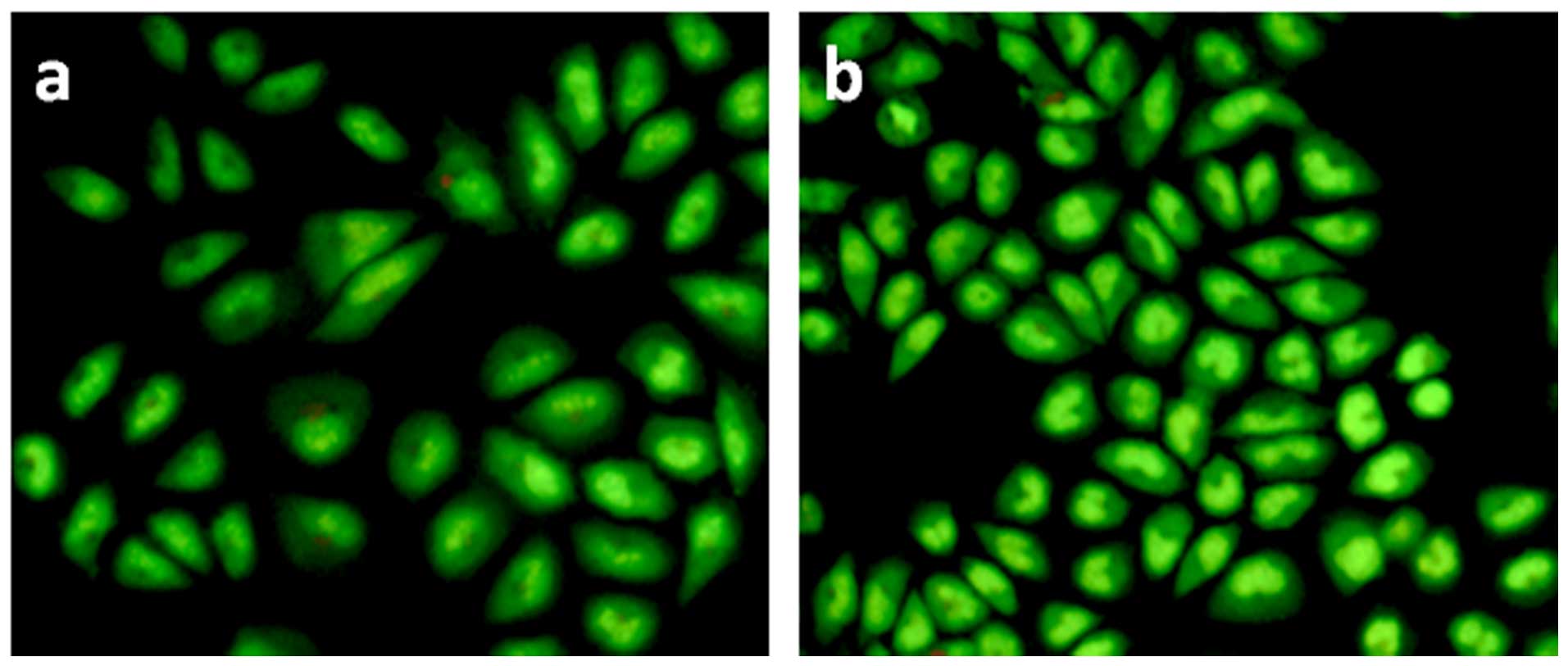
Apoptosis assay with AO/EB staining
method
Induction of apoptosis is one of the considerations
in drug development, as most cytotoxic anticancer drugs in current
use induce apoptosis in susceptible cells (10). In order to determine whether or not
daidzein induces chromatin condensation and fragmentation, both of
which are recognized morphological features of apoptosis, BEL-7402
cells were treated with 30 µM of daidzein for 24 h. As shown
in Fig. 2a, control BEL-7402 cells
were stained with uniform green fluorescence and no apoptotic
features were observed. Following treatment of BEL-7402 cells with
daidzein for 24 h, obvious morphological changes and green
apoptotic cells containing apoptotic characteristics such as cell
blebbing, nuclear shrinkage and chromatin condensation were
observed (Fig. 2b). The results
suggest that daidzein induced BEL-7402 cell apoptosis.
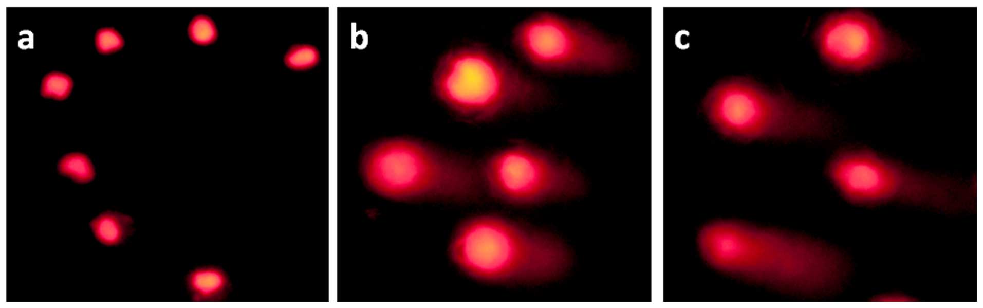
DNA damage assay
DNA fragmentation is a hallmark of apoptosis,
mitotic catastrophe, or both (11).
DNA damage is assayed by the use of single-cell gel electrophoresis
(comet assay) in agarose gel matrix. As shown in Fig. 3a, in the control cells, no comet
like appearance was observed. After BEL-7402 cells were exposed to
30 (Fig. 3b) and 60 µM
(Fig. 3c) of daidzein for 24 h, a
statistically significant number of well-formed comets were noted.
Furthermore, the length of the comet tails increased with
increasing concentrations of daidzein. These results indicate that
daidzein induced DNA fragmentation, which was further evidence of
apoptosis.
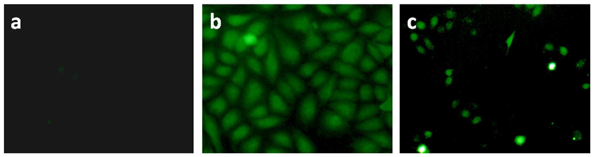
Detection of ROS levels by fluorescence
microscope
To determine the effect of daidzein on intracellular
ROS generation, DCHF-DA was used as a fluorescent probe. DCFH-DA is
a fluorescent dye that diffuses through cell membranes and is
hydrolyzed by intracellular esterases to DCFH. In the presence of
ROS, DCFH is oxidized to DCF, which is fluorescent and its level
corresponds to the level of generated ROS. As shown in Fig. 4a, in the control, no obvious
fluorescence images were found. Following treatment of BEL-7402
cells with Rosup (Fig. 4b, positive
control) and 30 µM of daidzein (Fig. 4c) for 24 h, the bright green
fluorescence images were observed. The results indicate that
daidzein increased the levels of ROS.
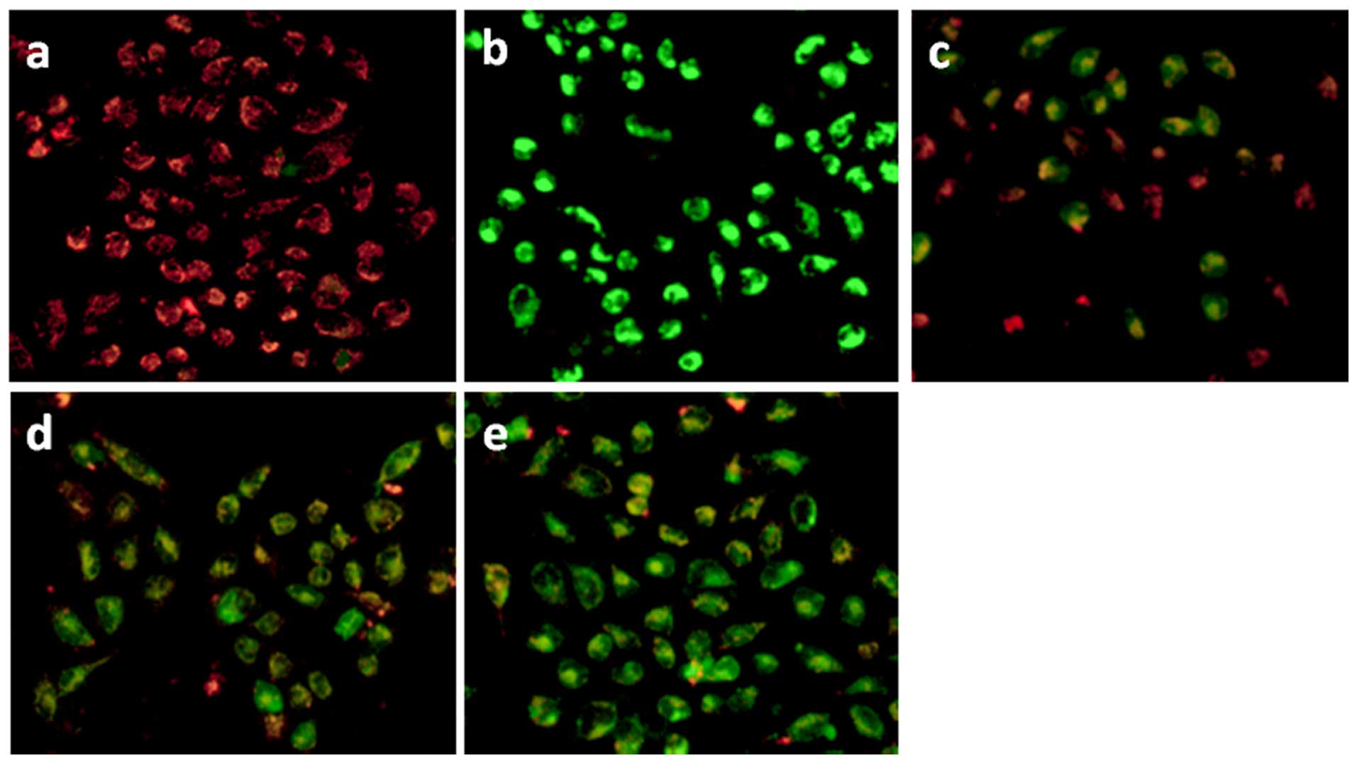
Changes in the mitochondrial membrane
potential
The changes in the mitochondrial membrane potential
were determined by fluorescence microscope. JC-1 was used as a
fluorescence probe for detecting the changes in the mitochondrial
membrane potential induced by daidzein. JC-1 forms aggregates,
which have a red fluorescence emission peak at high mitochondrial
membrane potential; JC-1 forms monomers, which emit a green
fluorescence peak at low mitochondrial membrane potential. As shown
in Fig. 5a, in the control, JC-1
exhibited a red fluorescence (JC-1 aggregates) confirming high
mitochondrial membrane potential. After BEL-7402 cells were exposed
to cccp (Fig. 5b) and 30 (Fig. 5c), 60 (Fig. 5d) and 90 µM (Fig. 5e) daidzein for 24 h, JC-1 showed
green fluorescence (JC-1 monomers) with little red fluorescence
corresponding to low mitochondrial membrane potential. The changes
from red to green fluorescence indicate a decrease in mitochondrial
membrane potential. Moreover, the green fluorescence increased and
the red fluorescence decreased with increasing concentrations of
daidzein. The results demonstrated that daidzein induced a decrease
in mitochondrial membrane potential and daidzein induced apoptosis
in the BEL-7402 cells through a mitochondrial signal transduction
pathway.
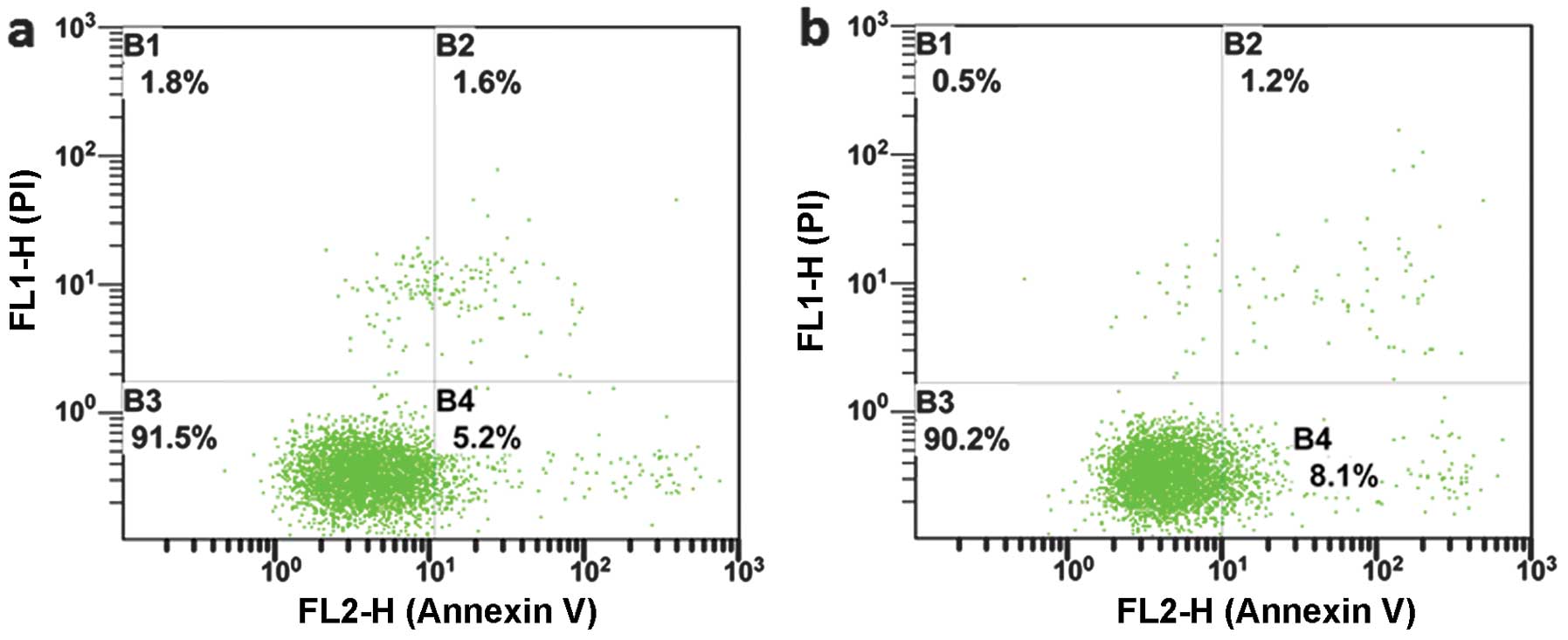
Apoptosis assay by flow cytometry
AO/EB staining studies showed that daidzein induced
the apoptosis of BEL-7402 cells. To determine the percentage of
apoptotic and necrotic BEL-7402 cells, the cells were treated with
30 µM of daidzein for 24 h and stained with Annexin V and PI
followed by cell apoptosis analyses using flow cytometry. The
percentages of apoptotic, necrotic and BEL-7402 living cells are
shown in Fig. 6. In the control
cells (Fig. 6a), the percentage of
apoptotic (B4) and necrotic (B2) cells were 5.2 and 1.6%,
respectively. Following treatment of BEL-7402 cells with daidzein
for 24 h (Fig. 6b), the percentages
of apoptotic and necrotic cells were 8.1 and 1.2%, respectively.
The increase in the percentage of apoptotic cells of 2.9% suggests
that daidzein induced apoptosis in the BEL-7402 cells.
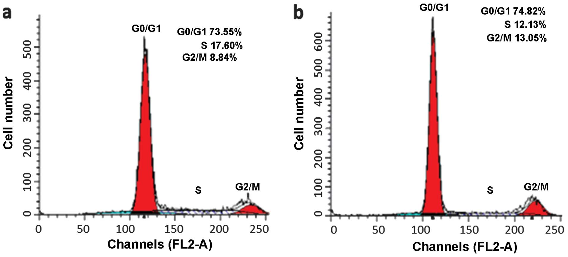
Cell cycle arrest studies
Flow cytometry was used to determine the effects of
daidzein on the cell cycle progression of BEL-7402 cells. As shown
in Fig. 7a, in the control cells,
the percentage of cells in the G2/M and S phases were 8.84 and
17.60%, respectively. After BEL-7402 cells were exposured to 30
µM of daidzein (Fig. 7b) for
24 h, the percentage of cells at the G2/M and S phases were 13.05
and 12.13%, respectively. An evident increase of 4.21% in the
percentage of cells at the G2/M phase was observed, which was
accompanied by a corresponding reduction of 5.47% in the percentage
of cells in the S phase. The data showed that daidzein induced cell
cycle arrest at the G2/M phase.
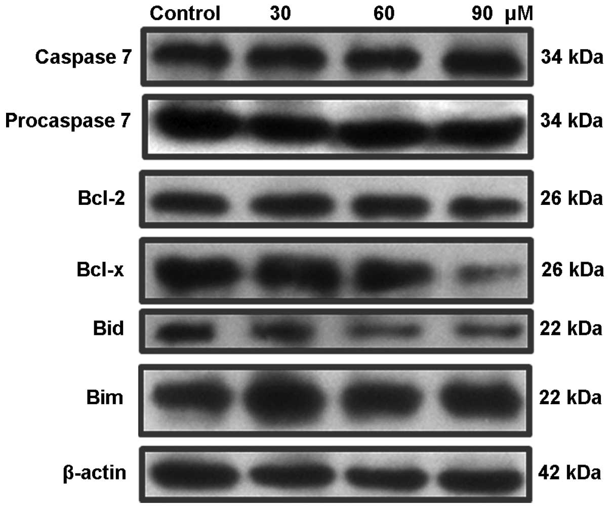
Expression of caspase and Bcl-2 family
proteins
Caspase 3 and 7 are executioners of apoptosis as the
processing of their substrates leads to morphological changes
associated with apoptosis, including DNA degradation, chromatin
condensation and membrane blebbing (12). The expression of caspase 7 and
procaspase 7 was assayed by western blot analysis. As shown in
Fig. 8, after the treatment of
BEL-7402 cells with different concentrations of daidzein, the
expression levels of caspase 7 increased, whereas the expression
levels of procaspase 7 were downregulated. To investigate the
effect of daidzein on the expression levels of antiapoptotic and
proapoptotic proteins, BEL-7402 cells were treated with different
concentrations of daidzein for 24 h. As expected, the expression
levels of antiapoptotic proteins Bcl-2 and Bcl-x were
downregulated. The levels of the proapoptotic protein Bid
decreased, whereas the expression of Bim was upregulated. These
results demonstrate that daidzein induced apoptosis in BEL-7402
cells through activation of caspase 7, downregulation of Bcl-2,
Bcl-x and Bid, upregulation of Bim and ROS-mediated mitochondrial
dysfunction pathways.
In conclusion, daidzein showed moderate cytotoxic
activity in BEL-7402 cells, while no cytotoxicity toward HeLa, A549
and MG-63 cells was noted. The compound induced the apoptosis of
BEL-7402 cells. Daidzein increased the levels of ROS and induced a
decrease in mitochondrial membrane potential. Additionally,
daidzein inhibited the cell cycle progression of BEL-7402 cells at
the G2/M phase. The compound activated caspase 7, downregulated the
expression of Bcl-2, Bcl-x and Bid, and upregulated the expression
of Bim. In summary, daidzein induced apoptosis in the BEL-7402
cells by an ROS-mediated mitochondrial dysfunction pathway, which
was accompanied by regulation of Bcl-2 family proteins.
Acknowledgments
The present study was supported by the High-Level
Personnel Project of Guangdong Province in 2013, and the Joint
Nature Science Fund of the Department of Science and Technology,
and the First Affiliated Hospital of Guangdong Pharmaceutical
University (no. GYFYLH201315).
References
|
1
|
Shu XO, Zheng Y, Cai H, Gu K, Chen Z,
Zheng W and Lu W: Soy food intake and breast cancer survival. JAMA.
302:2437–2443. 2009. View Article : Google Scholar : PubMed/NCBI
|
|
2
|
Ferlay J, Shin HR, Bray F, Forman D,
Mathers C and Parkin DM: Estimates of worldwide burden of cancer in
2008: GLOBOCAN 2008. Int J Cancer. 127:2893–2917. 2010. View Article : Google Scholar
|
|
3
|
Kreijkamp-Kaspers S, Kok L, Grobbee DE, de
Haan EH, Aleman A, Lampe JW and van der Schouw YT: Effect of soy
protein containing isoflavones on cognitive function, bone mineral
density, and plasma lipids in postmenopausal women: A randomized
controlled trial. JAMA. 292:65–74. 2004. View Article : Google Scholar : PubMed/NCBI
|
|
4
|
Park D, Huang T and Frishman WH:
Phytoestrogens as cardio-protective agents. Cardiol Rev. 13:13–17.
2005.
|
|
5
|
Vedavanam K, Srijayanta S, O'Reilly J,
Raman A and Wiseman H: Antioxidant action and potential
antidiabetic properties of an isoflavonoid-containing soyabean
phytochemical extract (SPE). Phytother Res. 13:601–608. 1999.
View Article : Google Scholar : PubMed/NCBI
|
|
6
|
Magee PJ, Allsopp P, Samaletdin A and
Rowland IR: Daidzein, R-(+)equol and S-(−)equol inhibit the
invasion of MDA-MB-231 breast cancer cells potentially via the
down-regulation of matrix metalloproteinase-2. Eur J Nutr.
53:345–350. 2014. View Article : Google Scholar
|
|
7
|
Hirota K, Morikawa K, Hanada H, Nonaka M,
Nakajima Y, Kobayashi M and Nakajima R: Effect of genistein and
daidzein on the proliferation and differentiation of human
preadipocyte cell line. J Agric Food Chem. 58:5821–5827. 2010.
View Article : Google Scholar : PubMed/NCBI
|
|
8
|
Mosmann T: Rapid colorimetric assay for
cellular growth and survival: Application to proliferation and
cytotoxicity assays. J Immunol Methods. 65:55–63. 1983. View Article : Google Scholar : PubMed/NCBI
|
|
9
|
Lo KK, Lee TK, Lau JS, Poon WL and Cheng
SH: Luminescent biological probes derived from ruthenium(II)
estradiol polypyridine complexes. Inorg Chem. 47:200–208. 2008.
View Article : Google Scholar
|
|
10
|
Hickman JA: Apoptosis induced by
anticancer drugs. Cancer Metastasis Rev. 11:121–139. 1992.
View Article : Google Scholar : PubMed/NCBI
|
|
11
|
Alapetite C, Wachter T, Sage E and
Moustacchi E: Use of the alkaline comet assay to detect DNA repair
deficiencies in human fibroblasts exposed to UVC, UVB, UVA and
gamma-rays. Int J Radiat Biol. 69:359–369. 1996. View Article : Google Scholar : PubMed/NCBI
|
|
12
|
Danial NN and Korsmeyer SJ: Cell death:
Critical control points. Cell. 116:205–219. 2004. View Article : Google Scholar : PubMed/NCBI
|