Introduction
Cervical cancer, a prevalent gynecological
malignancy, is the fourth leading cause of cancer-related death in
women worldwide, with an estimated 527,600 new cases and 265,700
deaths worldwide in 2012 (1). The
majority of cases occur in developing countries and shows a trend
for younger patients (2). Despite
the decreasing incidence and mortality rate of cervical cancer in
recent years owing to improved diagnosis and treatment, the
clinical outcome for patients with advanced-stage disease remains
bleak (2). Moreover, lymph node
metastasis (LNM) can cause higher mortality and recurrence rates,
even in patients with early-stage cervical cancer, and definite
information on lymph node status is essential for tailoring
adjuvant treatment (3,4). However, no sensitive biomarkers
specific for indication of LNM, and the early detection and
prognosis of cervical cancer are available to date. Therefore, it
is urgent to identify novel molecular markers of cervical cancer to
facilitate a more accurate prediction of clinical outcome and to
prescribe effective treatment.
GINS complex subunit 2 (GINS2), also known as PSF2,
encodes a protein with a molecular weight of ~21 kDa. GINS2 belongs
to the GINS complex family that also consists of GINS2, GINS3 and
GINS4 (5). The GINS complex has
been identified as playing a critical role in the initiation of DNA
replication and the cell cycle. Stably interacting with
minichromosome maintenance (MCM) 2–7 complex and CDC45, the GINS
family functions to correctly establish and maintain DNA
replication forks (5). Moreover,
GINS components may play a role in cell division, and more
accurately, in chromosome segregation (6). GINS2 was reported to be involved in
tumorigenesis in several types of cancers. For example, genome-wide
gene expression profile analysis revealed that GINS2 is highly
expressed in lung carcinoma (7).
Zheng et al indicated that GINS2 is correlated with
aggressive characteristics of breast cancer, and speculated that it
is involved in lung metastasis (8).
In addition, enhanced expression of GINS2 was found to promote
leukemia cell proliferation and desensitize cells to apoptosis
(9). These findings all suggest
that GINS2 plays an important role in cancer progression. However,
the clinical significance of GINS2 in cervical cancer has not been
investigated.
In the present study, we explored the GINS2
expression pattern and its clinical implication and prognostic
significance in early-stage cervical cancer. Furthermore, we gained
insight into the important functions of this protein in cervical
cancer development.
Materials and methods
Cell lines
Six cervical cancer cell lines, SiHa, HeLa, C33A,
Caski, MS751 and ME180, were purchased from the American Type
Culture Collection (ATCC; Rockville, MD, USA), and HCC94 and
HeLa299, were purchased from the Cell Bank of the Type Culture
Collection of the Chinese Academy of Sciences (Shanghai, China).
The cell lines were all cultured in RPMI-1640 medium (Gibco, Grand
Island, NY, USA) supplemented with 10% fetal bovine serum (FBS)
(HyClone Laboratories, Logan, UT, USA) and 1% antibiotics.
Patients and samples
In the present study, we enrolled 155 patients with
cervical cancer who underwent radical hysterectomy and
lymphadenectomy at The First Affiliated Hospital, Sun Yat-sen
University. All patients had stage IA2-IIA disease and received
treatment from January 2007 to December 2009. The clinical staging
and clinicopathological classifications were determined according
to the International Federation of Obstetrics and Gynecology
(FIGO), 2009. The clinicopathological characteristics of the
enrolled cases are summarized in Table
I. The follow-up duration for all patients was >5 years and
the last follow-up date was January 2014. Survival was counted from
the date of surgery to the date of death or the last follow-up.
Eight paired fresh cervical tumor tissues and the adjacent normal
tissues were collected for real-time PCR and western blotting. All
paraffin-embedded and fresh tissues used in the present study were
obtained with the consent of each patient and with institutional
research ethics committee approval.
 | Table I.Clinicopathological features and
GINS2 expression of patients (n=155) with early-stage cervical
cancer. |
Table I.
Clinicopathological features and
GINS2 expression of patients (n=155) with early-stage cervical
cancer.
|
Characteristics | No. of cases
(%) |
|---|
| Age (years) |
|
|
≤47 | 78 (50.3) |
|
>47 | 77 (49.7) |
| FIGO stage |
|
| Ia | 18 (11.6) |
| Ib | 83 (53.5) |
|
IIa | 54 (34.9) |
| Histological
type |
|
|
Squamous cell carcinoma | 149 (96.1) |
|
Adenocarcinoma | 6 (3.9) |
| Pelvic lymph node
metastasis (PLNM) |
|
| No | 99 (63.9) |
|
Yes | 56 (36.1) |
| Tumor size
(cm) |
|
| ≤4 | 132 (85.2) |
|
>4 | 23 (14.8) |
| SCC-Ag (ng/ml) |
|
|
≤1.5 | 91 (58.7) |
|
>1.5 | 64 (41.3) |
| Deep stromal
invasion |
|
| No | 49 (31.6) |
|
Yes | 106 (68.4) |
| Lymphovascular
space involvement |
|
| No | 146 (94.2) |
|
Yes | 9 (5.8) |
| Positive surgical
margin |
|
| No | 149 (96.1) |
|
Yes | 6 (3.9) |
| Positive
parametrium |
|
| No | 149 (96.1) |
|
Yes | 6 (3.9) |
| Tumor
recurrence |
|
| No | 137 (88.4) |
|
Yes | 18 (11.6) |
| Vital status (at
follow-up) |
|
|
Alive | 126 (81.3) |
|
Dead | 29 (18.7) |
| Expression of
GINS2 |
|
| Low or
no expression | 48 (31.0) |
| High
expression | 107 (69.0) |
Plasmids
To silence endogenous GINS2 expression, the
following 2 short hairpin RNAs (shRNAs) were synthesized and
purchased: GINS2 shRNA1, CCGGATCCCGAAGGCA
GACGAAATCCTCGAGGATTTCGTCTGCCTTCGGGAT TTTTTG; GINS2 shRNA2,
CCGGGAATGGATTCAGGAT GTTGTTCTCGAGAACAACATCCTGAATCCATTCTTT TTG. The
shRNA sequences were cloned into pSuper-retro-neo plasmids to
generate the respective pSuper-retro-GINS2-RNAi(s). After 48 h
infection, the SiHa and HeLa cell lines stably expressing the GINS2
shRNAs were selected with puromycin (0.5 µg/ml).
Real-time PCR
Total RNA from cervical cancer cells and fresh tumor
tissues was extracted using TRIzol (Invitrogen, Carlsbad, CA, USA)
according to the manufacturer's instructions. The isolated RNA was
pretreated with RNase-free DNase, and 2 µg RNA/sample was used for
complementary DNA (cDNA) synthesis. For the PCR amplication of
GINS2 cDNA, an initial amplification step using CINS2-specific
primers was performed with denaturation at 95̊C for 10 min,
followed by 28 denaturation cycles at 95̊C for 60 sec, primer
annealing at 58̊C for 30 sec, and a primer extension phase at 72̊C
for 30 sec. Upon completion of the cycling steps, a final extension
step at 72̊C for 5 min was performed before the reaction mixture
was stored at 4̊C. Quantitative PCR (qPCR) was then conducted to
determine the increase in GINS2 mRNA in each of the primary
cervical tumors relative to the paired normal cervical tissue from
the same patient and in the 8 cervical cancer cell lines relative
to that in normal cervical tissue. Expression data were normalized
to the geometric mean of the expression level of the housekeeping
gene glyceraldehyde-3-phosphate dehydrogenase (GAPDH). The primers
were designed using Primer Express v 2.0 software (Applied
Biosystems, Foster City, CA, USA). The GINS2 forward and reverse
primer sequences were: 5′-GCTGGCGATTAACCTGAAAC-3′ and
5′-TTCCTTTCGTTCATGATCCC-3′, respectively. The GAPDH forward and
reverse primers were: 5′-TTGAGGTCAATGAAGGGGTC-3′ and
5′-GAAGGTGAAGGTCGGAGTCA-3′, respectively.
Western blotting
Cells at 80–90% confluence were washed twice with
ice-cold phosphate-buffered saline (PBS) and lysed on ice in
radioimmunoprecipitation assay buffer (RIPA; Cell Signaling
Technology, Danvers, MA, USA) containing complete protease
inhibitor cocktail (Roche Applied Sciences, Mannheim, Germany).
Fresh tissue samples were ground to powder in liquid nitrogen and
lysed with SDS-PAGE sample buffer. Protein concentration was
determined by the Bradford assay (Bio-Rad Laboratories, Hercules,
CA, USA). Equal protein samples (30 µg) extracted from the cervical
cancer cell lines and tissues were electrophoretically separated on
10.5% sodium dodecyl sulfate (SDS)/polyacrylamide gels, and
transferred onto polyvinylidene difluoride (PVDF) membranes
(Immobilon-P; Millipore, Bedford, MA, USA). The membranes were
blocked with 5% fat-free milk in Tris-buffered saline containing
0.1% Tween-20 (TBST) for 1 h at room temperature. The membranes
were then incubated with anti-GINS2 rabbit monoclonal antibody
(1:2,000; HPA057285; Sigma, St. Louis, MO, USA) overnight at 4̊C.
After washing 3 times with TBST, the membranes were probed with
horseradish peroxidase-conjugated goat anti-rabbit immunoglobulin G
(1:2,000; SC-2004; Santa Cruz Biotechnology, Santa Cruz, CA, USA),
and protein expression was detected by enhanced chemiluminescence
(Amersham Pharmacia Biotech, Piscataway, NJ, USA) according to the
manufacturer's suggested protocols. An anti-α-tubulin mouse
monoclonal or an anti-GAPDH mouse monoclonal antibody (1:2,000;
Sigma) was used as a loading control.
Immunohistochemistry (IHC)
IHC was used to examine GINS2 expression in 155
human cervical cancer specimens. Briefly, the paraffin-embedded
specimens were cut into 4-µm sections and baked at 60̊C for 1 h,
deparaffinized with xylenes and rehydrated, submerged in EDTA
antigen retrieval buffer, and microwaved for antigen retrieval. The
samples were treated with 3% hydrogen peroxide in methanol to
quench the endogenous peroxidase activity, followed by incubation
with 1% bovine serum albumin to block non-specific binding. The
sections were then incubated with anti-GINS2 rabbit monoclonal
antibody (1:600; HPA057285; Sigma) overnight at 4̊C. Normal goat
serum was used as the negative control. After washing, the sections
were incubated with a biotinylated anti-rabbit secondary antibody,
followed by incubation with streptavidin-horseradish peroxidase
complex (both from Abcam, Cambridge, MA, USA). The sections were
immersed in 3-amino-9-ethylcarbazole and counterstained with 10%
Mayer's hematoxylin, dehydrated and mounted in crystal mount.
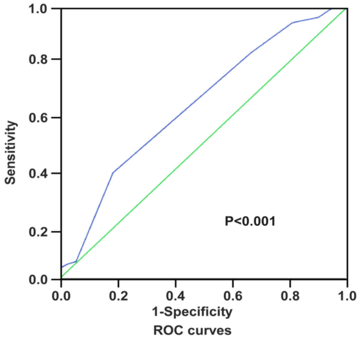
Two independent observers blinded to the
histopathological features and patient data of the samples
evaluated and scored the degree of immunostaining. The scores were
based on the proportion of positively stained tumor cells [graded
as: 1 (<10% positive), 2 (10–50% positive), 3 (51–75% positive)
or 4 (>75% positive)] and staining intensity [categorized as 1
(no staining), 2 (weak staining, light yellow), 3 (moderate
staining, yellow brown) or 4 (strong staining, brown)]. The
staining index was generated by multiplying the scores for staining
intensity and for the proportion of positive cells (scored as 1, 2,
3, 4, 6, 8, 9, 12 and 16). Staining index score ≥8 indicated tumors
with high GINS2 expression; a score of <8 defined low GINS2
expression. Receiver operating characteristics (ROC) curve analysis
was conducted using variables including GINS2 expression and
patient outcomes to determine the optimum cut-off values of the
scores (Fig. 1).
Colony formation and
anchorage-independent growth ability assays
Cells (5×10²/well) were plated in 6-well
plates and cultured for 2 weeks. The colonies were washed with PBS
3 times and fixed with 4% formaldehyde for 10 min. Then, the
colonies were stained with 1% crystal violet for 10 min. After
washing, the colonies were counted. For the anchorage-independent
growth ability assay, 500 cells were trypsinized and suspended in 2
ml complete medium plus 0.3% agar (Sigma). The agar-cell mixture
was plated on 1% agar complete medium mixture. For ~10 days, viable
colonies that were >0.1 mm in diameter were counted. The
experiment was carried out in triplicate for each cell line.
Wound healing and Transwell migration
and invasion assays
In the wound healing assay, cells
(2×10⁶/well) were seeded in 6-well plates. When the
cells were 90% confluent, they were serum-starved for 24 h. A
linear wound was created in the confluent monolayer using a 10-µl
pipette tip. The wounds were observed and photographed immediately
(time 0) and thereafter at 24 and 48 h (magnification, ×200). Each
experiment was repeated at least 3 times. For the Transwell
migration and invasion assays, 2×10⁴ cells were seeded
in 8-µm pore inserts coated with (for invasion) or without (for
migration) 50 µl Matrigel in triplicate wells. After 24 h
incubation at 37̊C, cells that had passed through the filter into
the bottom chamber were fixed with 1% paraformaldehyde, stained
with hematoxylin, and counted under a magnification of ×200 (10
random fields/well).
Statistical analysis
All statistical analyses were conducted using the
SPSS software package (standard version 16.0; IBM). The Chi-square
and Fisher's exact tests were used to evaluate the relationship
between GINS2 expression and the clinicopathological features.
Spearman's rank correlation coefficients were performed to
calculate bivariate correlations between the study variables.
Survival curves were estimated using the Kaplan-Meier method and
the log-rank test. The independent prognostic indicator in all of
the clinical parameters was determined using univariate and
multivariate analyses. P<0.05 was considered statistically
significant in all cases.
Results
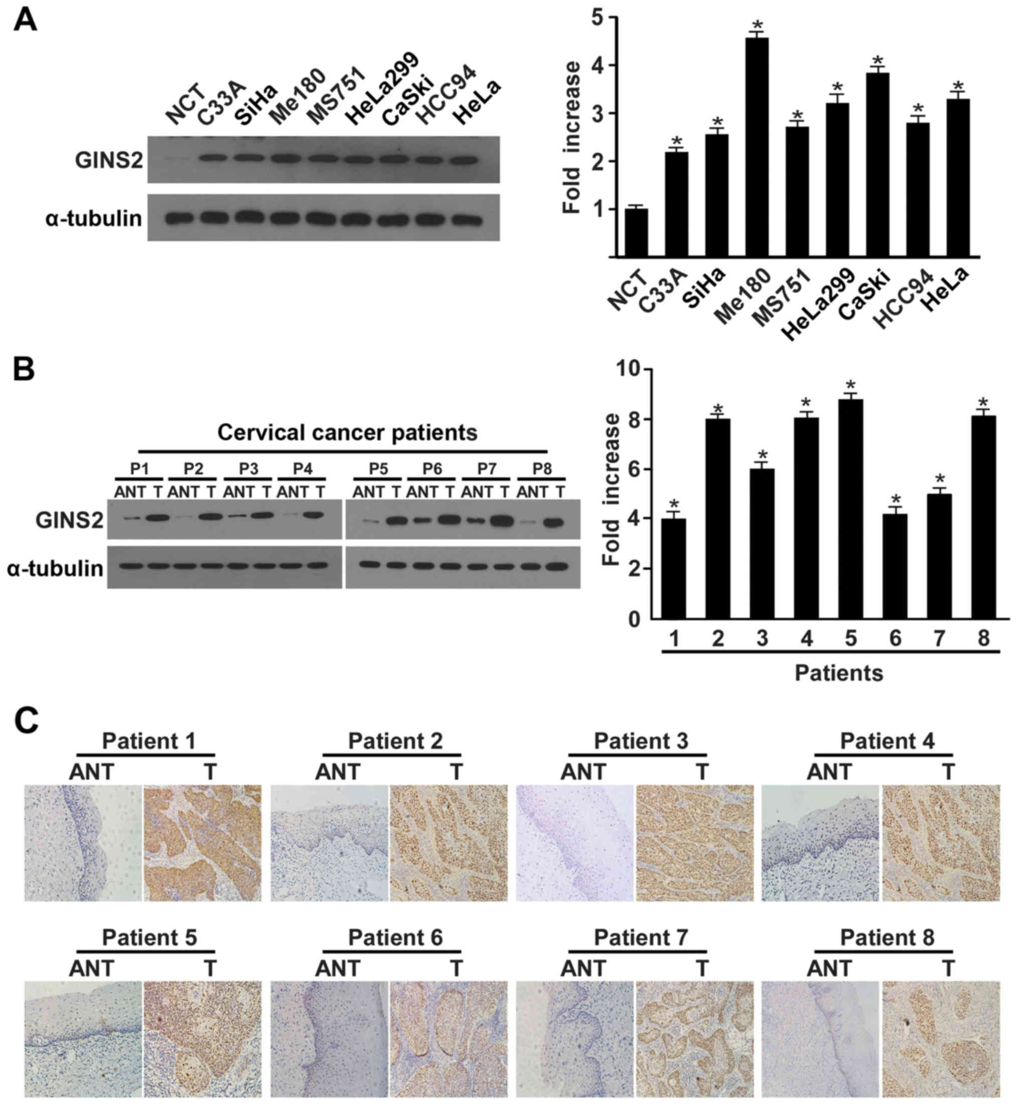
GINS2 is overexpressed in cervical
cancer
We used real-time PCR and western blotting to
determine the GINS2 expression pattern in cervical carcinoma cell
lines and samples. There were higher levels of both GINS2
transcription and translation in the cervical carcinoma cell lines
in comparison to normal cervical tissue (Fig. 2A). Consistent with the findings in
the cervical cancer cell lines, GINS2 mRNA and protein levels were
clearly differentially increased in the 8 cervical cancer tissue
samples than these levels in the matched adjacent non-cancerous
tissues (Fig. 2B). Furthermore, IHC
confirmed GINS2 overexpression in the cervical cancer clinical
samples (Fig. 2C).
GINS2 expression correlates with
clinical characteristics in early-stage cervical cancer
To investigate the clinical relevance of GINS2
expression and cervical cancer progression, IHC was performed on
155 paraffin-embedded, archived clinical cervical cancer samples,
which included 18, 83 and 54 cases of stage Ia2, Ib and IIa
disease, respectively. We detected strong positive expression of
GINS2 in 48 (31%) cervical cancer specimens, whereas there was no
or marginally detectable staining in the remaining 107 (69%;
Table I) clinical tumor samples.
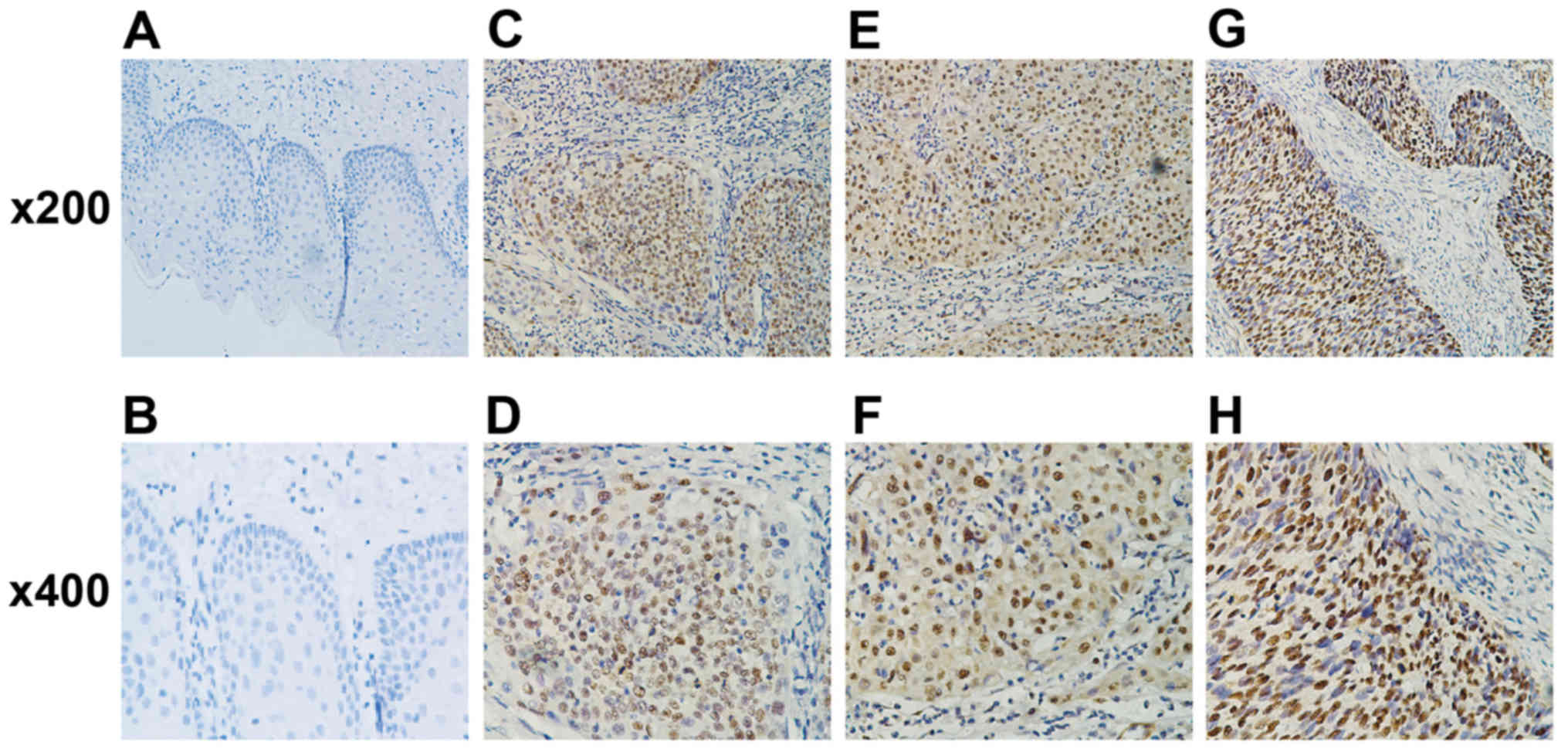
GINS2 primarily localized in the tumor cell nuclei and was absent
from the adjacent normal cervical tissues (Fig. 3).
Notably, GINS2 expression correlated with several
clinical features of cervical cancer, including SCC-Ag
(P<0.001), pelvic LNM (PLNM; P<0.001), deep stromal invasion
(P=0.021), vital status (P<0.001) and recurrence (P<0.001).
In contrast, GINS2 and other clinical characteristics, including
FIGO stage, surgical margin, lymphovascular invasion, parauterine
organ infiltration and age, were not obviously related (Table II). Spearman correlation analysis
(Table III) further confirmed the
strong association of GINS2 expression and significant prognostic
risk factors, including SCC-Ag (R=0.430; P<0.001), deep stromal
invasion (R=0.185; P=0.021), vital status (R=0.287; P<0.001),
recurrence (R=0.323; P<0.001), and in particular, PLNM (R=0.455;
P<0.001).
| Table II.Correlation of clinicopathological
characteristics and GINS2 expression in the early-stage cervical
cancer patients. |
Table II.
Correlation of clinicopathological
characteristics and GINS2 expression in the early-stage cervical
cancer patients.
|
|
| GINS2 |
|
|
|---|
|
|
|
|
|
|
|---|
|
Characteristics | Total (n=155) | Low expression | High
expression | Chi-square test
P-value | Fishers exact test
P-value |
|---|
| Age (years) |
|
|
| 0.688 | 0.730 |
|
≤47 | 78 | 55 (35.5) | 23 (14.8) |
|
|
|
>47 | 77 | 52 (33.5) | 25 (16.2) |
|
|
| FIGO stage |
|
|
| 0.571 |
|
| Ia | 18 | 14 (9.0) | 4 (2.6) |
|
|
| Ib | 83 | 58 (37.4) | 25 (16.1) |
|
|
|
IIa | 54 | 35 (22.6) | 19 (12.3) |
|
|
| Pelvic lymph node
metastasis (PLNM) |
|
|
| <0.001 | <0.001 |
| No | 99 | 84 (54.2) | 15 (9.7) |
|
|
|
Yes | 56 | 23 (14.8) | 33 (21.3) |
|
|
| Histological
types |
|
|
| 0.440 | 0.667 |
|
Squamous cell carcinoma | 149 | 102 (65.8) | 47 (30.3) |
|
|
|
Adenocarcinoma | 6 | 5 (3.2) | 1 (0.7) |
|
|
| SCC-Ag (ng/ml) |
|
|
| <0.001 | <0.001 |
|
≤1.5 | 91 | 78 (50.3) | 13 (8.4) |
|
|
|
>1.5 | 64 | 29 (18.7) | 35 (22.6) |
|
|
| Tumor size
(cm) |
|
|
| 0.058 | 0.085 |
| ≤4 | 132 | 95 (61.3) | 37 (23.9) |
|
|
|
>4 | 23 | 12 (7.7) | 11 (7.1) |
|
|
| Positive surgical
margin |
|
|
| 0.304 | 0.374 |
| No | 149 | 104 (67.1) | 45 (29.0) |
|
|
|
Yes | 6 | 3 (1.9) | 3 (1.9) |
|
|
| Deep stromal
invasion |
|
|
| 0.021 | 0.025 |
| No | 49 | 40 (25.8) | 9 (5.8) |
|
|
|
Yes | 106 | 67 (43.2) | 39 (25.2) |
|
|
| Positive
parametrium |
|
|
| 0.898 | 1.000 |
| No | 149 | 103 (66.5) | 46 (29.7) |
|
|
|
Yes | 6 | 4 (2.6) | 2 (1.3) |
|
|
| Lymphovascular
space involvement |
|
|
| 0.874 | 1.000 |
| No | 146 | 101 (65.2) | 45 (29.0) |
|
|
|
Yes | 9 | 6 (3.9) | 3 (1.9) |
|
|
| Vital status (at
follow-up) |
|
|
| <0.001 | 0.001 |
|
Alive | 126 | 95 (61.3) | 31 (20.0) |
|
|
|
Dead | 29 | 12 (7.7) | 17 (11.0) |
|
|
| Recurrence |
|
|
| <0.001 | <0.001 |
| No | 137 | 102 (65.8) | 35 (22.6) |
|
|
|
Yes | 18 | 5 (3.2) | 13 (8.4) |
|
|
| Table III.Spearman analysis of the correlation
of GINS2 expression and clinicopathological features. |
Table III.
Spearman analysis of the correlation
of GINS2 expression and clinicopathological features.
|
| GINS2 expression
level |
|---|
|
|
|
|---|
| Variables | Spearman
correlation | P-value |
|---|
| PLNM | 0.455 | <0.001 |
| SCC-Ag (ng/ml) | 0.430 | <0.001 |
| Vital status (at
follow-up) | 0.287 | <0.001 |
| Deep stromal
invasion | 0.185 | 0.021 |
| Recurrence | 0.323 | <0.001 |
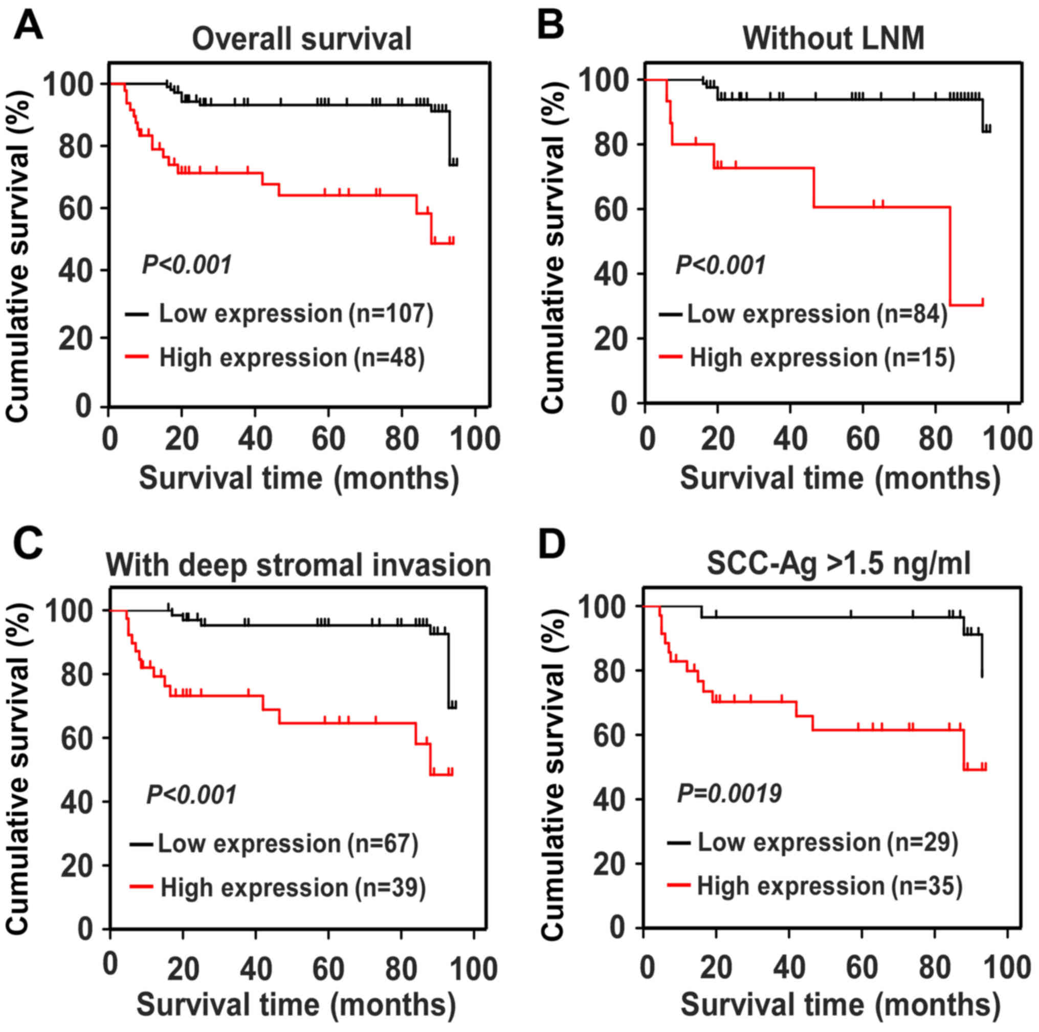
GINS2 expression is associated with
poor survival outcomes in early-stage cervical cancer
We used Kaplan-Meier survival curves and the
log-rank test to evaluate the impact of GINS2 on predicting the
prognosis of patients with early-stage cervical cancer. Fig. 4A shows that patients with higher
levels of GINS2 had shorter OS (P<0.001), whereas those with
lower GINS2 expression survived longer. For the patients with low
GINS2 expression, the cumulative OS rates were 89.36% [95%
confidence interval (CI), 85.68–93.05%], but were drastically
decreased to 65.19% (95% CI, 53.89–76.49%) in the high GINS2 group.
In addition, we calculated the prognostic value of GINS2 expression
in specific patient subgroups, which were stratified based on age,
FIGO stage, PLNM, SCC-Ag, tumor size, differentiation grade, deep
stromal invasion, properties of the surgical margin, parauterine
organ infiltration and lymphovascular space involvement. Fig. 4B-D shows that there was a markedly
negative association between GINS2 expression and OS in the
patients without LNM (log-rank test; P<0.001), with SCC-Ag
>1.5 ng/ml (log-rank test; P=0.0019), and with deep stromal
invasion (log-rank test; P<0.001). The Cox regression model
indicated that GINS2 expression and recurrence were independent
prognostic risk factors of cervical cancer (Table IV). Taken together, our results
indicate that GINS2 plays an important role in cervical cancer
progression and may serve as a valuable prognostic predictor for
patients with early-stage cervical cancer.
| Table IV.Univariate and multivariate analysis
of the prognostic parameters in early-stage cervical cancer using
Cox-regression model. |
Table IV.
Univariate and multivariate analysis
of the prognostic parameters in early-stage cervical cancer using
Cox-regression model.
|
| Univariate
analysis | Multivariate
analysis |
|---|
|
|
|
|
|---|
|
| P-value | Hazard ratio (95%
CI) | P-value | Hazard ratio (95%
CI) |
|---|
| GINS2
expression | <0.001 | 5.160 | 0.011 | 3.643 |
|
Low |
| (2.438–10.925) |
| (1.347–9.850) |
|
High |
|
|
|
|
| Pelvic lymph node
metastasis (PLNM) | 0.008 | 2.716 | 0.127 | 1.827 |
| No |
| (1.299–5.680) |
| (0.843–3.962) |
|
Yes |
|
|
|
|
| SCC-Ag (ng/ml | 0.099 | 1.854 | 0.862 | 0.922 |
|
≤1.5 |
| (0.891–3.855) |
| (0.369–2.303) |
|
>1.5 |
|
|
|
|
| Recurrence | <0.001 | 10.201 | <0.001 | 8.523 |
| No |
| (4.883–21.352) |
| (3.944–18.416) |
|
Yes |
|
|
|
|
| Deep stromal
invasion | 0.543 | 1.304 | 0.669 | 0.823 |
| No |
| (0.555–3.063) |
| (0.337–2.008) |
|
Yes |
|
|
|
|
| Positive
parametrium | 0.840 | 1.228 | 0.510 | 2.076 |
| No |
| (0.166–9.092) |
| (0.236–18.222) |
|
Yes |
|
|
|
|
| Lymphovascular
space involvement | 0.735 | 1.282 | 0.971 | 1.032 |
| No |
| (0.304–5.400) |
| (0.190–5.623) |
|
Yes |
|
|
|
|
| Tumor size
(cm) | 0.523 | 1.371 | 0.481 | 0.675 |
| ≤4 |
| (0.521–3.609) |
| (0.227–2.013) |
|
>4 |
|
|
|
|
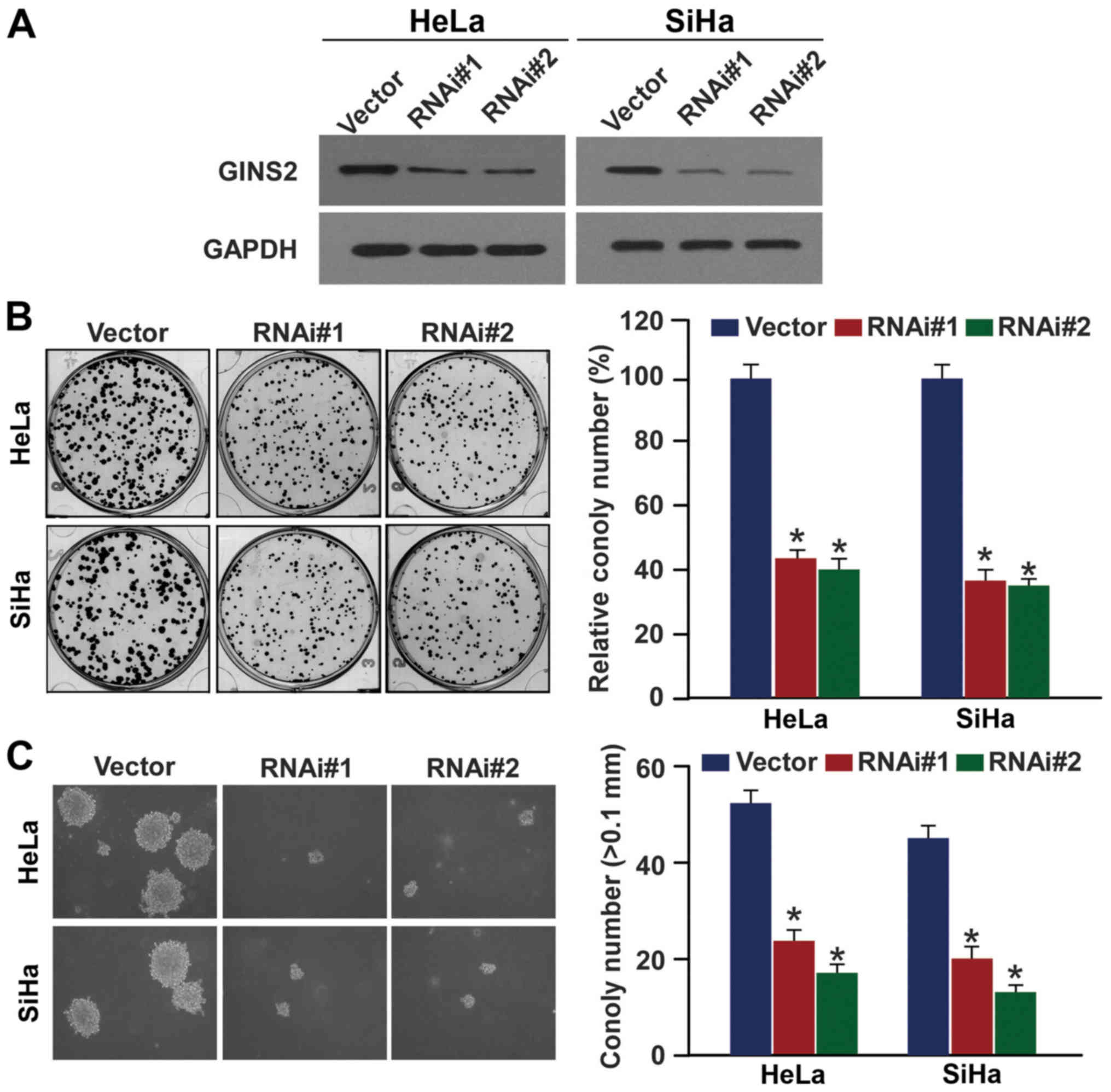
GINS2 downregulation inhibits cervical
cancer cell proliferation and tumorigenic ability
Given the involvement of GINS2 in cervical cancer
progression and its potential as a biomarker for identifying
patients with a more aggressive cervical cancer phenotype, we
explored the effects of GINS2 on the proliferation and
tumorigenicity of cervical cancer cell lines. Two shRNAs against
GINS2 were transduced into the SiHa and HeLa cervical cancer cell
lines to stably suppress endogenous GINS2 expression. The
downregulation efficiency was verified using western blotting
(Fig. 5A). As expected, the colony
formation assay revealed that GINS2 depletion induced the formation
of much smaller and fewer colonies as compared to the vector
control cells (Fig. 5B).
Furthermore, silencing of GINS2 markedly decreased the
anchorage-independent growth ability of the two cell lines in soft
agar (Fig. 5C). These results
suggest that GINS2 promotes proliferation and tumorigenesis of
cervical cancer cells.
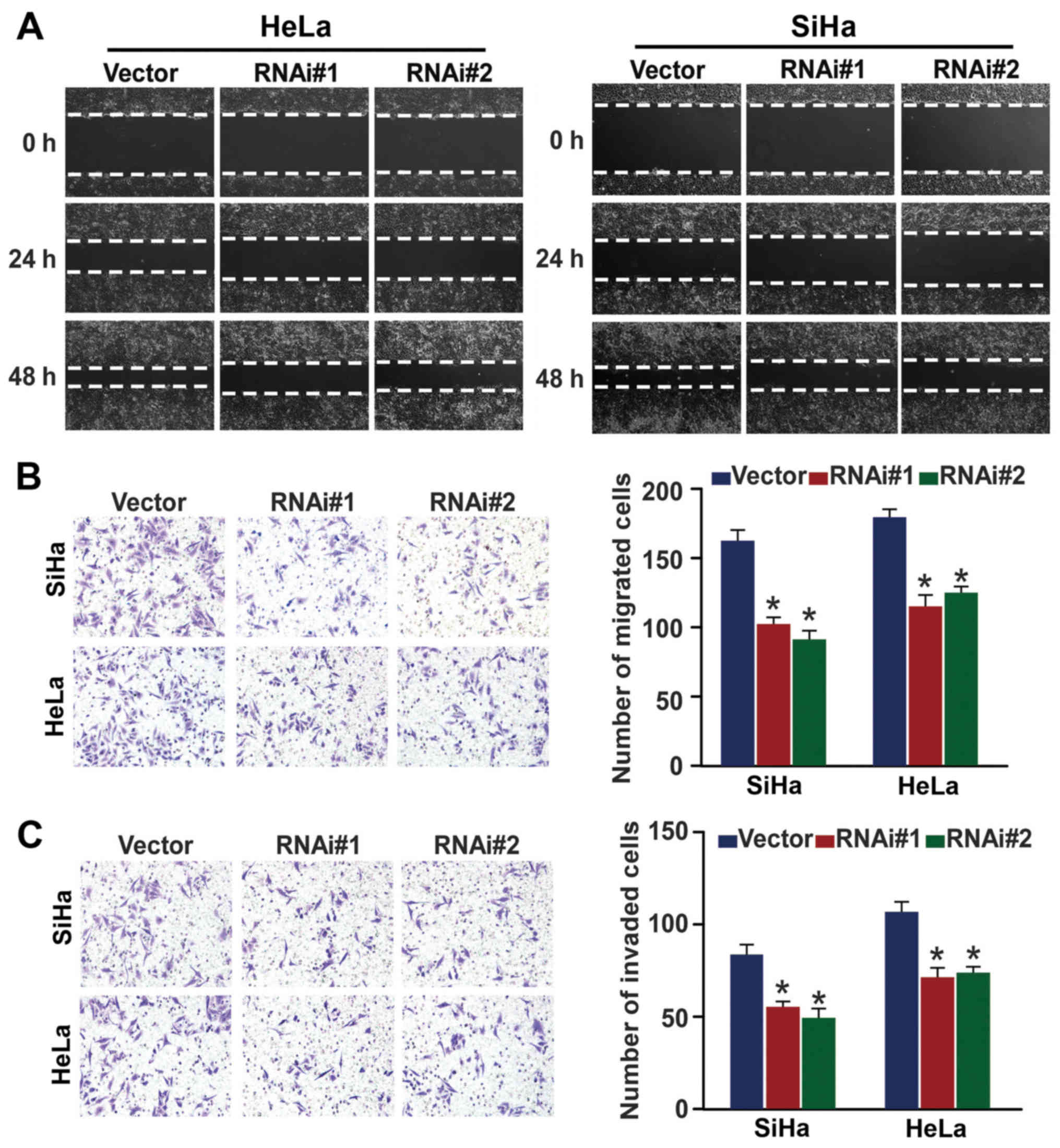
GINS2 inhibition reduces cervical
cancer cell migration and invasion
After demonstrating that GINS2 is closely connected
with PLNM, we then investigated whether GINS2 regulates cervical
cancer cell motility and invasiveness. Fig. 6A shows that in the wound healing
assay, GINS2 ablation slowed the speed with which the SiHa and HeLa
cells filled the gap in comparison to the control in an obvious
manner. The Transwell migration and matrix invasion assays yielded
similar results (Fig. 6B and C), as
indicated by the smaller number of migrated and invaded cells that
passed through the filter in the GINS2 downregulation group as
compared to the negative control group. Collectively, our findings
confirmed that GINS2 enhances the migratory and invasive properties
of cervical cancer cells.
Discussion
In the present study, we demonstrated for the first
time that GINS2 upregulation correlated with poor prognosis and
reduced survival in early-stage cervical cancer. In addition, GINS2
downregulation hampered the cellular capabilities of proliferation,
tumorigenesis, migration and invasion in an obvious manner. These
findings are convincing evidence that GINS2 plays a significant
role in the progression of cervical cancer and has the potential to
be a neoteric prognostic biomarker of early-stage cervical
cancer.
Clinical evidence has confirmed the role of GINS2 as
an oncogene in various malignancies. GINS2 is elevated in
intrahepatic cholangiocarcinoma, lung adenocarcinoma and breast
cancer, and induces intrahepatic cholangiocarcinoma cell
proliferation (7,8,10).
Moreover, GINS2 overexpression contributes to maintenance of the
cancer stem cell population and has been correlated with
progression and poor prognosis in breast cancer (8). The above mentioned studies indicated
that GINS2 plays a critical role in carcinogenesis and cancer
progression. Herein, we investigated its role in cervical cancer.
Consistent with the above studies, GINS2 mRNA and protein
expression was higher in the 8 cervical cancer cell lines and
clinical samples. IHC analysis indicated that GINS2 expression was
associated with well-known prognostic parameters: SCC-Ag
(P<0.001), deep stromal invasion (P=0.021), vital status
(P<0.001) and recurrence (P<0.001), particularly PLNM
(P<0.001), providing strong evidence that GINS2 plays an
oncogenic role in cervical cancer development and that it may act
as a biomarker for assessing patients with a more aggressive form
of the disease. More importantly, the present study demonstrated
that GINS2 overexpression correlates with poor clinical outcome and
is potentially an independent prognostic factor of poor OS in
cervical cancer. Taken together, our results implicate GINS2 as a
crucial contributing factor in tumor progression.
Clearly, the fundamental characteristic of tumor
cells is their malignant uncontrolled growth, the key process of
which is DNA replication (11,12).
Correct DNA replication requires the proper combination of
replication-associated proteins and prereplication complexes.
Defects in the proper function of these proteins are responsible
for chromosome instability and the subsequent generation of
neoplasms (13–16). Indeed, aberrant expression of
several DNA replication-related proteins has been demonstrated in
certain cancers. Gan et al proposed MCM genes, which
represent primary helicase for unwinding DNA during replication, as
diagnostic or prognostic markers in cervical carcinoma (17). Tane et al showed that
elevated levels of PSF3, a GINS family member, predicted poor
prognosis of non-small cell lung cancer (18). In addition, silencing of PSF3
inhibited cell proliferation by inducing G1-S arrest, suggesting
that PSF3 may be required for the development of lung cancer. In
accordance with these previous studies, we established the clinical
significance of GINS2 in cervical cancer and demonstrated that
GINS2 knockdown markedly impaired the proliferative and tumorigenic
properties of cervical cancer cells. GINS2 was previouly found to
alter the percentages of the apoptosis-relevant genes BAX and BCL2,
and the levels of the cell cycle regulators ataxia-telangiectasia
mutated (ATM), checkpoint kinase 2 (CHK2), and P53 to stimulate
G2/M transition (9). Accordingly,
we similarly hypothesized that GINS2 accelerated G2/M transitional
entry by circumventing cell cycle checkpoints, consequently
exerting its oncogenic potential in cervical cancer. However,
further investigations are needed to verify the hypothesis and to
clarify the carcinogenesis mechanisms in greater detail.
Currently, radical hysterectomy plus pelvic lymph
node dissection or chemoradiation is the criterion standard of
treatment for patients with early-stage cervical cancer (19,20).
Lymph node status is critical for determining the appropriate
treatment strategies. Patients with LNM require chemoradiation,
which may render the initial surgical intervention unnecessary in
retrospect (19,21). Meanwhile, unnecessary
lymphadenectomy in patients with negative LNM can cause unfavorable
complications such as infection, lymphocysts, and lymphedema
(22). However, imaging methods and
sentinel lymph node biopsy both fail to accurately detect LNM
(21,23,24).
Worse, there is no existing preoperative marker for sensitive and
efficient LNM diagnosis. Surprisingly, our data showed that GINS2
was closely associated with LNM; moreover, inhibition of GINS2
significantly suppressed the aggressive cervical cancer cell
phenotype. Therefore, we speculated that GINS2 is a valuable marker
for predicting PLNM. However, a larger cohort of patients with LNM
should be enrolled in future studies, and the definite regulatory
mechanisms involved warrant further investigation.
SCC-Ag is currently most widely applied in clinical
diagnosis, evaluation of therapeutic effects, and for predicting
the clinical outcomes of patients with cervical cancer (25,26).
SCC-Ag levels are associated with the extent of cervical carcinoma
(27–33). Sustained high or continuous
elevation of SCC-Ag represents persistent disease or recurrence
(34,35). However, the clinical practical value
of serum SCC-Ag is still controversial. The numerical changes of
SCC-Ag do not specifically reflect cervical cancer; increased
SCC-Ag levels have also been observed in patients with lung cancer,
esophageal SCC and other benign diseases (36–38).
More unfortunately, the autofit cut-off level of SCC-Ag is still a
matter of debate partially due to the different disease stages of
patient groups (39). Therefore, it
appears that SCC-Ag is not an ideal indicator for predicting
outcomes in cervical cancer.
Notably, we reported that GINS2 plays an important
role in the development of cervical cancer and may serve as a
valuable prognostic marker for assessing survival in cervical
cancer. Therefore, determining the expression of GINS2 in cervical
tissues via biopsy can provide significant guidance for deciding
preferred treatment modality for patients with early-stage cervical
cancer.
To date, persistent human papillomavirus (HPV)
infection is recognized as the main cause of cervical cancer, and
HPV 16 and HPV 18 are considered the most carcinogenic HPVs. The
HPV oncoproteins E6 and E7 target the tumor-suppressor gene P53 and
retinoblastoma protein (Rb), respectively, simultaneously with the
activation of certain oncogenes and pathways, thereby coordinately
contributing to the initiation and progression of cervical cancer
(40–42). Clinical evidence has demonstrated
that GINS2 targets P53, and silencing of GINS2 upregulated P53
expression (9). We hypothesized
that GINS2 cooperates with the HPV oncoprotein E6 jointly to
promote tumor transformation and development. However, we did not
investigate their interaction in the present study, and it warrants
further investigation.
While the prognostic value of GINS2 in early-stage
cervical cancer was validated in our research. However, there still
existed limitations, one of which was the small sample size.
Furthermore, as all the patients involved were all in the
early-stage of cervical cancer, their prognosis was relatively
favorable and few developed pathological risk features including
LNM, positive parametrium, positive surgical margin, large tumor
size and recurrence. Thus, to validate our results, it is urgent to
incorporate a larger cohort of patients with aggressive clinical
factors in our subsequent study.
In summary, the present study, confirms the aberrant
expression and clinical significance of GINS2 in early-stage
cervical cancer. Additionally, we found that GINS2 knockdown
suppressed cervical cancer cell proliferation, tumorigenicity,
migration and invasion. GINS2 may represent a novel indicator for
identifying patients at high-risk, and potentially serve as a
clinically relevant biomarker for predicting the patient outcome of
early-stage cervical cancer, which could aid gynecologists in
determining more appropriate therapeutic strategies.
Acknowledgements
The present study was funded by the Natural Science
Foundation of Guangdong Province (S2013010015552), the National
Natural Science Foundation of China (nos. 81602723, 81302550 and
81671805), the Postdoctoral Science Foundation of China
(2016A020215214), and the Guangdong Medical Research Foundation
(A2015130).
Glossary
Abbreviations
Abbreviations:
|
GINS2
|
GINS complex subunit 2
|
|
OS
|
overall survival
|
|
PLNM
|
pelvic lymph node metastasis
|
|
LNM
|
lymph node metastasis
|
|
MCM
|
minichromosome maintenance
|
|
shRNAs
|
short hairpin RNAs
|
|
GAPDH
|
glyceraldehyde-3-phosphate
dehydrogenase
|
|
PBS
|
phosphate-buffered saline
|
|
PVDF
|
polyvinylidene difluoride
|
|
IHC
|
immunohistochemistry
|
|
SCC-Ag
|
squamous cell carcinoma antigen
|
|
HPV
|
human papilloma virus
|
|
ATM
|
ataxia-telangiectasia mutated
|
|
CHK2
|
checkpoint kinase 2
|
|
Rb
|
retinoblastoma protein
|
|
FIGO
|
International Federation of Obstetrics
and Gynecology
|
References
|
1
|
Jemal A, Bray F, Center MM, Ferlay J, Ward
E and Forman D: Global cancer statistics. CA Cancer J Clin.
61:69–90. 2011. View Article : Google Scholar : PubMed/NCBI
|
|
2
|
Torre LA, Bray F, Siegel RL, Ferlay J,
Lortet-Tieulent J and Jemal A: Global cancer statistics, 2012. CA
Cancer J Clin. 65:87–108. 2015. View Article : Google Scholar : PubMed/NCBI
|
|
3
|
Noordhuis MG, Fehrmann RS, Wisman GB,
Nijhuis ER, van Zanden JJ, Moerland PD, Ver Loren van Themaat E,
Volders HH, Kok M, ten Hoor KA, et al: Involvement of the TGF-beta
and beta-catenin pathways in pelvic lymph node metastasis in
early-stage cervical cancer. Clin Cancer Res. 17:1317–1330. 2011.
View Article : Google Scholar : PubMed/NCBI
|
|
4
|
Yang L, Jia X, Li N, Chen C, Liu Y and
Wang H: Comprehensive clinic-pathological characteristics of
cervical cancer in southwestern China and the clinical significance
of histological type and lymph node metastases in young patients.
PLoS One. 8:e758492013. View Article : Google Scholar : PubMed/NCBI
|
|
5
|
Gambus A, Jones RC, Sanchez-Diaz A,
Kanemaki M, van Deursen F, Edmondson RD and Labib K: GINS maintains
association of Cdc45 with MCM in replisome progression complexes at
eukaryotic DNA replication forks. Nat Cell Biol. 8:358–366. 2006.
View Article : Google Scholar : PubMed/NCBI
|
|
6
|
Rantala JK, Edgren H, Lehtinen L, Wolf M,
Kleivi K, Vollan HK, Aaltola AR, Laasola P, Kilpinen S, Saviranta
P, et al: Integrative functional genomics analysis of sustained
polyploidy phenotypes in breast cancer cells identifies an
oncogenic profile for GINS2. Neoplasia. 12:877–888. 2010.
View Article : Google Scholar : PubMed/NCBI
|
|
7
|
Liu M, Pan H, Zhang F, Zhang Y, Zhang Y,
Xia H, Zhu J, Fu W and Zhang X: Identification of TNM
stage-specific genes in lung adenocarcinoma by genome-wide
expression profiling. Oncol Lett. 6:763–768. 2013.PubMed/NCBI
|
|
8
|
Zheng M, Zhou Y, Yang X, Tang J, Wei D,
Zhang Y, Jiang JL, Chen ZN and Zhu P: High GINS2 transcript level
predicts poor prognosis and correlates with high histological grade
and endocrine therapy resistance through mammary cancer stem cells
in breast cancer patients. Breast Cancer Res Treat. 148:423–436.
2014. View Article : Google Scholar : PubMed/NCBI
|
|
9
|
Zhang X, Zhong L, Liu BZ, Gao YJ, Gao YM
and Hu XX: Effect of GINS2 on proliferation and apoptosis in
leukemic cell line. Int J Med Sci. 10:1795–1804. 2013. View Article : Google Scholar : PubMed/NCBI
|
|
10
|
Obama K, Ura K, Satoh S, Nakamura Y and
Furukawa Y: Up-regulation of PSF2, a member of the GINS
multiprotein complex, in intrahepatic cholangiocarcinoma. Oncol
Rep. 14:701–706. 2005.PubMed/NCBI
|
|
11
|
Mazurek A, Luo W, Krasnitz A, Hicks J,
Powers RS and Stillman B: DDX5 regulates DNA replication and is
required for cell proliferation in a subset of breast cancer cells.
Cancer Discov. 2:812–825. 2012. View Article : Google Scholar : PubMed/NCBI
|
|
12
|
Costa A, Hood IV and Berger JM: Mechanisms
for initiating cellular DNA replication. Annu Rev Biochem.
82:25–54. 2013. View Article : Google Scholar : PubMed/NCBI
|
|
13
|
Oehlmann M, Score AJ and Blow JJ: The role
of Cdc6 in ensuring complete genome licensing and S phase
checkpoint activation. J Cell Biol. 165:181–190. 2004. View Article : Google Scholar : PubMed/NCBI
|
|
14
|
Weaver BA and Cleveland DW: Does
aneuploidy cause cancer? Curr Opin Cell Biol. 18:658–667. 2006.
View Article : Google Scholar : PubMed/NCBI
|
|
15
|
Dutta A: Chaotic license for genetic
instability and cancer. Nat Genet. 39:10–11. 2007. View Article : Google Scholar : PubMed/NCBI
|
|
16
|
Hermand D and Nurse P: Cdc18 enforces
long-term maintenance of the S phase checkpoint by anchoring the
Rad3-Rad26 complex to chromatin. Mol Cell. 26:553–563. 2007.
View Article : Google Scholar : PubMed/NCBI
|
|
17
|
Gan N, Du Y, Zhang W and Zhou J: Increase
of Mcm3 and Mcm4 expression in cervical squamous cell carcinomas.
Eur J Gynaecol Oncol. 31:291–294. 2010.PubMed/NCBI
|
|
18
|
Tane S, Sakai Y, Hokka D, Okuma H, Ogawa
H, Tanaka Y, Uchino K, Nishio W, Yoshimura M and Maniwa Y:
Significant role of Psf3 expression in non-small-cell lung cancer.
Cancer Sci. 106:1625–1634. 2015. View Article : Google Scholar : PubMed/NCBI
|
|
19
|
Benedet JL, Bender H, Jones H III, Ngan HY
and Pecorelli S: FIGO Committee on Gynecologic Oncology: FIGO
staging classifications and clinical practice guidelines in the
management of gynecologic cancers. Int J Gynaecol Obstet.
70:209–262. 2000. View Article : Google Scholar : PubMed/NCBI
|
|
20
|
Koh WJ, Greer BE, Abu-Rustum NR, Apte SM,
Campos SM, Chan J, Cho KR, Cohn D, Crispens MA, DuPont N, et al:
National Comprehensive Cancer Network: Cervical cancer. J Natl
Compr Canc Netw. 11:320–343. 2013.PubMed/NCBI
|
|
21
|
Selman TJ, Mann C, Zamora J, Appleyard TL
and Khan K: Diagnostic accuracy of tests for lymph node status in
primary cervical cancer: A systematic review and meta-analysis.
CMAJ. 178:855–862. 2008. View Article : Google Scholar : PubMed/NCBI
|
|
22
|
Matsuura Y, Kawagoe T, Toki N, Tanaka M
and Kashimura M: Long-standing complications after treatment for
cancer of the uterine cervix - clinical significance of medical
examination at 5 years after treatment. Int J Gynecol Cancer.
6:294–297. 2006. View Article : Google Scholar
|
|
23
|
Altgassen C, Hertel H, Brandstädt A,
Köhler C, Dürst M and Schneider A: AGO Study Group: Multicenter
validation study of the sentinel lymph node concept in cervical
cancer: AGO Study Group. J Clin Oncol. 26:2943–2951. 2008.
View Article : Google Scholar : PubMed/NCBI
|
|
24
|
Slama J, Dundr P, Dusek L and Cibula D:
High false negative rate of frozen section examination of sentinel
lymph nodes in patients with cervical cancer. Gynecol Oncol.
129:384–388. 2013. View Article : Google Scholar : PubMed/NCBI
|
|
25
|
Bonfrer JM, Gaarenstroom KN, Korse CM, Van
Bunningen BN and Kenemans P: Cyfra 21–1 in monitoring cervical
cancer: A comparison with tissue polypeptide antigen and squamous
cell carcinoma antigen. Anticancer Res. 17:2329–2334.
1997.PubMed/NCBI
|
|
26
|
Esajas MD, Duk JM, de Bruijn HW, Aalders
JG, Willemse PH, Sluiter W, Pras B, ten Hoor K, Hollema H and van
der Zee AG: Clinical value of routine serum squamous cell carcinoma
antigen in follow-up of patients with early-stage cervical cancer.
J Clin Oncol. 19:3960–3966. 2001. View Article : Google Scholar : PubMed/NCBI
|
|
27
|
Avall-Lundqvist EH, Sjovall K, Nilsson BR
and Eneroth PH: Prognostic significance of pretreatment serum
levels of squamous cell carcinoma antigen and CA 125 in cervical
carcinoma. Eur J Cancer. 28A:1695–1702. 1992. View Article : Google Scholar : PubMed/NCBI
|
|
28
|
Duk JM, Groenier KH, de Bruijn HW, Hollema
H, ten Hoor KA, van der Zee AG and Aalders JG: Pretreatment serum
squamous cell carcinoma antigen: A newly identified prognostic
factor in early-stage cervical carcinoma. J Clin Oncol. 14:111–118.
1996. View Article : Google Scholar : PubMed/NCBI
|
|
29
|
Yuan CC, Wang PH, Ng HT, Tsai LC, Juang CM
and Chiu LM: Both TPA and SCC-Ag levels are prognostic even in
high-risk stage Ib-IIa cervical carcinoma as determined by a
stratification analysis. Eur J Gynaecol Oncol. 23:17–20.
2002.PubMed/NCBI
|
|
30
|
Strauss HG, Laban C, Lautenschläger C,
Buchmann J, Schneider I and Koelbl H: SCC antigen in the serum as
an independent prognostic factor in operable squamous cell
carcinoma of the cervix. Eur J Cancer. 38:1987–1991. 2002.
View Article : Google Scholar : PubMed/NCBI
|
|
31
|
Molina R, Filella X, Lejarcegui JA, Pahisa
J, Torné A, Rovirosa A, Mellado B, Ordi J, Puig-Tintore LM,
Alicarte J, et al: Prospective evaluation of squamous cell
carcinoma and carcinoembryonic antigen as prognostic factors in
patients with cervical cancer. Tumour Biol. 24:156–164. 2003.
View Article : Google Scholar : PubMed/NCBI
|
|
32
|
Reesink-Peters N, van der Velden J, Hoor
Ten KA, Boezen HM, de Vries EG, Schilthuis MS, Mourits MJ, Nijman
HW, Aalders JG, Hollema H, et al: Preoperative serum squamous cell
carcinoma antigen levels in clinical decision making for patients
with early-stage cervical cancer. J Clin Oncol. 23:1455–1462. 2005.
View Article : Google Scholar : PubMed/NCBI
|
|
33
|
Gaarenstroom KN, Kenter GG, Bonfrer JM,
Korse CM, Van de Vijver MJ, Fleuren GJ and Trimbos JB: Can initial
serum cyfra 21–1, SCC antigen, and TPA levels in squamous cell
cervical cancer predict lymph node metastases or prognosis? Gynecol
Oncol. 77:164–170. 2000. View Article : Google Scholar : PubMed/NCBI
|
|
34
|
Montag TW: Tumor markers in gynecologic
oncology. Obstet Gynecol Surv. 45:94–105. 1990. View Article : Google Scholar : PubMed/NCBI
|
|
35
|
Duk JM, de Bruijn HW, Groenier KH, Hollema
H, ten Hoor KA, Krans M and Aalders JG: Cancer of the uterine
cervix: Sensitivity and specificity of serum squamous cell
carcinoma antigen determinations. Gynecol Oncol. 39:186–194. 1990.
View Article : Google Scholar : PubMed/NCBI
|
|
36
|
Li J, Chen P, Mao CM, Tang XP and Zhu LR:
Evaluation of diagnostic value of four tumor markers in
bronchoalveolar lavage fluid of peripheral lung cancer. Asia Pac J
Clin Oncol. 10:141–148. 2014. View Article : Google Scholar : PubMed/NCBI
|
|
37
|
Cao X, Zhang L, Feng GR, Yang J, Wang RY,
Li J, Zheng XM and Han YJ: Preoperative Cyfra21-1 and SCC-Ag serum
titers predict survival in patients with stage II esophageal
squamous cell carcinoma. J Transl Med. 10:1972012. View Article : Google Scholar : PubMed/NCBI
|
|
38
|
Torre GC: SCC antigen in malignant and
nonmalignant squamous lesions. Tumour Biol. 19:517–526. 1998.
View Article : Google Scholar : PubMed/NCBI
|
|
39
|
Ryu HK, Baek JS, Kang WD and Kim SM: The
prognostic value of squamous cell carcinoma antigen for predicting
tumor recurrence in cervical squamous cell carcinoma patients.
Obstet Gynecol Sci. 58:368–376. 2015. View Article : Google Scholar : PubMed/NCBI
|
|
40
|
Moody CA and Laimins LA: Human
papillomavirus oncoproteins: Pathways to transformation. Nat Rev
Cancer. 10:550–560. 2010. View Article : Google Scholar : PubMed/NCBI
|
|
41
|
Ferenczy A and Franco E: Persistent human
papillomavirus infection and cervical neoplasia. Lancet Oncol.
3:11–16. 2002. View Article : Google Scholar : PubMed/NCBI
|
|
42
|
Schiffman M, Castle PE, Jeronimo J,
Rodriguez AC and Wacholder S: Human papillomavirus and cervical
cancer. Lancet. 370:890–907. 2007. View Article : Google Scholar : PubMed/NCBI
|