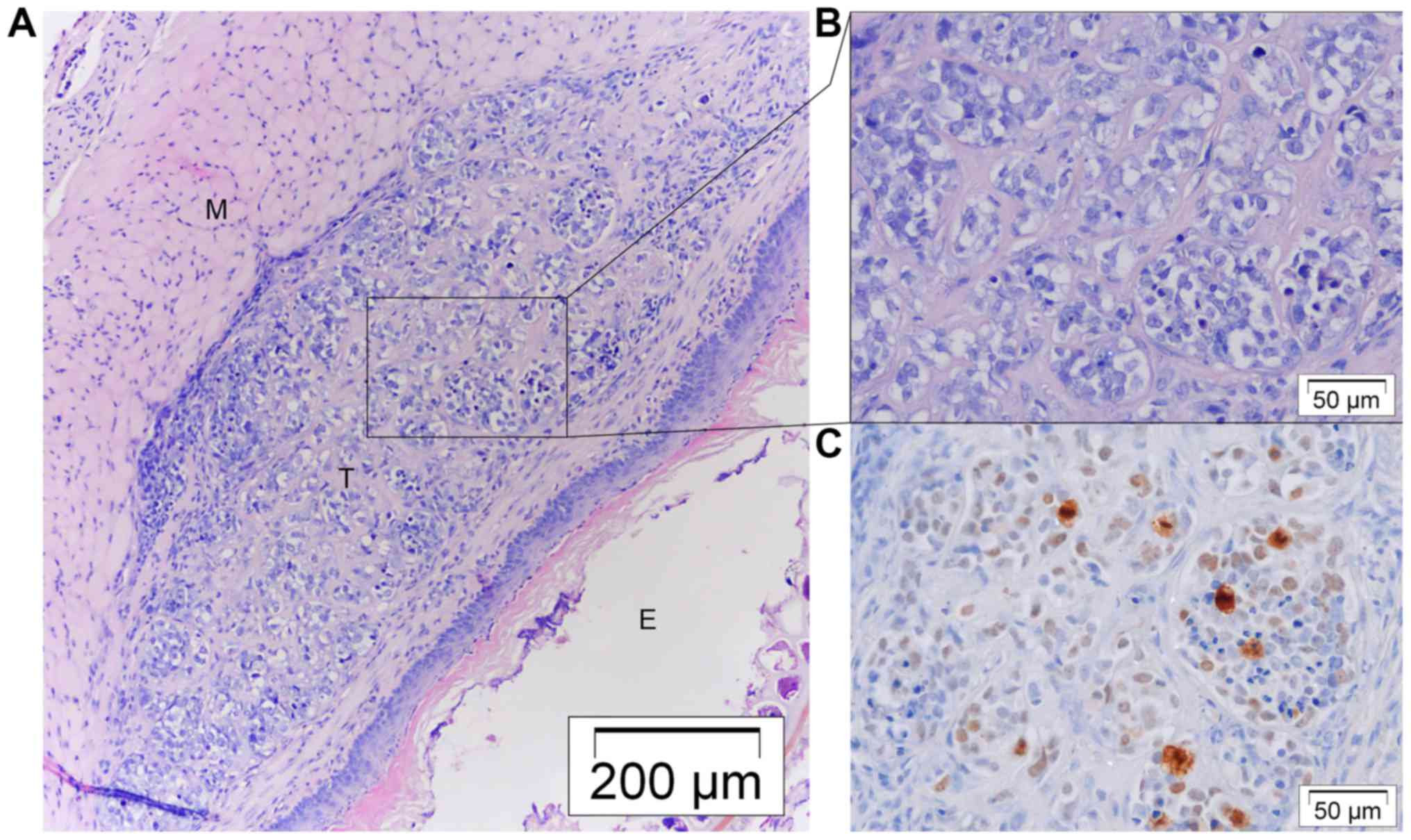
|
1
|
Napier KJ, Scheerer M and Misra S:
Esophageal cancer: A Review of epidemiology, pathogenesis, staging
workup and treatment modalities. World J Gastrointest Oncol.
6:112–120. 2014. View Article : Google Scholar : PubMed/NCBI
|
|
2
|
Stahl M, Mariette C, Haustermans K,
Cervantes A and Arnold D: ESMO Guidelines Working Group:
Oesophageal cancer: ESMO Clinical Practice Guidelines for
diagnosis, treatment and follow-up. Ann Oncol. 24 Suppl
6:vi51–vi56. 2013. View Article : Google Scholar : PubMed/NCBI
|
|
3
|
Gavin AT, Francisci S, Foschi R, Donnelly
DW, Lemmens V, Brenner H and Anderson LA: EUROCARE-4 Working Group:
Oesophageal cancer survival in Europe: A EUROCARE-4 study. Cancer
Epidemiol. 36:505–512. 2012. View Article : Google Scholar : PubMed/NCBI
|
|
4
|
Tétreault MP: Esophageal cancer: Insights
from mouse models. Cancer Growth Metastasis. 8 Suppl 1:S37–S46.
2015. View Article : Google Scholar
|
|
5
|
Bibby MC: Orthotopic models of cancer for
preclinical drug evaluation: Advantages and disadvantages. Eur J
Cancer. 40:852–857. 2004. View Article : Google Scholar : PubMed/NCBI
|
|
6
|
Hanahan D and Weinberg RA: Hallmarks of
cancer: The next generation. Cell. 144:646–674. 2011. View Article : Google Scholar : PubMed/NCBI
|
|
7
|
Kuroda S, Kubota T, Aoyama K, Kikuchi S,
Tazawa H, Nishizaki M, Kagawa S and Fujiwara T: Establishment of a
non-invasive semi-quantitative bioluminescent imaging method for
monitoring of an orthotopic esophageal cancer mouse model. PLoS
One. 9:e1145622014. View Article : Google Scholar : PubMed/NCBI
|
|
8
|
Song S, Chang D, Cui Y, Hu J, Gong M, Ma
K, Ding F, Liu ZH and Wang TY: New orthotopic implantation model of
human esophageal squamous cell carcinoma in athymic nude mice.
Thorac Cancer. 5:417–424. 2014. View Article : Google Scholar : PubMed/NCBI
|
|
9
|
Habibollahi P, Figueiredo JL, Heidari P,
Dulak AM, Imamura Y, Bass AJ, Ogino S, Chan AT and Mahmood U:
Optical imaging with a cathepsin B activated probe for the enhanced
detection of esophageal adenocarcinoma by dual channel fluorescent
upper GI endoscopy. Theranostics. 2:227–234. 2012. View Article : Google Scholar : PubMed/NCBI
|
|
10
|
Gros SJ, Dohrmann T, Peldschus K, Schurr
PG, Kaifi JT, Kalinina T, Reichelt U, Mann O, Strate TG, Adam G, et
al: Complementary use of fluorescence and magnetic resonance
imaging of metastatic esophageal cancer in a novel orthotopic mouse
model. Int J Cancer. 126:2671–2681. 2010.PubMed/NCBI
|
|
11
|
Drenckhan A, Kurschat N, Dohrmann T, Raabe
N, Koenig AM, Reichelt U, Kaifi JT, Izbicki JR and Gros SJ:
Effective inhibition of metastases and primary tumor growth with
CTCE-9908 in esophageal cancer. J Surg Res. 182:250–256. 2013.
View Article : Google Scholar : PubMed/NCBI
|
|
12
|
Kuroda S, Fujiwara T, Shirakawa Y,
Yamasaki Y, Yano S, Uno F, Tazawa H, Hashimoto Y, Watanabe Y, Noma
K, et al: Telomerase-dependent oncolytic adenovirus sensitizes
human cancer cells to ionizing radiation via inhibition of DNA
repair machinery. Cancer Res. 70:9339–9348. 2010. View Article : Google Scholar : PubMed/NCBI
|
|
13
|
Furihata T, Sakai T, Kawamata H, Omotehara
F, Shinagawa Y, Imura J, Ueda Y, Kubota K and Fujimori T: A new in
vivo model for studying invasion and metastasis of esophageal
squamous cell carcinoma. Int J Oncol. 19:903–907. 2001.PubMed/NCBI
|
|
14
|
Ip JC, Ko JM, Yu VZ, Chan KW, Lam AK, Law
S, Tong DK and Lung ML: A versatile orthotopic nude mouse model for
study of esophageal squamous cell carcinoma. Biomed Res Int 910715.
2015. View Article : Google Scholar
|
|
15
|
Ohara T, Takaoka M, Sakurama K, Nagaishi
K, Takeda H, Shirakawa Y, Yamatsuji T, Nagasaka T, Matsuoka J,
Tanaka N, et al: The establishment of a new mouse model with
orthotopic esophageal cancer showing the esophageal stricture.
Cancer Lett. 293:207–212. 2010. View Article : Google Scholar : PubMed/NCBI
|
|
16
|
Hori T, Yamashita Y, Ohira M, Matsumura Y,
Muguruma K and Hirakawa K: A novel orthotopic implantation model of
human esophageal carcinoma in nude rats: CD44H mediates cancer cell
invasion in vitro and in vivo. Int J Cancer. 92:489–496. 2001.
View Article : Google Scholar : PubMed/NCBI
|
|
17
|
Gros SJ, Kurschat N, Drenckhan A, Dohrmann
T, Forberich E, Effenberger K, Reichelt U, Hoffman RM, Pantel K,
Kaifi JT, et al: Involvement of CXCR4 chemokine receptor in
metastastic HER2-positive esophageal cancer. PLoS One.
7:e472872012. View Article : Google Scholar : PubMed/NCBI
|
|
18
|
Gros SJ, Kurschat N, Dohrmann T, Reichelt
U, Dancau AM, Peldschus K, Adam G, Hoffman RM, Izbicki JR and Kaifi
JT: Effective therapeutic targeting of the overexpressed HER-2
receptor in a highly metastatic orthotopic model of esophageal
carcinoma. Mol Cancer Ther. 9:2037–2045. 2010. View Article : Google Scholar : PubMed/NCBI
|
|
19
|
Gros SJ, Dohrmann T, Rawnaq T, Kurschat N,
Bouvet M, Wessels J, Hoffmann RM, Izbicki JR and Kaifi JT:
Orthotopic fluorescent peritoneal carcinomatosis model of
esophageal cancer. Anticancer Res. 30:3933–3938. 2010.PubMed/NCBI
|
|
20
|
Castro C, Bosetti C, Malvezzi M, Bertuccio
P, Levi F, Negri E, La Vecchia C and Lunet N: Patterns and trends
in esophageal cancer mortality and incidence in Europe (1980–2011)
and predictions to 2015. Ann Oncol. 25:283–290. 2014. View Article : Google Scholar : PubMed/NCBI
|
|
21
|
Almhanna K, Meredith KL, Hoffe SE,
Shridhar R and Coppola D: Targeting the human epidermal growth
factor receptor 2 in esophageal cancer. Cancer Control. 20:111–116.
2013.PubMed/NCBI
|
|
22
|
De Wever O, Hendrix A, De Boeck A,
Westbroek W, Braems G, Emami S, Sabbah M, Gespach C and Bracke M:
Modeling and quantification of cancer cell invasion through
collagen type I matrices. Int J Dev Biol. 54:887–896. 2010.
View Article : Google Scholar : PubMed/NCBI
|
|
23
|
Raggi M, Langer R, Feith M, Friess H,
Schauer M and Theisen J: Successful evaluation of a new animal
model using mice for esophageal adenocarcinoma. Langenbecks Arch
Surg. 395:347–350. 2010. View Article : Google Scholar : PubMed/NCBI
|
|
24
|
Quante M, Bhagat G, Abrams JA, Marache F,
Good P, Lee MD, Lee Y, Friedman R, Asfaha S, Dubeykovskaya Z, et
al: Bile acid and inflammation activate gastric cardia stem cells
in a mouse model of Barrett-like metaplasia. Cancer Cell. 21:36–51.
2012. View Article : Google Scholar : PubMed/NCBI
|
|
25
|
De Vlieghere E, Carlier C, Ceelen W,
Bracke M and De Wever O: Data on in vivo selection of SK-OV-3 Luc
ovarian cancer cells and intraperitoneal tumor formation with low
inoculation numbers. Data Brief. 6:542–549. 2016. View Article : Google Scholar : PubMed/NCBI
|
|
26
|
Minn AJ, Gupta GP, Siegel PM, Bos PD, Shu
W, Giri DD, Viale A, Olshen AB, Gerald WL and Massagué J: Genes
that mediate breast cancer metastasis to lung. Nature. 436:518–524.
2005. View Article : Google Scholar : PubMed/NCBI
|