Introduction
Lung cancer is the leading cause of cancer-related
deaths worldwide. The five-year survival of patients diagnosed with
NSCLC remains poor (1). Currently,
researchers and physicians are committed to the development of
molecular targets for cancer. With the increasing understanding of
the molecular basis of lung cancer, some targeted treatments have
been revealed to be effective in clinical practice (2). However, the targeted therapies for
other common somatic mutations such as p53 and KRAS in lung cancer
are still far from optimal. Therefore, the identification of the
potential targets for lung cancer is crucial for the development of
novel molecular-targeting agents.
Long non-coding RNAs (lncRNAs) are
non-protein-coding transcripts longer than 200 nucleotides, which
represent novel biomarkers for prognosis (3). The expression of lncRNAs is frequently
dysregulated in cancer (4) and
participates in the proliferation, survival, migration, and
invasion of cancer cells by modulating transcriptional,
post-transcriptional, and epigenetic molecular events. Studies have
revealed that targeting cancer-associated lncRNAs can impair cancer
cell growth and metastasis (5).
Since lncRNAs are critical for the carcinogenic process, they could
serve as novel therapeutic targets for cancer treatment (6).
p53 is one of the most common sites of genetic
alterations of NSCLC (7). p53
mutations have been associated with lung cancer progression and
treatment responses (8), however
the molecular mechanism underlying their tumor-suppressing effects
requires further studies (9).
Recently, the tumor-suppressor gene p53 was found to be associated
with lncRNAs (10). lncRNA
LOC285194 has been revealed to inhibit the growth of tumor cells as
a tumor suppressor in osteosarcoma (11). Liu et al (12) demonstrated that LOC285194 was a
transcriptional target of p53, and that its ectopic expression
functioned as an inhibitor of tumor cell growth in vitro and
in vivo. Whether LOC285194 is associated with the loss of
p53 tumor suppression in lung cancer has not been studied.
In the present study, we first focused on LOC285194
expression patterns in NSCLC tissues and cell lines as compared to
that in normal tissues or cell lines. Further functional
experiments revealed that LOC285194 may act as a tumor-suppressing
non-coding RNA through its interaction with LOC285194 and p53 in
NSCLC cells. Finally, based on examinations of the relationship
between the expression levels of LOC285194 in tumor tissues and the
clinicopathological features of clinical samples, LOC285194 may be
a potential non-coding predictor for diagnosis.
Materials and methods
Analysis of TCGA data
RNA sequencing data and the accompanying clinical
data were downloaded from the lung adenocarcinoma (LUAD) dataset of
the TCGA sequencing project (13)
and used for signaling pathway analysis. The survival data for
LOC285194 and p53 were obtained from cBioPortal (http://www.cbioportal.org/) (14,15).
Overall survival was analyzed by Kaplan-Meier curves and log-rank
tests for all LUAD patients. P-values <0.05 were considered to
indicate a statistically significant result.
Patients and tissue samples
A total of 56 paired NSCLC tissues and adjacent
non-tumor tissues were obtained from the Affiliated Cancer Hospital
and Institute of Guangzhou Medical University (Guangzhou, China)
between January 2006 and January 2011. All specimens were resected
by surgery, and a pathological examination confirmed the diagnosis
of lung adenocarcinoma. Patients with tuberculosis, diabetes,
pneumonia, and COPD were excluded from the study. None of patients
had received preoperative chemotherapy or radiotherapy. The tissues
were cut into specimens 0.5 cm × 0.5 cm in size and immediately
placed in cryogenic vials with 1.5 ml RNAlater (Thermo Fisher
Scientific, Inc., Waltham, MA, USA). NSCLC and normal tissues were
stored at −80°C until total RNA was extracted. This study was
performed in accordance with a protocol approved and reviewed by
the Medical Ethics Committee of the Affiliated Cancer Hospital and
Institute of Guangzhou Medical University.
Cell lines and culture conditions
Five human NSCLC adenocarcinoma cell lines (A549,
H1299, PC9, H460 and Calu3) and a normal human bronchial epithelial
cell line (16HBE) were obtained from American Type Culture
Collection (ATTC; Manassas, VA, USA). All cell lines were cultured
in Dulbecco's modified Eagle's medium (DMEM; Invitrogen GmbH,
Karlsruhe, Germany), supplemented with 10% fetal bovine serum
(FBS), 100 U/ml penicillin and 100 g/ml streptomycin, and left to
grow at 37°C in a 5% CO2 atmosphere.
RNA Preparation, reverse transcription
and quantitative real-time PCR
Total RNAs were extracted with TRIzol (Invitrogen;
Thermo Fisher Scientific, Inc., Carlsbad, CA, USA), following the
manufacturer's protocol. The reverse transcription reactions were
performed by Reverse Transcriptase M-MLV (Takara Biotechnology Co.,
Ltd., Dalian, China), and the samples were incubated for 60 min at
42°C, 15 min at 72°C, and stored at −20°C. For real-time PCR, 1 µl
of diluted RT products were mixed with 10 µl of SYBR®
Premix Ex Taq™ (Takara Biotechnology Co., Ltd.), 0.5 µl of forward
and reverse primers, and 4 µl of nuclease-free water for a final
volume of 20 µl, according to the manufacturer's instructions. The
primers used in the present study were 5′-TGTGCCTGTTTGACCTCTGA-3′
(forward) and 5′-AGGAAGGATAAAAGACCGACCA-3′ (reverse). The qPCR
reactions were run on the ABI-7500 real-time PCR system using the
following conditions: 95°C for 30 sec, followed by 45 cycles at
95°C for 3 sec and 60°C for 34 sec. The relative expression of
LOC285194 was calculated using the comparative cycle threshold (CT)
(2−∆∆CT), with small nucleolar RNA U6 as the internal
control to normalize the data.
Plasmid constructs and transfection of
NCSCL cells
The LOC285194 shRNA sequence was synthesized and
cloned into pLVX-Puro (Clontech; Takara Biotechnology Co., Ltd.).
The pLVX Puro and package plasmids were then transfected into 293T
cells. Next, the 293T cell line generated the lentivirus particles,
and a supernatant containing the virus was centrifuged to
concentrate the virus. The virus was then used to transduce the
cell lines of interest: A549 and H1299. The expression level of
LOC285194 was assessed by qPCR.
Cell proliferation assays
Cells were digested using the common passage method
to cause cell suspension, and stably-transfected plasmids and empty
vector cells (3000 cells/well) were seeded in 96-well plates. When
harvested, the cells were stained with
methylthiazolyldiphenyl-tetrazolium bromide (MTT) and dissolved in
dimethyl sulphoxide (DMSO). Cell growth was assessed every 12 h.
All experiments were performed in triplicate.
Flow cytometric analysis of
apoptosis
Stably-transfected A549 and H1299 cells and empty
vectors were harvested by trypsinization. Following the standard
FITC-Annexin V and propidium iodide (PI) double staining procedure,
the cells were analyzed by flow cytometry (BD Accuri C6; BD
Biosciences, San Jose, CA, USA). The percentage of apoptotic cells
was assessed (GuavaSoft 3.2). All of the samples were assayed in
triplicate.
Cell invasion assay
After 48 h transfection, 1×10⁵ cells in
serum-free media were placed into the upper chamber pre-coated with
Matrigel (8.0 µm; BD Biosciences). The chambers were then incubated
for 0, 24 and 48 h, respectively, in culture medium with 10% FBS in
the lower chambers before cell counting. The cells on the upper
surface were removed with a cotton swab, whereas the invaded cells
on the lower surface were fixed with 4% paraformaldehyde and
stained with 0.05% crystal violet. Finally, the invaded cells were
counted using a inverted microscope (BDS200) in triplicate. The
experiments were independently repeated in triplicate.
Wound healing assay
The cells were seeded in 6-well plates and cultured
until 100% confluence. A scratch was performed using 200-µl pipette
tips, and then the serum-free medium was replaced. The scratch
healing process was observed at 0, 24 and 48 h after
incubation.
RNA pull down and protein analysis of
the LOC285194-associated protein
The lncRNA cDNA sequence was cloned to the pGMT
vector, and the linear cDNA was transcribed in vitro using a
MEGAscript T7 Transcription kit (cat. no. AM1333; Life
Technologies; Thermo Fisher Scientific, Inc.), following the
manufacturer's instructions. Protein products (8–10 µg/µl; 50–60 µg
total) were separated with 1% SDS-PAGE on a 10% gel and
subsequently transferred overnight onto a polyvinylidene difluoride
membrane (EMD Millipore, Billerica, MA, USA) using SDS transfer
buffer (Bio-Rad Laboratories, Inc., Hercules, CA, USA). The
membrane was blocked for 1 h by a western-blocking reagent (Bio-Rad
Laboratories, Inc., Hercules, CA, USA) at room temperature prior to
protein detection by specific monoclonal p53 antibody at 1:1,000
dilution (cat. no. ab131442; Abcam, Cambridge, MA, USA) overnight
at 4°C. This was followed by incubation with a horseradish
peroxidase-conjugated anti-mouse secondary antibody (1:1,000; cat.
no. 6120-05; SouthernBiotech, Birmingham, AL, USA). The Amersham™
ECL™ Prime Western Blotting Detection Reagent (GE Healthcare Life
Sciences, Shanghai, China) was used to visualize the blots,
following the manufacturer's protocol, with a 5–10 min exposure to
SuperRX X-ray film (Fujifilm Investment Co., Ltd., Shanghai,
China).
RNA immunoprecipitation
An EZ-Magna RIP™ kit (EMD Millipore, Bedford, MA,
USA) was used (according to the manufacturer's instructions) to
perform RNA-binding protein immunoprecipitation. The anti-p53
antibody co-precipitated RNAs were purchased from Abcam and the
primers used for the detection of LOC285194 were: H-LOC285194-F
forward, 5′-CCTGTGCCTGTTTGACCTCT-3′ and reverse,
5′-CTGGTTTGCAGTTTGGCCTC-3′; LOC285194 P2 forward,
5′-CCCTCTTGTAGAGCCACAGG-3′ and reverse,
5′-CGAACACTGGCATTCATTGAGGG-3′; LOC285194 P3 forward,
5′-CAGTTCCTCAAATTTGACCCC-3′ and reverse,
5′-TTTGAAGGTTTTCCACATGG-3′.
Western blot analysis
Briefly, the cells were washed with PBS and lysed.
Protein products (8–10 µg/µl; 50–60 µg total) were separated using
10% SDS-PAGE and subsequently transferred overnight onto a
polyvinylidene difluoride membranes (EMD Millipore). The membranes
were blocked for 1 h with a Blotting-Grade Blocker (no. 1706404,
Bio-Rad Laboratories, Inc.). The specific monoclonal p53 antibody
(diluted 1:1,000; cat. no. ab1101; Abcam) was incubated overnight
at 4°C, followed by incubation with a horseradish
peroxidase-conjugated anti-mouse secondary antibody (1:1,000; cat.
no. 6120-05; SouthernBiotech, Birmingham, AL, USA). Amersham™ ECL™
Prime Western Blotting Detection Reagent (GE Healthcare Life
Sciences) was used to visualize the blots. The protein bands were
exposed onto SuperRX X-ray film (Fujifilm Investment Co., Ltd.).
Anti-GAPDH was used as a loading control (1:1,000; cat. no. ab9485;
Abcam, Cambridge, UK).
Statistical analysis
All data were presented as the means ± standard
error of the mean (SEM). The mean values of the two groups were
compared using the Student's t-test. Differences between the groups
were analyzed with a one-way analysis of variance (ANOVA). The
survival data were compared using the Kaplan-Meier analysis and
log-rank test. SPSS 19 software (IBM Corp., Armonk NY, USA) was
used for statistical analysis.
Results
LOC285194 is downregulated in cancer
cell lines and tissues
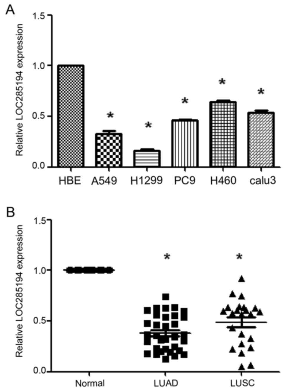
First, we aimed to investigate whether LOC285194 was
detectable and aberrantly expressed in NSCLC and bronchial
epithelial cell lines. Among the five NSCLC cell lines, the
expression level of LOC285194 was lower in these selected NSCLC
cell lines when compared with normal bronchial epithelial cells
(HBE) (P<0.05; Fig. 1A).
Furthermore, we examined the expression of LOC285194 in NSCLC
cancer tissues and adjacent normal tissues. We detected that
LOC285194 expression was significantly downregulated in both the
lung adenocarcinoma and the squamous tumor tissues when compared to
the adjacent normal tissues (P<0.001; Fig. 1B).
LOC285194 expression and
clinicopathological factors in NSCLC
The clinicopathological data of 56 patients are
shown in Table I. We divided the 56
patients into low or high expression groups (average ∆∆Ct
expression value of 0.44), with the median expression level of
LOC285194 as a cutoff. As indicated in Table I, the low LOC285194 group was
significantly associated with increased tumor size (P=0.027), but
no significant association was found between LOC285194 expression
and other clinicopathological data including sex, age, tumor
location, histological subtype, lymph node metastasis, distant
organ metastases, and TNM stage (P>0.05; Table I). Thus, our results indicated that
the reduced expression of LOC285194 was correlated with tumor
growth in NSCLC.
 | Table I.LOC285194 expression and
clinicopathological characteristics of 56 NSCLC patients. |
Table I.
LOC285194 expression and
clinicopathological characteristics of 56 NSCLC patients.
|
| LOC285194
subgroupsa |
|
|---|
|
|
|
|
|---|
|
| Low | High | P-value |
|---|
| Sex |
|
| 0.576 |
|
Male | 19 | 18 |
|
|
Female | 10 | 9 |
|
| Age |
|
| 0.202 |
|
<60 | 14 | 17 |
|
|
≥60 | 15 | 10 |
|
| Location |
|
| 0.169 |
|
LUL | 4 | 5 |
|
|
LLL | 3 | 9 |
|
|
RUL | 13 | 10 |
|
|
RML | 3 | 1 |
|
|
RLL | 6 | 2 |
|
| Histology |
|
| 0.595 |
|
LUAD | 16 | 15 |
|
|
LUSC | 13 | 12 |
|
| Size |
|
| 0.027c |
|
T1-T2 | 12 | 19 |
|
|
T3-T4 | 17 | 8 |
|
| Lymph node |
|
| 0.469 |
|
Negative | 18 | 18 |
|
|
Positive | 11 | 9 |
|
| Metastasis |
|
| 0.535 |
|
Negative | 26 | 25 |
|
|
Positive | 3 | 2 |
|
| TNM
stageb |
|
| 0.485 |
| I+
II | 16 | 16 |
|
| III+
IV | 13 | 11 |
|
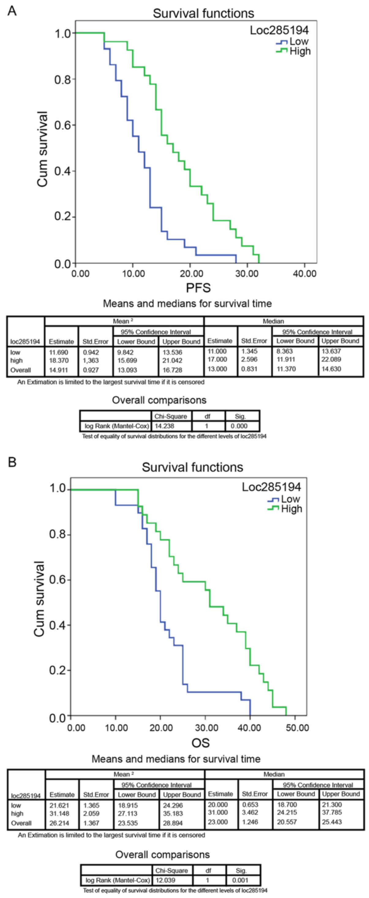
Kaplan-Meier analysis revealed that patients with
lower LOC285194 expression levels had a significantly worse
progression-free survival (PFS) (P<0.01; Fig. 2A) and overall survival (OS)
(P<0.01; Fig. 2B) than those
with high LOC285194 expression. Moreover, multivariate Cox
proportional hazard regression analysis revealed that low LOC285194
expression was an independent prognostic factor in NSCLC patients
(P<0.01; Table II).
Additionally, the survival data from cBioPortal also revealed that
the deletion of LOC285194 led to poor patient survival compared to
patients with normal LOC285194 expression, similar to the loss of
p53 in NSCLC patients compared with patients without a loss of p53
expression. These data inferred that LOC285194 expression was
associated with prognosis after lobectomy and shared the prognostic
pattern of p53 loss in NSCLC populations.
| Table II.Multivariate Cox regression analysis
of the 5-year progression-free survival and overall survival of 56
NSCLC patients. |
Table II.
Multivariate Cox regression analysis
of the 5-year progression-free survival and overall survival of 56
NSCLC patients.
|
| PFS | OS |
|---|
|
|
|
|
|---|
| Variables | HR | 95% CI | P-value | HR | 95% CI | P-value |
|---|
| Age |
| (<60/≥60
years) | 0.177 | 0.085–0.368 |
<0.001a | 0.241 | 0.118–0.490 |
<0.001a |
| Sex |
| (Male/Female) | 0.427 | 0.201–0.908 | 0.027a | 0.681 | 0.334–1.388 | 0.291 |
| Location |
|
(LUL/LLL/RUL/RML/RLL) | 1.138 | 0.887–1.458 | 0.309 | 1.250 | 0.968–1.614 | 0.087 |
| Histology |
| (LUAD/LUSC) | 1.427 | 0.116–17.499 | 0.781 | 3.253 | 0.251–42.207 | 0.367 |
| Size |
| (T1+T2/T3+T4) | 1.132 | 0.594–2.160 | 0.706 | 0.821 | 0.439–1.536 | 0.821 |
| Lymph node |
|
(Absence/Presence) | 0.819 | 0.287–2.341 | 0.710 | 0.385 | 0.125–1.185 | 0.096 |
| Metastasis |
|
(Absence/Presence) | 0.734 | 0.212–2.539 | 0.625 | 1.194 | 0.353–4.039 | 0.775 |
| Stage |
| (I+II/III+IV) | 6.535 | 0.521–82.002 | 0.146 | 5.304 | 0.375–74.950 | 0.217 |
| LOC285194 |
| (Low/High) | 0.153 | 0.073–0.318 |
<0.001a | 0.199 | 0.094–0.423 |
<0.001a |
LOC285194 inhibits proliferation and
promotes apoptosis of lung cancer cells in vitro
To further study the effect of LOC285194 in NSCLC,
lentiviral vectors of pLVX-LOC285194 and shLOC285194 were produced
and then separately transfected into A549 and H1299 cells. The qPCR
results confirmed that the expression level of LOC285194 was
significantly knocked down or overexpressed in both cells
transfected with shLOC285194 or pLVX-LOC285194 (Fig. 3A and B). We also found that the
ectopic expression of LOC285194 inhibited the proliferation ability
of both A549 and H1299 cells (Fig.
3D), while the knockdown of LOC285194 did not increase the
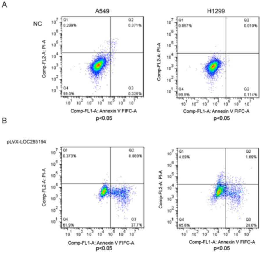
proliferation ability of H1299 cells (Fig. 3C). Additionally, the flow cytometric
analysis of A549 and H1299 cells revealed that overexpression of
LOC285194 induced apoptosis in comparison with the control cells
(Fig. 4A and B).
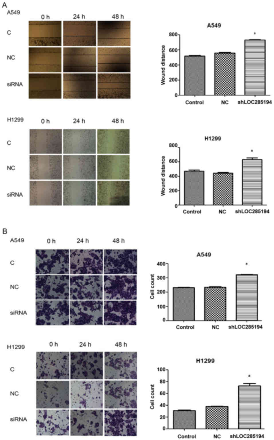
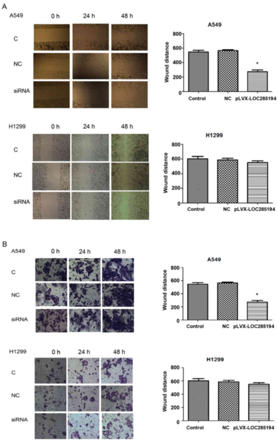
Effect of LOC285194 on cell migration
and invasiveness
To determine whether LOC285194 was involved in the
migration of tumor cells and facilitated cell invasion in
vitro, we evaluated cancer cell migration and invasion by wound
healing assay and matrigel invasion assay. Knockdown of LOC285194
expression by shRNA increased A549 and H1299 cell migration and
invasion (Fig. 5A and B). Ectopic
expression of LOC285194 led to significantly decreased migration
and invasion abilities of A549 cells. (P<0.05; Fig. 6A and B). Collectively, these results
revealed that the alteration of LOC285194 expression could affect
NSCLC cell migration and invasion abilities compared with the
control group.
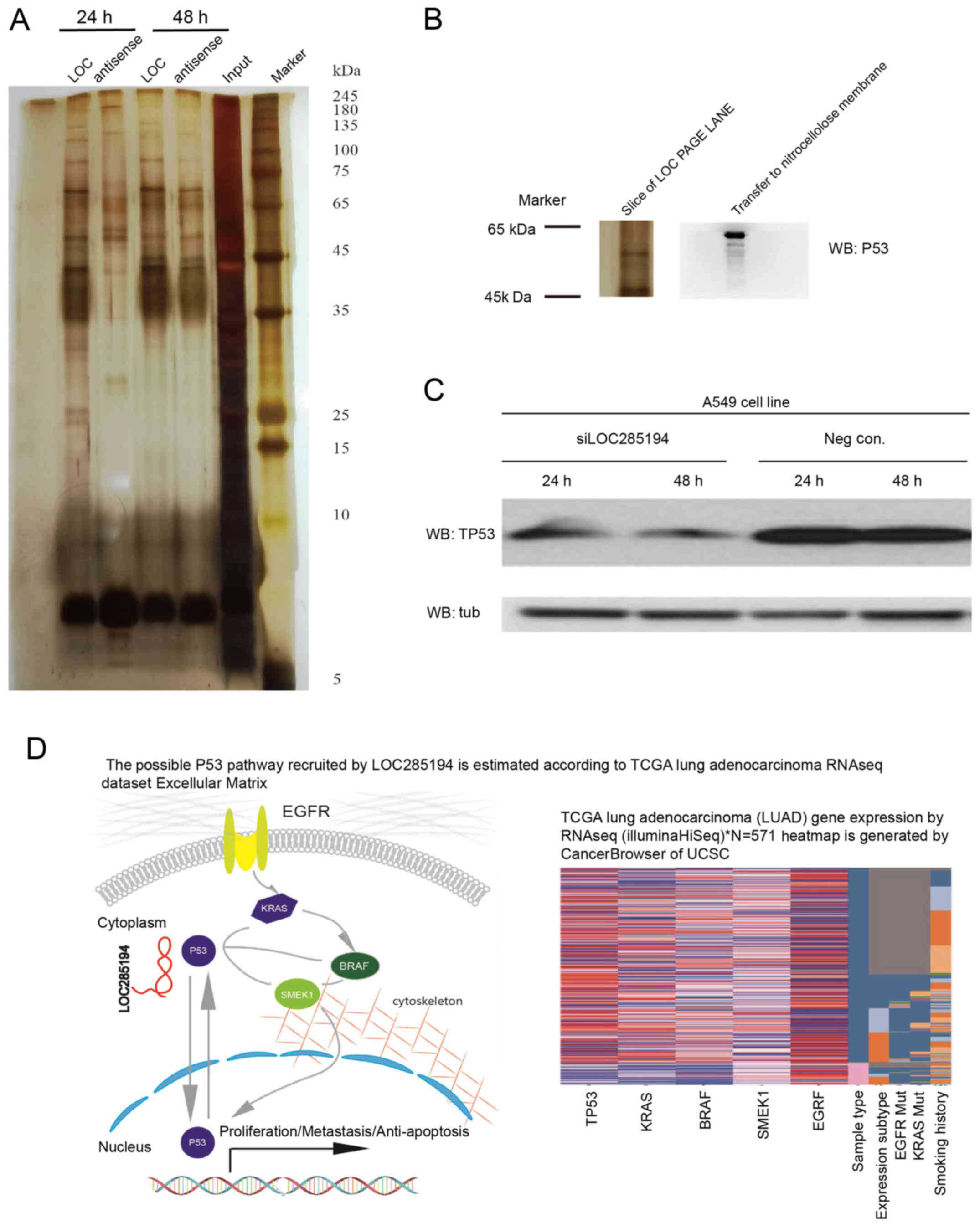
LOC285194 binds to the tumor supressor
p53 protein
Since lncRNAs may perform their molecular functions
by binding to specific proteins and regulate cell growth by
modulating the p53 pathway (10),
we hypothesized that LOC285194 may inhibit tumor growth and
metastasis through this mechanism. The RNA pull-down experiment and
western blot analysis were performed to identify proteins that are
associated with LOC285194 (Fig.
7A). In brief, we resolved the RNA-enriched proteins on an
SDS-PAGE gel, cut out the bands specific to p53, and subjected them
to western blot analysis (Fig. 7B).
We then detected p53 expression in LOC285194 siRNA-interfering
NSCLC cells. The results revealed that the expression of p53 was
low as shown in Fig. 7C. By
analyzing the LUAD dataset from the TCGA sequencing project by UCSC
cancer browser, we discovered that the p53 expression pattern was
correlated with KRAS, BRAF and SMEK, but not EGFR and PI3K. These
data confirmed that LOC285194 functions as a tumor suppressor
non-coding RNA by interfering with EGFR/KRAS/BRAF signaling in
NSCLC (Fig. 7D).
Discussion
Lung cancer is one of leading causes of
cancer-related deaths worldwide, with very high incidence and
mortality rates (16). Early
detection, diagnosis, and treatment are vital to the management of
lung cancer patients. The metastasis of distant organs contributes
to the decline in the quality of life and to poor disease
prognosis. Increasing evidence suggests that lncRNAs may be
involved in the regulation of cancer invasion and metastasis. Some
lncRNAs are more aberrantly expressed in cancer tissues than in
normal tissues (17,18), and thus some of them can be used as
novel molecular markers for tumor diagnosis and treatment. Other
studies have revealed that lncRNAs play a key role in EGFR exon 19
deletions (19), drug resistance
(20), and chemical carcinogenesis
(21–24). Additionally, lncRNAs such as MALAT1
can be used as markers of lung cancer, but with low sensitivity
(25).
lncRNA LOC285194, also known as antisense RNA
LSAMP3, is a long non-coding RNA located at the 3q13.31 loci with a
length of 2 kb. It has been revealed to be a tumor suppressor
(11), inhibiting the growth of
osteosarcoma cells. Liu et al (12) revealed that LOC285194 was a
transcriptional target of p53, and that the ectopic expression of
LOC285194 inhibited the growth of tumor cells in vitro and
in vivo. Qi et al (26) determined that LOC285194 expression
was downregulated in colon cancer specimens compared to normal
tissues. Similar findings were found in pancreatic ductal
adenocarcinoma (27).
In the present study, we first investigated the
relationship between LOC285194 expression and the prognosis of
NSCLC and found that the expression of LOC285194 was significantly
lower in lung cancer tissues than in the adjacent lung tissues.
Similar to previous studies, the lack of LOC285194 expression in
patients with lung adenocarcinoma led to poor prognosis compared to
patients with normal expression (28). In addition, the ectopic expression
of LOC285194 significantly inhibited the growth, migration, and
invasion abilities of lung cancer cells in vitro. Inversely,
the knockdown of LOC285194 promoted cell growth, migration, and
invasion. These results revealed that LOC285194 may play an
important role in the progression of lung cancer.
The presence of TP53 tumor suppressor gene mutation
occurs in 50–80% of human cancers. However, it remains unclear
whether the lack of its related tumor suppressor lncRNA can also
promote tumor occurrence and development. Recent studies reported
that TP53 was involved in tumorigenesis and progression through
interaction with lncRNAs. For example, Zhang et al (29) found that lncRNA TUG1 was the direct
transcriptional target of p53 genes. Moreover, Han et al
(30) confirmed that the lncRNA
PANDAR was the direct transcriptional target of the p53 gene in
NSCLC. Recently, the two binding sites of the HOTAIR promoter
region were reported to be bound by p53, with p53 binding
inhibiting the transcription of HOTAIR mRNA. When the p53 gene was
overexpressed in A549 cells, the expression of lncRNA HOTAIR was
downregulated and the cell proliferation rate and invasion
abilities were decreased (31). In
the present study, we revealed that LOC285194 can interact with p53
and interfere with lung cancer cell carcinogenesis. By analyzing
the LUAD dataset of the TCGA sequencing project and the
corresponding clinical data and incorporating the UCSC cancer
browser for visualization, we discovered that the p53 expression
pattern was correlated with KRAS, BRAF, and SMEK but not with EGFR
and PI3K. Therefore, we proposed that in the NSCLC model, the loss
of p53 by LOC285194 recruitment may impair antitumor function
through the KRAS/BRAF/SMEK pathway.
The main difference between this study and previous
research (12) is that we studied
the function of LOC285194 on lung cancer cells, and confirmed the
direct binding of LOC285194 and p53 through molecular biology
experiments. The related research of this study demonstrated that
LOC285194 could be used as a biomarker for prognosis (32).
Our results revealed that LOC285194 may be a tumor
suppressor that may regulate p53. However, further experi-ments are
warranted, such as the western blot analysis of the KRAS/BRAF/SMEK
pathway downstream proteins. RNA-targeting drugs based on RNAi and
lipid nanoparticles (LNPs) are under development and have been
tested in humans (33). Restoring
the function of the p53 protein by targeting lncRNAs could lead to
a new method of treatment for malignant tumors.
To the best of our knowledge, no study has been
previously published with respect to the association between
LOC285194 expression levels and lung cancer prognosis. Our results
revealed that LOC285194 expression was significantly decreased in
NSCLC tissues and cell lines. Lower expression of LOC285194 was
associated with poor prognosis. LOC285194 may play a role in
suppressing the progression of lung cancer by recruiting p53. Thus,
approaches for targeting LOC285194 to block NSCLC proliferation are
needed for further investigation.
Acknowledgements
We would like to extend our sincere gratitude to
Jianzhao and Wenfan Fu for the collection of lung cancer samples
and to Xiaobing Le for investigating the bioinformatics
analysis.
Funding
The present study was supported by the Youth
Foundation of Guangzhou Medical University (2015A21).
Availability of data and materials
The datasets used during the present study are
available from the corresponding author upon reasonable
request.
Authors' contributions
JH designed and supervised the project. XS took part
in the fund raising, experimental design, data acquisition, and
article writing. HZh performed the majority of the experiments and
drafted the manuscript. JL and JC designed and supervised the in
vitro functional study. HZo, YZ and QD constructed the
LOC285194 overexpressed and knocked down stable cell lines and
performed the RNA pull-down and western blot experiments. MH
confirmed the pathological diagnosis. HZo supervised the RNA
pull-down and western blot experiments and also performed the
bioinformatics analysis. AC supervised the results. KS performed
the preliminary experiments. JS and XZ contributed in the design of
surgical approaches and were major contributors in revising the
manuscript. All authors were involved in the conception of the
study, read and approved the manuscript and agree to be accountable
for all aspects of the research in ensuring that the accuracy or
integrity of any part of the work are appropriately investigated
and resolved.
Ethics approval and consent to
participate
All clinical data have been reviewed and approved by
the Medical Ethics Committee of the Affiliated Cancer Hospital and
Institute of Guangzhou Medical University (no. 2016-170).
Patient consent for publication
Not applicable.
Competing interests
The authors state that they have no competing
interests.
Glossary
Abbreviations
Abbreviations:
|
lncRNA
|
long non-coding RNA
|
|
RNA-seq
|
RNA sequencing
|
|
TCGA
|
The Cancer Genome Atlas
|
|
LUAD
|
lung adenocarcinoma
|
|
NSCLC
|
non-small cell lung cancer
|
|
p53
|
tumor protein p53
|
|
qPCR
|
quantitative real-time polymerase
chain reaction
|
|
SDS-PAGE
|
sodium dodecyl sulfate/polyacrylamide
gel electrophoresis
|
|
OS
|
overall survival
|
|
PFS
|
progression-free survival
|
References
|
1
|
Siegel R, Naishadham D and Jemal A: Cancer
statistics, 2013. CA Cancer J Clin. 63:11–30. 2013. View Article : Google Scholar : PubMed/NCBI
|
|
2
|
Tan CS, Gilligan D and Pacey S: Treatment
approaches for EGFR-inhibitor-resistant patients with
non-small-cell lung cancer. Lancet Oncol. 16:e447–e459. 2015.
View Article : Google Scholar : PubMed/NCBI
|
|
3
|
Yarmishyn AA and Kurochkin IV: Long
noncoding RNAs: A potential novel class of cancer biomarkers. Front
Genet. 6:1452015. View Article : Google Scholar : PubMed/NCBI
|
|
4
|
Gibb EA, Brown CJ and Lam WL: The
functional role of long non-coding RNA in human carcinomas. Mol
Cancer. 10:382011. View Article : Google Scholar : PubMed/NCBI
|
|
5
|
Li CH and Chen Y: Targeting long
non-coding RNAs in cancers: Progress and prospects. Int J Biochem
Cell Biol. 45:1895–1910. 2013. View Article : Google Scholar : PubMed/NCBI
|
|
6
|
Prensner JR and Chinnaiyan AM: The
emergence of lncRNAs in cancer biology. Cancer Discov. 1:391–407.
2011. View Article : Google Scholar : PubMed/NCBI
|
|
7
|
Semenova EA, Nagel R and Berns A: Origins,
genetic landscape, and emerging therapies of small cell lung
cancer. Genes Dev. 29:1447–1462. 2015. View Article : Google Scholar : PubMed/NCBI
|
|
8
|
Muller PA and Vousden KH: p53 mutations in
cancer. Nat Cell Biol. 15:2–8. 2013. View
Article : Google Scholar : PubMed/NCBI
|
|
9
|
Bieging KT, Mello SS and Attardi LD:
Unravelling mechanisms of p53-mediated tumour suppression. Nat Rev
Cancer. 14:359–370. 2014. View
Article : Google Scholar : PubMed/NCBI
|
|
10
|
Chaudhary R and Lal A: Long noncoding RNAs
in the p53 network. Wiley Interdiscip Rev RNA. 8:32017. View Article : Google Scholar
|
|
11
|
Pasic I, Shlien A, Durbin AD, Stavropoulos
DJ, Baskin B, Ray PN, Novokmet A and Malkin D: Recurrent focal
copy-number changes and loss of heterozygosity implicate two
noncoding RNAs and one tumor suppressor gene at chromosome 3q13.31
in osteosarcoma. Cancer Res. 70:160–171. 2010. View Article : Google Scholar : PubMed/NCBI
|
|
12
|
Liu Q, Huang J, Zhou N, Zhang Z, Zhang A,
Lu Z, Wu F and Mo YY: LncRNA loc285194 is a p53-regulated tumor
suppressor. Nucleic Acids Res. 41:4976–4987. 2013. View Article : Google Scholar : PubMed/NCBI
|
|
13
|
Rahman M, Jackson LK, Johnson WE, Li DY,
Bild AH and Piccolo SR: Alternative preprocessing of RNA-Sequencing
data in The Cancer Genome Atlas leads to improved analysis results.
Bioinformatics. 31:3666–3672. 2015. View Article : Google Scholar : PubMed/NCBI
|
|
14
|
Gao J, Aksoy BA, Dogrusoz U, Dresdner G,
Gross B, Sumer SO, Sun Y, Jacobsen A, Sinha R, Larsson E, et al:
Integrative analysis of complex cancer genomics and clinical
profiles using the cBioPortal. Sci Signal. 6:pl12013. View Article : Google Scholar : PubMed/NCBI
|
|
15
|
Cerami E, Gao J, Dogrusoz U, Gross BE,
Sumer SO, Aksoy BA, Jacobsen A, Byrne CJ, Heuer ML, Larsson E, et
al: The cBio cancer genomics portal: An open platform for exploring
multidimensional cancer genomics data. Cancer Discov. 2:401–404.
2012. View Article : Google Scholar : PubMed/NCBI
|
|
16
|
Siegel RL, Miller KD and Jemal A: Cancer
statistics, 2015. CA Cancer J Clin. 65:5–29. 2015. View Article : Google Scholar : PubMed/NCBI
|
|
17
|
Yang J, Lin J, Liu T, Chen T, Pan S, Huang
W and Li S: Analysis of lncRNA expression profiles in non-small
cell lung cancers (NSCLC) and their clinical subtypes. Lung Cancer.
85:110–115. 2014. View Article : Google Scholar : PubMed/NCBI
|
|
18
|
Xu G, Chen J, Pan Q, Huang K, Pan J, Zhang
W, Chen J, Yu F, Zhou T and Wang Y: Long noncoding RNA expression
profiles of lung adenocarcinoma ascertained by microarray analysis.
PLoS One. 9:e1040442014. View Article : Google Scholar : PubMed/NCBI
|
|
19
|
Wang Y, Chen W, Chen J, Pan Q and Pan J:
LncRNA expression profiles of EGFR exon 19 deletions in lung
adenocarcinoma ascertained by using microarray analysis. Med Oncol.
31:1372014. View Article : Google Scholar : PubMed/NCBI
|
|
20
|
Yang Y, Li H, Hou S, Hu B, Liu J and Wang
J: The noncoding RNA expression profile and the effect of lncRNA
AK126698 on cisplatin resistance in non-small-cell lung cancer
cell. PLoS One. 8:e653092013. View Article : Google Scholar : PubMed/NCBI
|
|
21
|
Gao L, Mai A, Li X, Lai Y, Zheng J, Yang
Q, Wu J, Nan A, Ye S and Jiang Y: LncRNA-DQ786227-mediated cell
malignant transformation induced by benzo(a)pyrene. Toxicol Lett.
223:205–210. 2013. View Article : Google Scholar : PubMed/NCBI
|
|
22
|
Yang Q, Zhang S, Liu H, Wu J, Xu E, Peng B
and Jiang Y: Oncogenic role of long noncoding RNA AF118081 in
anti-benzo[a]pyrene-trans-7,8-dihydrodiol-9,10-epoxide-transformed
16HBE cells. Toxicol Lett. 229:430–439. 2014. View Article : Google Scholar : PubMed/NCBI
|
|
23
|
Thai P, Statt S, Chen CH, Liang E,
Campbell C and Wu R: Characterization of a novel long noncoding
RNA, SCAL1, induced by cigarette smoke and elevated in lung cancer
cell lines. Am J Respir Cell Mol Biol. 49:204–211. 2013. View Article : Google Scholar : PubMed/NCBI
|
|
24
|
Kaplan R, Luettich K, Heguy A, Hackett NR,
Harvey BG and Crystal RG: Monoallelic up-regulation of the
imprinted H19 gene in airway epithelium of phenotypically normal
cigarette smokers. Cancer Res. 63:1475–1482. 2003.PubMed/NCBI
|
|
25
|
Weber DG, Johnen G, Casjens S, Bryk O,
Pesch B, Jöckel KH, Kollmeier J and Brüning T: Evaluation of long
noncoding RNA MALAT1 as a candidate blood-based biomarker for the
diagnosis of non-small cell lung cancer. BMC Res Notes. 6:5182013.
View Article : Google Scholar : PubMed/NCBI
|
|
26
|
Qi P, Xu MD, Ni SJ, Huang D, Wei P, Tan C,
Zhou XY and Du X: Low expression of LOC285194 is associated with
poor prognosis in colorectal cancer. J Transl Med. 11:1222013.
View Article : Google Scholar : PubMed/NCBI
|
|
27
|
Ding YC, Yu W, Ma C, Wang Q, Huang CS and
Huang T: Expression of long non-coding RNA LOC285194 and its
prognostic significance in human pancreatic ductal adenocarcinoma.
Int J Clin Exp Pathol. 7:8065–8070. 2014.PubMed/NCBI
|
|
28
|
Collisson EA, Campbell JD, Brooks AN,
Berger AH, Lee W, Chmielecki J, Beer DG, Cope L, Creighton CJ,
Danilova L, et al: ; Cancer Genome Atlas Research Network:
Comprehensive molecular profiling of lung adenocarcinoma. Nature.
511:543–550. 2014. View Article : Google Scholar : PubMed/NCBI
|
|
29
|
Zhang EB, Yin DD, Sun M, Kong R, Liu XH,
You LH, Han L, Xia R, Wang KM, Yang JS, et al: P53-regulated long
non-coding RNA TUG1 affects cell proliferation in human non-small
cell lung cancer, partly through epigenetically regulating HOXB7
expression. Cell Death Dis. 5:e12432014. View Article : Google Scholar : PubMed/NCBI
|
|
30
|
Han L, Zhang EB, Yin DD, Kong R, Xu TP,
Chen WM, Xia R, Shu YQ and De W: Low expression of long noncoding
RNA PANDAR predicts a poor prognosis of non-small cell lung cancer
and affects cell apoptosis by regulating Bcl-2. Cell Death Dis.
6:e16652015. View Article : Google Scholar : PubMed/NCBI
|
|
31
|
Zhai N, Xia Y, Yin R, Liu J and Gao F: A
negative regulation loop of long noncoding RNA HOTAIR and p53 in
non-small-cell lung cancer. OncoTargets Ther. 9:5713–5720. 2016.
View Article : Google Scholar
|
|
32
|
Shi X, Chen Y, Chen AM, Le X, Huang K,
Chen J, Wen S, Zeng H, Chen C and Li J: LncRNA TUSC7 affects
malignant tumor prognosis by regulating protein ubiquitination: A
genome-wide analysis from 10,237 pan-cancer patients. Transl Cancer
Res. 6:834–842. 2017. View Article : Google Scholar
|
|
33
|
Zatsepin TS, Kotelevtsev YV and
Koteliansky V: Lipid nanoparticles for targeted siRNA delivery -
going from bench to bedside. Int J Nanomedicine. 11:3077–3086.
2016. View Article : Google Scholar : PubMed/NCBI
|