Introduction
Cancer is one of the leading causes of human
diseases and death due to the high rates of morbidity and mortality
(1–4). Currently, chemotherapy, one of the
primary therapeutic strategies, exerts its efficacy by multiple
mechanisms, such as induction of apoptosis and inhibition of tumor
growth (5–8). Numerous studies have been focused on
natural products (9–12) as they are an important source of
novel anti-cancer drugs, targeting various key proteins and the DNA
of cancer cells (5,13,14).
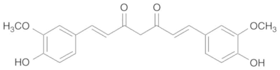
Curcumin
[(1,7-bis(4-hydroxy-3-methoxyphenyl)-1,6-heptane-3,5-dione] is a
yellow pigment present in the rhizome of Curcuma longa Linn
(15). Curcumin (Fig. 1) is the principal constituent of
turmeric, a popular Indian spice. Being reported to have
pharmacological properties, curcumin may be useful for treating
cancer, Dejerine-Sottas disease, inflammation, ulcer, depression,
contraception, diabetes, and viral diseases, among others (16).
Accumulating data has suggested that curcumin
decreases the proliferation of various cancer cells by inhibiting
cell growth, migration, and invasion. In addition, curcumin has
been reported to induce apoptosis and growth repression of cancer
cells in vivo and in vitro. Recent studies have
suggested that curcumin exerts epigenetic regulatory effects on
non-coding RNAs in various cancers (17,18).
Depending on their length, non-coding RNAs are classified as short
non-coding (snc) or long non-coding (lnc) RNAs (19,20).
Curcumin regulates cancer microRNAs
(miRNAs)
Biogenesis and function of miRNA
Endogenous miRNAs are non-coding RNAs that are ~22
nucleotides in length, which play important roles in
post-transcriptional regulation, inhibiting target messenger RNA
(mRNA) translation by complementary binding to the 3′-untranslated
region (3′-UTR) of mRNAs (21,22).
Consequently, miRNAs either repress translation or initiate mRNA
degradation (23,24). It has been reported that one miRNA
may bind to different mRNAs, and each mRNA can be targeted by many
different miRNAs, emphasizing the important regulatory role of
miRNAs (25,26). The first miRNA was discovered in
1993, and the term ‘microRNA’ was first used in 2001. Currently, at
least 2,000 miRNAs associated with the human genome are known, the
majority of which are abnormal in tumors (27,28).
Numerous miRNAs may be classified as oncogenic miRNAs (oncomiRNAs)
as they facilitate the proliferation and progression of certain
cancers (e.g., lung cancer, breast cancer, and esophageal
carcinoma) by downregulating genes via translational repression and
mRNA destabilization mechanisms (10). Furthermore, miRNAs are also
important prognostic biomarkers of chronic lymphocytic leukemia
(CLL), pancreatic cancer, neuroblastoma, and colorectal cancer
(29,30). It is well established that miRNAs
are central mediators in cancer biology, mediating the network
communication between cancer cells and their microenvironments
(13).
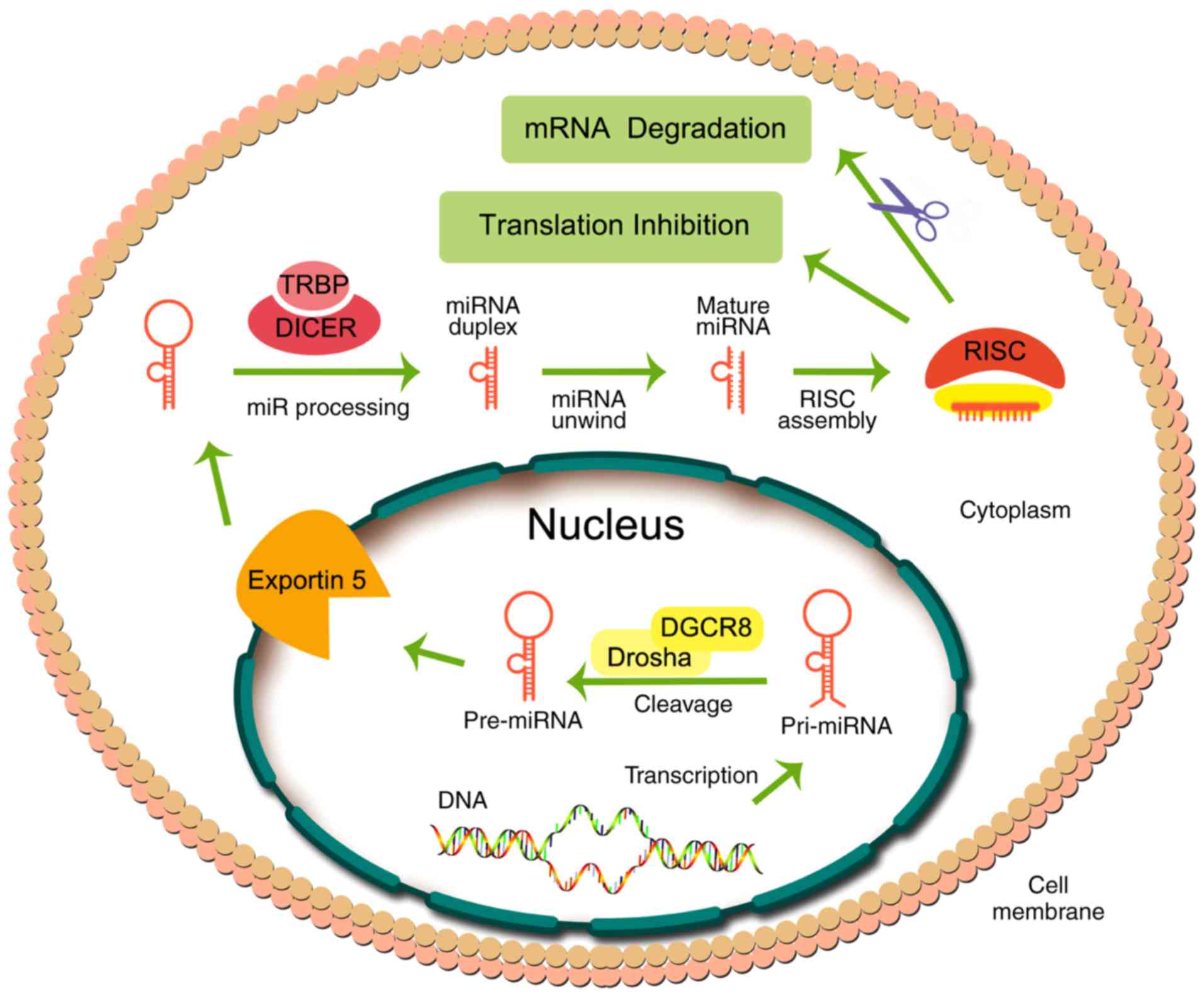
miRNA biogenesis consists of several steps (Fig. 2), starting in the nucleus, where
miRNAs are transcribed into a hairpin-shaped primary miRNA
(pri-miRNA), catalyzed by RNA polymerase II. Subsequently,
pri-miRNAs, which are hundreds to thousands of nucleotides in
length, are biotransformed into pre-miRNAs (~70 nucleotides in
length) in the nucleus by the microprocessor complex containing the
protein, Pasha/DGCR8, and the RNase III enzyme, Drosha.
Subsequently, pre-miRNA in the nucleus is transported to the
cytoplasm through nuclear pores via the protein, exportin 5. In the
cytoplasm, pre-miRNA is cleaved by a helicase containing an RNase
motif (known as Dicer) to produce the functional miRNA, with a
length of ~21–23 nucleotides (31–33).
The functional miRNA is recruited by Argonaute
proteins into the RNA-induced silencing complex (RISC). Finally,
the single-stranded mature miRNA combines with RISC, producing
inhibition of translation or degradation of the mRNA by binding to
the 3′-UTR of the target mRNA (34,35).
Recent evidence indicates that miRNAs can be extracellularly bound
to lipoproteins (36). Also, miRNAs
may be loaded into extracellular vesicles and transferred to
receptor cells, inducing remote effects as a form of cell-to-cell
communication (37).
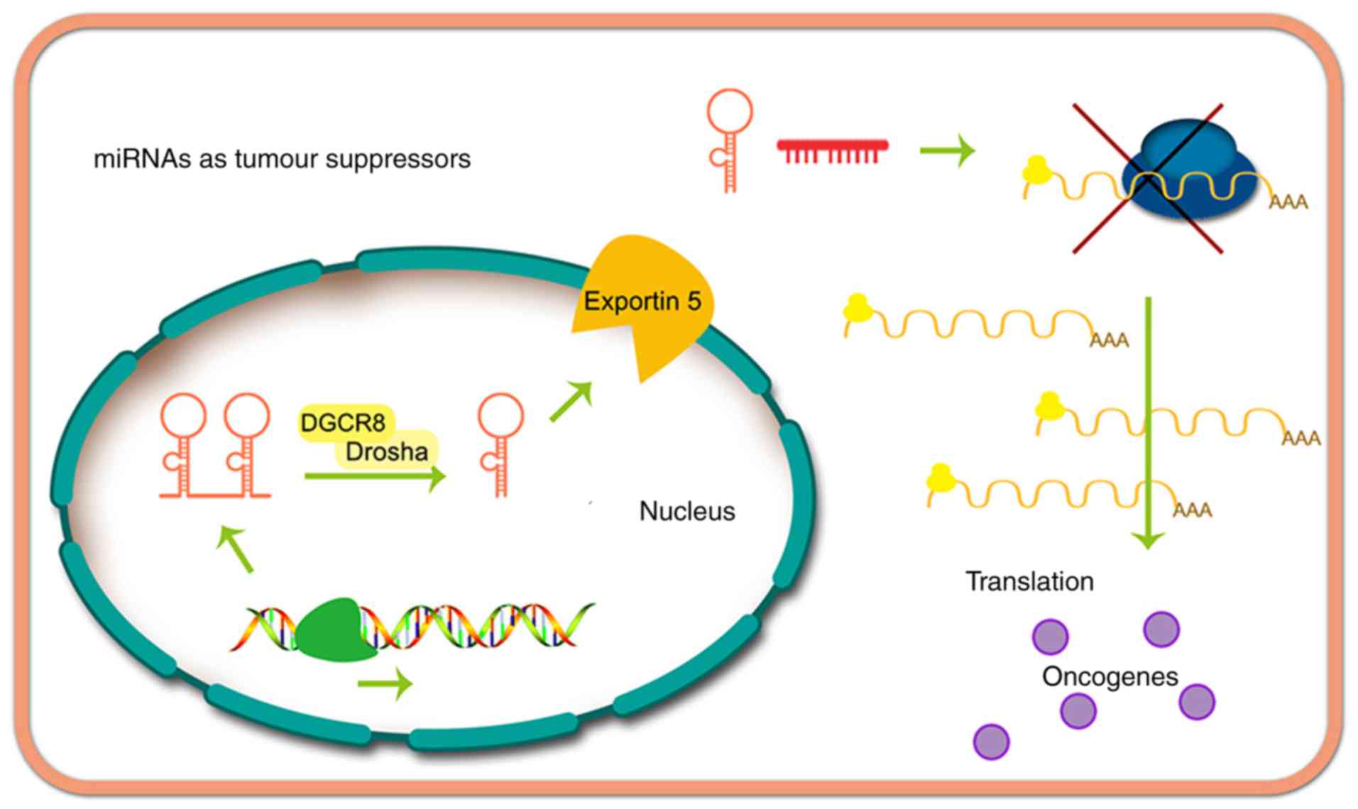
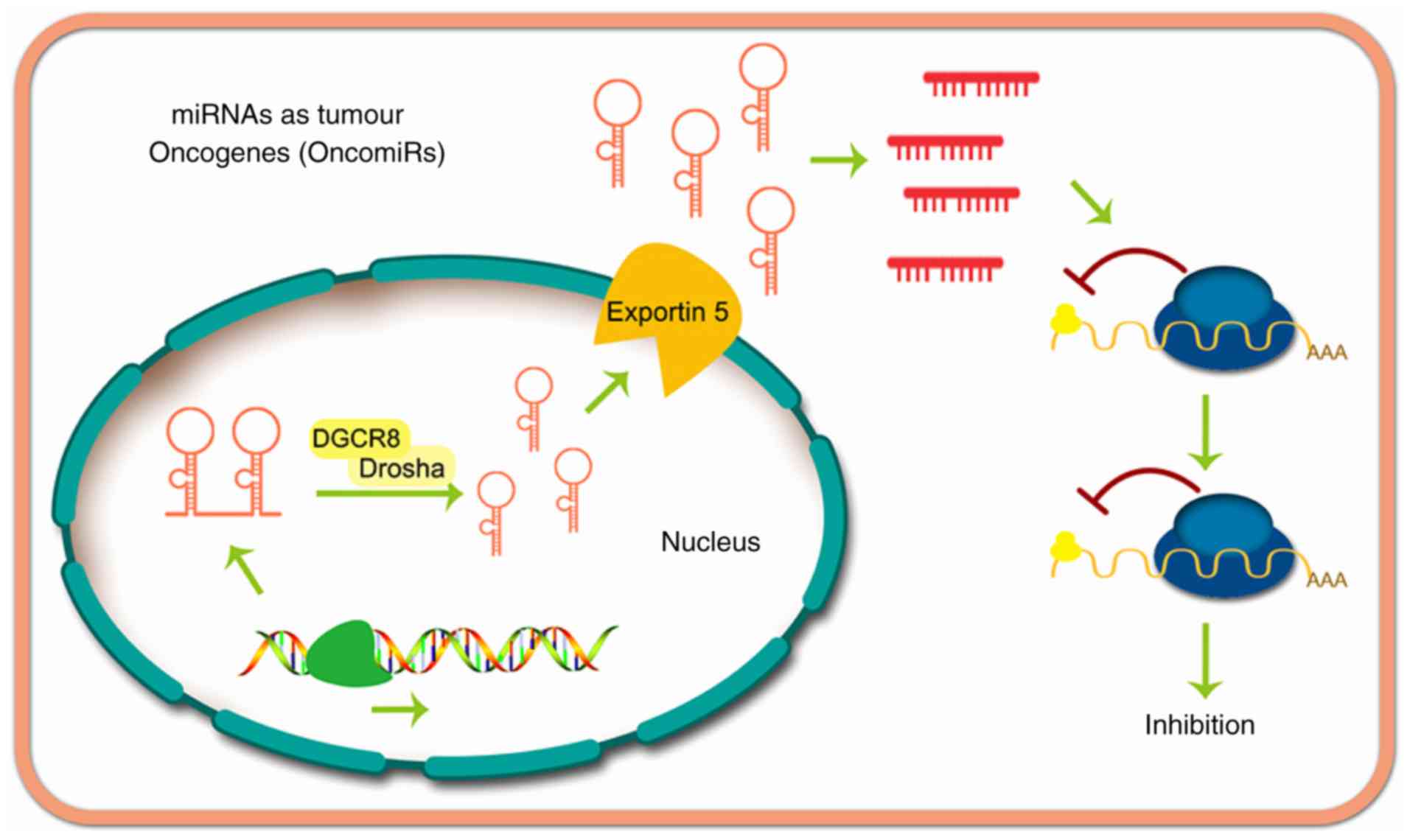
miRNAs may suppress cancer (Fig. 3) or exert oncogenic effects
(Fig. 4). The function of miRNAs in
tumor progression was first reported by Calin et al
(38), whose research demonstrated
that miR-16 and miR-15 are downregulated in patients with CLL.
Currently, thousands of miRNAs have been proven to be dysregulated
in other types of human cancer, and some of these miRNAs appear to
serve an important role in carcinogenesis by altering the
expression of oncogenes and cancer suppressor genes (16,39).
Curcumin effects on miRNAs in lung
cancer
Lung cancer is the leading cause of mortality among
cancer-associated deaths globally, resulting in at least 1.5
million deaths each year (40,41).
Lung cancer is categorized into two fundamental histological
subtypes: Small cell lung cancer (SCLC; 15–20 % incidence) and
non-small cell lung cancer (NSCLC; 80–85 % incidence) (42). Recent evidence has indicated that an
association exists between lung cancer and miRNA expression,
suggesting that this could be used as a novel treatment strategy
(43,44). The miRNAs of lung cancer modulated
by curcumin are summarized in Table
I.
 | Table I.Curcumin modulates miRNAs in lung
cancer. |
Table I.
Curcumin modulates miRNAs in lung
cancer.
| Author, year | Cell line | miRNA | Downstream
target(s) | (Refs.) |
|---|
| Jin et al,
2015 | NCL-H460, A549 | miR-192-5p (↑) | PI3K/Akt | (45) |
| Liu et al,
2017 | A549 | miRNA-98 (↑) | LIN28A | (46) |
|
|
|
| MMP2 |
|
|
|
|
| MMP9 |
|
| Zhang et al,
2010 | A549/DDP | miR-186* (↓) | Caspase-10 | (47) |
| Ye et al,
2015 | H460, A427 | miR-192-5p (↑) | XIAP | (48) |
|
|
| miR-215 (↑) |
|
|
| Wu et al,
2016 | A549,
NCI-H520, | let 7c (↑) | EZH2 | (50) |
|
| NCI-H1373, | miR-101 (↑) | NOTCH1 |
|
|
| NCI-H2170 |
|
|
|
| Zhang et al,
2014 | A549 | miR-21 (↓) | PTEN | (51) |
Curcumin has been shown to markedly increase
miR-192-5p expression, and suppress the phosphoinositide 3-kinase
(PI3K)/Akt signaling pathway, in A549 cells. Furthermore,
miR-192-5p mimics may significantly increase the efficacy of
curcumin in cell viability inhibition, apoptosis induction, and
suppression of the PI3K/Akt signaling pathway. Furthermore,
anti-miR-192-5p mimics abolish the cytotoxicity of curcumin and
PI3K/Akt pathway suppression produced by curcumin in A549 cells
(44,45).
The metastasis of lung cancer has been shown to be
positively correlated with suboptimal clinical results and
increased mortality. In A549 cells, curcumin markedly enhances the
expression of miR-98, and the expression of its downstream target,
the LIN28A gene, which is correlated with the self-renewal capacity
of stem cells, is decreased. The matrix metalloproteinases (MMPs)
MMP2 and MMP9, which are downstream proteins of LIN28A, are
downregulated in vitro and in vivo upon treatment
with curcumin. Furthermore, curcumin at a concentration of 100 µM
was shown to induce LIN28A-induced migration and invasion in A549
cells (46).
Curcumin at concentrations of 5, 10, 20, 30, and 40
µM led to a marked induction of apoptosis, also exhibiting
anti-cancer efficacy in drug-sensitive A549 cells and
multidrug-resistant A549/DDP cells. Curcumin markedly downregulated
miR-186* expression in A549 and A549/DDP cells, whereas
transfection with an miR-186* inhibitor was shown to increase
apoptosis of the A549 and A549/DDP cells. By contrast, the
overexpression of miR-186* markedly inhibited curcumin-induced
apoptosis in A549 and A549/DDP cells (47).
Curcumin has been reported to affect different
miRNAs in various cell lines. For example, RT-qPCR and miRNA
microarray data indicated that miR-215 and miR-192-5p are the most
responsive miRNAs in A427 and H460 cells following incubation with
curcumin (48). Curcumin was also
shown to regulate the levels of key miRNAs in A549 cells that were
detected by microarray analysis, such as miR-330-5p, which was
maximally upregulated by curcumin in vitro (49).
Curcumin inhibits the growth and metastasis of lung
cancer cells, including the NCI-H2170, NCI-H520, NCI-H1373, and
A549 cell lines. Furthermore, curcumin can suppress the expression
of the mRNA that codes for the protein, enhancer of zeste homolog 2
(EZH2), by increasing the levels of miR-101 and let-7c. Curcumin
has been shown to downregulate the expression of NOTCH1 by
inhibiting EZH2. Interestingly, reciprocal interactions have been
identified between NOTCH1 and EZH2 in A549, NCI-H520, NCI-H1373,
and NCI-H2170 lung cancer cells (50).
It has been reported that curcumin leads to a marked
inhibition of cell growth, and induces apoptosis in A549 cells.
Curcumin produces a concentration-dependent repression of miRNA-21
expression. Phosphatase and tensin homolog (PTEN) is the downstream
target gene of miRNA-21, which is markedly increased in A549 cells
upon incubation with curcumin. The transfection of A549 cells with
PTEN small interfering RNA or a microRNA-21 mimic significantly
reversed curcumin-induced growth inhibition and apoptosis (51).
Curcumin and miRNAs in colorectal
cancer (CRC)
CRC is a commonly occurring type of cancer that
produces substantial morbidity and mortality rates globally in both
males and females. Despite the significant progress has been
achieved in the treatment of CRC, the prognosis in a large number
of cases remains poor. Therefore, understanding the underlying
molecular genesis of CRC is important for precise diagnosis,
treatment, and prognosis of the disease. It has been reported that
these processes are associated with miRNAs to varying degrees
(52,53). The miRNAs of CRC that are modulated
by curcumin are summarized in Table
II.
| Table II.Curcumin modulates miRNAs in
colorectal cancer. |
Table II.
Curcumin modulates miRNAs in
colorectal cancer.
| Author, year | Cell line | miRNA | Downstream
target(s) | (Refs.) |
|---|
| Toden et al,
2015; | HCT116, | miR-200b (↑) | EZH2 | (54,55) |
| Goel, 2017 | HCT116-5FUR | miR-200c (↑) | BMI1 |
|
|
|
| miR-141 (↑) | SUZ12 |
|
|
|
| miR-429 (↑) | Ring1B |
|
|
|
| miR-101 (↑) |
|
|
| Mudduluru et
al, 2011; | Rko, HCT116 | miR-21 (↓) | Pdcd4 | (56,57) |
| Riaz Rajoka et
al, 2018 |
|
|
|
|
| Toden et al,
2015 | HCT116, | miR-34a (↑) | FBXW7 | (58) |
|
| SW480 | miR-27a (↓) | CDK4, 6 |
|
|
|
|
| Cyclin D, E |
|
|
|
|
| c-Myc |
|
| Li et al,
2018 | HCT116 | miR-491 (↑) | PEG10 | (59) |
| Gandhy et
al, 2012 | RKO, SW480 | miR-27a (↓) | Sp1 | (60) |
|
|
| miR-20a (↓) | Sp3 |
|
|
|
| miR-17-5p (↓) | Sp4 |
|
| Dou et al,
2017 | SW480 | miR-130a (↓) | Wnt/β-catenin | (61) |
Curcumin mediates sensitization to 5-fluorouracil in
CRC cells by inhibition of the epithelial-to-mesenchymal transition
(EMT) and polycomb repressive complexes (PRCs) via the regulation
of certain miRNAs. Specifically, curcumin was shown to upregulate
the expression of miR-429, miR-200b, miR-200c, miR-141, and
miR-101, whereas 5-fluorouracil did not significantly alter the
expression of these miRNAs (54,55).
miR-21 fulfills an important role in cancer
development, and curcumin is able to inhibit tumor growth,
invasion, and in vivo metastasis by regulating miR-21 in
CRC. Furthermore, two novel transcriptional start sites of the
miR-21 gene have been identified in HCT116 and Rko cells. Curcumin
was shown to significantly reduce miR-21 expression and promoter
activity in a dose-dependent manner by inhibiting the binding of
activator protein 1 to the promoter. Subsequently, the expression
of programmed cell death protein 4 (PDCD4), which is a target of
miR-21, was shown to be increased, leading to tumor suppression
(56,57).
Previously published in vitro data have
indicated that, in CRC cells, curcumin, in combination with
3-acetyl-11-keto-β-boswellic acid (AKBA), downregulated the
expression of miR-27a, and upregulated the expression of
tumor-suppressive miR-34a. Furthermore, both curcumin and AKBA were
shown to markedly decrease tumor growth in a mouse xenograft model,
and these effects were identified with alterations in the
expression of miR-34a and miR-27a (58).
Paternally expressed gene-10 (PEG10) may be
regulated by miR-491 in certain types of cancer. Furthermore,
miR-491 regulates the sensitivity of anti-cancer drugs when used to
treat CRC. Curcumin was revealed to upregulate miR-491 expression,
which suppressed the PEG10 and Wnt/β-catenin growth pathways,
thereby inhibiting the proliferation of HCT116 cells (59).
Curcumin was also shown to inhibit the growth of RKO
and SW480 colon cancer cells and induce apoptosis, as well as
downregulate the specificity protein (Sp) transcription factors,
Sp1, Sp3, and Sp4. Therefore, genes regulated by Sp, including
those encoding epidermal growth factor receptor, c-MET, survivin,
bcl-2, cyclin D1 and nuclear factor-κB, have been shown to be
downregulated. The mechanism of curcumin-induced inhibition of Sp
transcription factors is mediated via an increase in the expression
of the Sp inhibitors, ZBTB10 and ZBTB4, and the downregulation of
miR-27a, miR-20a and miR-17-5p (60). Curcumin has also been shown to
inhibit the proliferation of colon cancer cells in a mouse model by
suppressing the Wnt/β-catenin pathway via inhibition of miR-130a.
These results suggested that curcumin may have potential in terms
of developing novel therapies to treat CRC (61).
Curcumin and miRNAs in prostate
cancer
Annually, approximately 1.1 million men are
diagnosed with prostate cancer (62). Moreover, worldwide, prostate cancer
is the second most commonly occurring type of cancer. Accumulating
data has suggested that miRNAs may be used as predictive,
diagnostic, and prognostic biomarkers (63,64).
The miRNAs of prostate cancer known to be modulated by curcumin are
summarized in Table III.
| Table III.Curcumin modulates miRNAs in prostate
cancer. |
Table III.
Curcumin modulates miRNAs in prostate
cancer.
| Author, year | Cell line | miRNA | Downstream
target(s) | (Refs.) |
|---|
| Cao et al,
2017 | DU145 | miR-143 (↑) | PGK1 | (65) |
| Liu et al,
2017 | PC3 | miR-143 (↑) | ATG2B | (66) |
|
| DU145 |
|
|
|
|
| LNCaP |
|
|
|
| Liu et al,
2017 | Du145 | miR-145 (↑) | – | (67) |
|
| 22RV1 |
|
|
|
| Zhang et al,
2018 | Du145 | miR-770-5p | – | (68) |
|
| 22RV1 | miR-1247 |
|
|
Curcumin was shown to markedly upregulate miR-143
expression and inhibit the proliferation and migration of prostate
cancer cells, which could be blocked by transfection with
anti-miR-143. Both miR-143 overexpression and curcumin
downregulated the expression of phosphoglycerate kinase-1 (PGK1),
which upregulated the protein, forkhead box D3 (FOXD3).
Furthermore, the ectopic expression of FOXD3 synergized with
curcumin to upregulate the expression of miR-143 (65).
Interestingly, curcumin elicited a radiosensitizing
effect in prostate cancer by increasing the expression of miR-143,
enhancing apoptosis, and decreasing cancer cell growth induced by
radiation. Curcumin was shown to restore the expression of miR-143
in PC3, DU145, and LNCaP prostate cancer cells. Curcumin, similar
to the compound 5-A2a-2′-deoxycytidine (5-AZA-dC), was shown to
decrease the methylation of CpG dinucleotides in the miR-143
promoter. In addition, curcumin decreased the expression of DNA
(cytosine-5-)-methyltransferase 1 (DNMT1) and DNMT3B, an effect
that elicited hypermethylation of the miR-143/miR-145 cluster
(66).
Six miRNAs (namely, miR-145, miR-1275, miR-1908,
miR-3127, miR-3178, and miR-3198) were shown to be markedly
upregulated in human prostate cancer stem cells (HuPCaSCs)
incubated with curcumin compared with cells that were incubated
with vehicle, or untreated cells. Conversely, eight miRNAs (i.e.,
miR-671-5p, miR-664*, miR-494, miR-222*, miR-210, miR-193b*,
miR-183, and miR-23b*) were markedly downregulated in HuPCaSCs
incubated with curcumin (67).
Curcumin (at a concentration of 46.5 µM) significantly inhibited
the proliferation and invasion of HuPCaSCs in vitro. The
expression levels of miR-770-5p and miR-1247 in the DLK1-DIO3
imprinted gene cluster were significantly increased in HuPCaSCs
incubated with curcumin, compared with cells incubated with vehicle
(68).
Curcumin and miRNAs of breast
cancer
In the United States, breast cancer is one of the
most commonly occurring cancers among females. Molecular profiling
of breast cancer has revealed a dysregulation of miRNAs, and miRNAs
may be putative diagnostic and prognostic markers in the treatment
of breast cancer (69,70). The miRNAs of breast cancer modulated
by curcumin are summarized in Table
IV.
| Table IV.Curcumin modulates miRNAs in breast
cancer. |
Table IV.
Curcumin modulates miRNAs in breast
cancer.
| Author, year | Cell line | miRNA | Downstream
target(s) | (Refs.) |
|---|
| Wang et al,
2017 | MCF-7 | miR-21 (↓) | PTEN/Akt | (71) |
| Li et al,
2014 | MCF-7 | miR-19a (↓) | PTEN/Akt/p53 | (72) |
|
|
| miR-19b (↓) |
|
|
| Yang et al,
2010; | MCF-7 | miR-15a (↑) | Bcl-2 | (73,74) |
| Norouzi et
al, 2018 |
| miR-16 (↑) |
|
|
| Guo et al,
2013 | MDA-MB-231 | miR-34a (↑) | Bcl-2 | (75) |
|
| MDA-MB-435 |
| Bmi-1 |
|
| Kronski et
al, 2014 | MDA-MB-231 | miR-181b (↑) | CXCL1 | (76) |
|
|
|
| CXCL2 |
|
Curcumin has been shown to attenuate the malignancy
of breast cancer cells by inhibiting the miR-21/PTEN/Akt signaling
pathway. Curcumin elicited a significant concentration-dependent
decrease in the expression level of miR-21 in MCF-7 cells. The
overexpression of miR-21 markedly inhibited the anti-cancer
efficacy of curcumin in MCF-7 cells by suppressing the expression
of PTEN and increasing the expression of the protein,
phosphorylated (p-)Akt (71).
In MCF-7 cells, curcumin was shown to reverse
bisphenol A (BPA)-induced upregulation of the oncogenic miRNAs,
miR-19a, miR-19b, and it also upregulated their downstream targets,
including proliferating cell nuclear antigen, p-Akt, p-MDM2, PTEN,
and p53 (72). Additional results
published in that study suggested that curcumin regulated the
miR-19/PTEN/Akt/p53 pathway to reverse BPA-induced breast cancer
progression (72). Furthermore, in
MCF-7 cells that were incubated with curcumin, curcumin was shown
to increase the expression levels of miR-15a and miR-16, resulting
in the downregulation of Bcl-2. Additionally, the silencing of
miR-15a and miR-16 via specific inhibitors restored the expression
of Bcl-2, whereas a decrease in the expression levels of Bcl-2
induced apoptosis of the MCF-7 cells (73,74).
Curcumin and emodin have been shown to produce
synergistic effects in breast cancer MDA-MB-435 and MDA-MB-231
cells, which are classified as triple-negative breast cancer cells.
Curcumin and emodin increased the expression of miR-34a in breast
cancer cells. Curcumin and emodin also significantly inhibited
Bcl-2 and Bmi-1 expression in MDA-MB-231 and MDA-MB-435 breast
cancer cells, and this decrease could be reversed by a miR-34a
inhibitor (75). In MDA-MB-231
cells, curcumin modulated the expression of various miRNAs; for
example, miR-181b was upregulated. Interestingly, miR-181b
downregulated the levels of the chemokines CXCL1 and CXCL2 by
binding to their 3′-UTR. The overexpression of miR-181b led to a
marked decrease in the levels of CXCL1 and CXCL2, influencing the
efficacy of curcumin on CXCL1 and CXCL2 (76).
Curcumin and miRNAs in nasopharyngeal
carcinoma (NPC)
The majority of NPC cases (75–90%) are diagnosed at
an advanced stage, which contributes to its high risk of
recurrence, and metastasis with poor clinical outcomes. Several
miRNAs have been shown to be potential biomarkers and therapeutic
targets in NPC (77,78). The miRNAs of NPC known to be
modulated by curcumin are summarized in Table V.
| Table V.Curcumin modulates miRNAs in
nasopharyngeal cancer. |
Table V.
Curcumin modulates miRNAs in
nasopharyngeal cancer.
| Author, year | Cell line | miRNA | Downstream
target(s) | (Refs.) |
|---|
| Gao et al,
2014 | HONE1 | miR-125a-5p
(↓) | TP53 | (79) |
|
|
| miR-574-3p (↓) |
|
|
|
|
| miR-210 (↓) |
|
|
| Feng et al,
2017 | CNE1 | miR-7 (↑) | Skp2 | (80) |
|
| CNE2 |
|
|
|
| Fan et al,
2016 | CNE2 | miR-593 (↑) | MDR1 | (81) |
RT-qPCR and miRNA microarray analysis suggested that
curcumin decreases the expression levels of miR-574-3p, miR-210,
and miR-125a-5p in NPC cells. The overexpression of miR-125a-5p
facilitated the proliferation, migration, and invasion of HONE1
cancer cells. Moreover, curcumin increased the expression of tumor
protein 53 (TP53), a downstream target of miR-125a-5p (79). Curcumin has been shown to inhibit
the growth, migration, and invasion of human NPC CNE1 and CNE2
cells by inducing cell cycle arrest and apoptosis following
irradiation. Curcumin was shown to increase the expression of
miR-7, which inhibits the expression of its direct target, S-phase
kinase-associated protein 2 (Skp2) (80). Curcumin, in combination with 4 Gy
irradiation, inhibited the proliferation of transplanted tumors
in vivo, producing a greater efficacy of curcumin compared
with radiotherapy treatment alone. The upregulation of miR-593
mediated by curcumin led to a decrease inexpression of multidrug
resistance 1 protein (MDR1), a downstream target of miR-593
(81).
Curcumin and miRNAs in pancreatic
cancer
The diagnosis and treatment of pancreatic cancer
remains a major clinical challenge. Due to the nature of early
metastasis, at least 80% of patients with pancreatic cancer have an
invasive lesion upon diagnosis (82). Thus, surgical and medical
interventions for pancreatic cancer are fundamentally unsuccessful,
resulting in high mortality rates and a poor clinical prognosis. It
has been hypothesized that there is a correlation between altered
miRNA expression and pancreatic cancer (83,84).
The miRNAs of pancreatic cancer modulated by curcumin are
summarized in Table VI.
| Table VI.Curcumin modulates miRNAs in
pancreatic cancer. |
Table VI.
Curcumin modulates miRNAs in
pancreatic cancer.
| Author, year | Cell line | miRNA | Downstream
target(s) | (Refs.) |
|---|
| Yang et al,
2017 | PANC-1 | miR-340 (↑) | XIAP | (85) |
| Ma et al,
2014 | AsPC-1 | miR-7 (↑) | SET8 | (86) |
|
| BxPC-3 |
|
|
|
| Sun et al,
2008 | BxPC-3 | miR-22 (↑) | SP1 | (87) |
|
|
|
| ESR1 |
|
Previously published data have suggested that
curcumin-induced apoptosis is associated with the miR-340/X-linked
inhibitor of apoptosis (XIAP) signaling pathway in PANC-1
pancreatic cancer cells. In addition, incubation with curcumin or
miR-340 induced apoptosis of pancreatic cancer cells, whereas
silencing the endogenous miR-340 significantly decreased the
apoptotic efficacy of curcumin. Western blotting and luciferase
reporter assays revealed that the oncogene, XIAP, is a direct
downstream target of miR-340. Furthermore, curcumin led to a
significant decrease in the expression of XIAP, a phenomenon that
was reversed by anti-miR-340 (85).
In the pancreatic cancer cell lines, BxPC-3 and
AsPC-1, curcumin has been shown to inhibit cell proliferation,
migration, and invasion, and to induce apoptosis. These effects
were correlated with an increase in the expression of miR-7 and
subsequent downregulation of SET domain-containing lysine
methyltransferase 8 (SET8), a downstream target of miR-7. These
results suggested that miR-7 is modulated by curcumin, and that
this may represent a novel therapeutic strategy for the treatment
of pancreatic cancer (86).
In addition, curcumin has been shown to modulate
miRNA expression of BxPC-3 human pancreatic cancer cells,
downregulating miRNA-199a* and upregulating miR-22. Upregulation of
miR-22 by curcumin, or transfection with miR-22 mimics in BxPC-3
cells, was shown to suppress the expression of downstream target
genes, namely the transcription factor Sp1 and estrogen receptor 1
(87).
Curcumin and miRNAs of leukemia
Globally, leukemia accounts for approximately 2.5%
of all new cancer cases, and 3.5% of all cancer-associated deaths
(88). Chronic myeloid leukemia
(CML), an acquired malignant disorder of hematopoietic stem cells,
is one of three common types of leukemia. Similarly to solid
cancers, it has been reported that miRNAs are dysregulated in
hematological malignancies (89,90).
The miRNAs of leukemia modulated by curcumin are summarized in
Table VII.
| Table VII.Curcumin modulates miRNAs in
leukemia. |
Table VII.
Curcumin modulates miRNAs in
leukemia.
| Author, year | Cell line | miRNA | Downstream
target(s) | (Refs.) |
|---|
| Taverna et
al, 2016; | K562 | miR-21 (↓) | PTEN | (91,92) |
| Taverna et
al, 2015 | LAMA84 | miR-196b (↑) | Bcr-Abl |
|
| Gao et al,
2012 | K562 | miR-15a (↑) | WT1 | (93) |
|
| HL-60 | miR-16-1 (↑) |
|
|
The incubation of CML K562 and LAMA84 cells with
curcumin (at concentrations of 10, 20 and 40 µM) was found to
produce a concentration-dependent upregulation of PTEN, one of the
targets of miR-21. Curcumin, in vitro, decreased the
expression of vascular endothelial growth factor and the
phosphorylation of Akt. Colony formation experiments indicated that
curcumin inhibits the viability of CML cells. Curcumin also
decreased the expression of miR-21 in CML cells, and the secretion
of exosomes. Furthermore, curcumin was shown to increase the
expression of miR-196b, which regulates the CML-associated protein
Bcr-Abl (91,92).
Curcumin-mediated overexpression of miR-15a and
miR-16-1 occurs prior to the downregulation of the protein, Wilms'
tumor 1 (WT1). Furthermore, anti-miR-15a and anti-miR-16-1
oligonucleotides are able to partially restore the decreased
expression of WT1 mediated by curcumin in leukemic K562 and HL-60
cells. In addition, anti-miR-15a/16-1 oligonucleotides increased
the proliferation of K562 and HL-60 cells that is elicited by
curcumin (93).
Curcumin and miRNAs in ovarian
cancer
Ovarian cancer is typically diagnosed at an advanced
stage without symptoms, thereby making it difficult to treat.
Emerging data have shown that miRNAs elicit oncogenesis or activate
tumor suppressors in ovarian tumors (94,95).
The miRNAs of ovarian cancer modulated by curcumin are summarized
in Table VIII.
| Table VIII.Curcumin modulates miRNAs in ovarian
cancer. |
Table VIII.
Curcumin modulates miRNAs in ovarian
cancer.
| Author, year | Cell line | miRNA | Downstream
target(s) | (Refs.) |
|---|
| Zhang et al,
2017 | OVCAR-3 | miR-214 (↓) | MEG3 | (96) |
|
| SKOV3 |
|
|
|
|
| A2780cp |
|
|
|
|
| A2780 |
|
|
|
| Zhao et al,
2017 | SKOV3 | miR-124 (↑) | Midkine | (97) |
| Zhao et al,
2014 | SKOV3 | miR-9 (↑) | p-Akt | (98) |
|
|
|
| p-FOXO1 |
|
miR-214 significantly modulates the effects of
chemotherapy in ovarian cancer. In vitro, miR-214 has been
shown to affect cisplatin resistance and cell survival by
regulating the PTEN/Akt signaling pathway. Furthermore, miR-214
induced stem cell properties by interacting with the p53/Nanog
pathway in ovarian cancer. Curcumin was shown to decrease miR-214
expression, which, in turn, led to a decrease in cisplatin
resistance in OVCAR-3 and SKOV3 cells (96).
In SKOV3 cells, the combination of
dihydroartemisinin and curcumin has been shown to synergistically
inhibit cell growth and induce cell cycle arrest and apoptosis.
Moreover, this led to a marked decrease in the expression of the
oncogene, midkine, and synergistically increased the expression of
miR-124. Furthermore, miR-124 was shown to directly bind to the
3′-UTR of midkine mRNA, causing its degradation, thereby decreasing
the expression of the midkine protein (97).
In addition, curcumin significantly inhibits the
proliferation of SKOV3 cells and elicits apoptosis, increasing the
expression of miR-9. The depletion of miR-9 attenuated the
anti-cancer efficacy of curcumin, whereas its overexpression
increases the levels of apoptosis in SKOV3 cells. Overexpression of
miR-9 and curcumin led to a marked decrease in the levels of p-Akt
and p-FOXO1 compared with cells incubated with vehicle (98).
Curcumin and miRNAs in other
cancers
Dendrosomal curcumin has been shown to potently
inhibit the proliferation of U87MG cells by i) arresting the cell
cycle during the G1 phase; and ii) inducing apoptosis.
Dendrosomal curcumin significantly increased the expression of
miR-145, leading to the subsequent downregulation of its downstream
proteins, including octamer-binding transcription factor 4A
(OCT4A), OCT4B1, (sex determining region Y)-box 2 (SOX-2), and
Nanog (99). Curcumin significantly
reduces the expression of miR-222, miR-221, miR-146b, and miR-21 in
SW1736 and 8505C cells. Furthermore, the abovementioned miRNAs, as
well as miR-204, were shown to mediate the efficacy of
nutraceuticals in thyroid cancer progression (100).
The expression level of miR-9 in oral squamous cell
carcinoma is lower compared with that in the adjacent non-tumor
tissue. Curcumin has been shown to inhibit the cell growth of SCC-9
oral squamous cell carcinoma cells by increasing the expression
levels of miR-9 and disrupting the Wnt/β-catenin pathway.
Specifically, the expression levels of glycogen synthase kinase-3β
(GSK-3β), p-GSK-3β and β-catenin were increased, whereas that of
cyclin D1 was decreased (34).
Clinically, the expression of the tumor suppressor,
miR-203, is epigenetically downregulated in bladder cancer.
Curcumin upregulates miR-203 expression in TCCSUP, T24, and J82
bladder cancer cells by producing DNA hypomethylation of the
miR-203 promoter. The overexpression of miR-203 downregulated the
target oncogenes Akt2/Src, thereby decreasing cell survival,
migration, and invasion (101).
A nanoparticle formulation of curcumin (Nano-CUR),
based on polylactic-co-glycolic acid, was developed and tested in
cervical cancer cells in vitro and in a pre-clinical
orthotopic mouse model. Nano-CUR has been shown to significantly
suppress cell proliferation, induce apoptosis, cause cell cycle
arrest in the cervical cancer cell lines Ca Ski and Si Ha,
downregulate the expression of oncogenic miRNA-21, repress
β-catenin levels in the nucleus, and decrease the expression level
of the oncoprotein, E6/E7 HPV. Furthermore, miRNA-21 decreased the
expression of E6/E7 and interleukin-6, whose levels are increased
by benzo[a]pyrene (102).
In U2OS and MG63 osteosarcoma cells, curcumin was
shown to significantly suppress the expression of estrogen-related
receptor-α (ERRα) by upregulating miR-125a. The overexpression of
ERRα decreased the induction of apoptosis mediated by curcumin,
whereas silencing ERRα led to a sensitization of the osteosarcoma
cells to curcumin (103). By
contrast, curcumin inhibited miR-125a expression in
undifferentiated NPC cells (79).
The differing tumor characteristics may be a factor underlying the
contrasting effects produced by curcumin in regulating
miR-125a.
Curcumin has been shown to repress cell growth,
increase caspase-3 activity, and stimulate apoptosis in the
laryngeal squamous cell carcinoma cell line, AMC-HN-8. Curcumin
increased miR-15a expression, and downregulated the protein levels
of Bcl-2, PI3K, and p-Akt. The suppression of miR-15a is able to
reverse the anti-proliferative efficacy of curcumin and increase
the expression levels of Bcl-2 and PI3K/Akt in AMC-HN-8 cells
(104).
Curcumin regulates cancer lncRNAs
Biogenesis and function of
lncRNAs
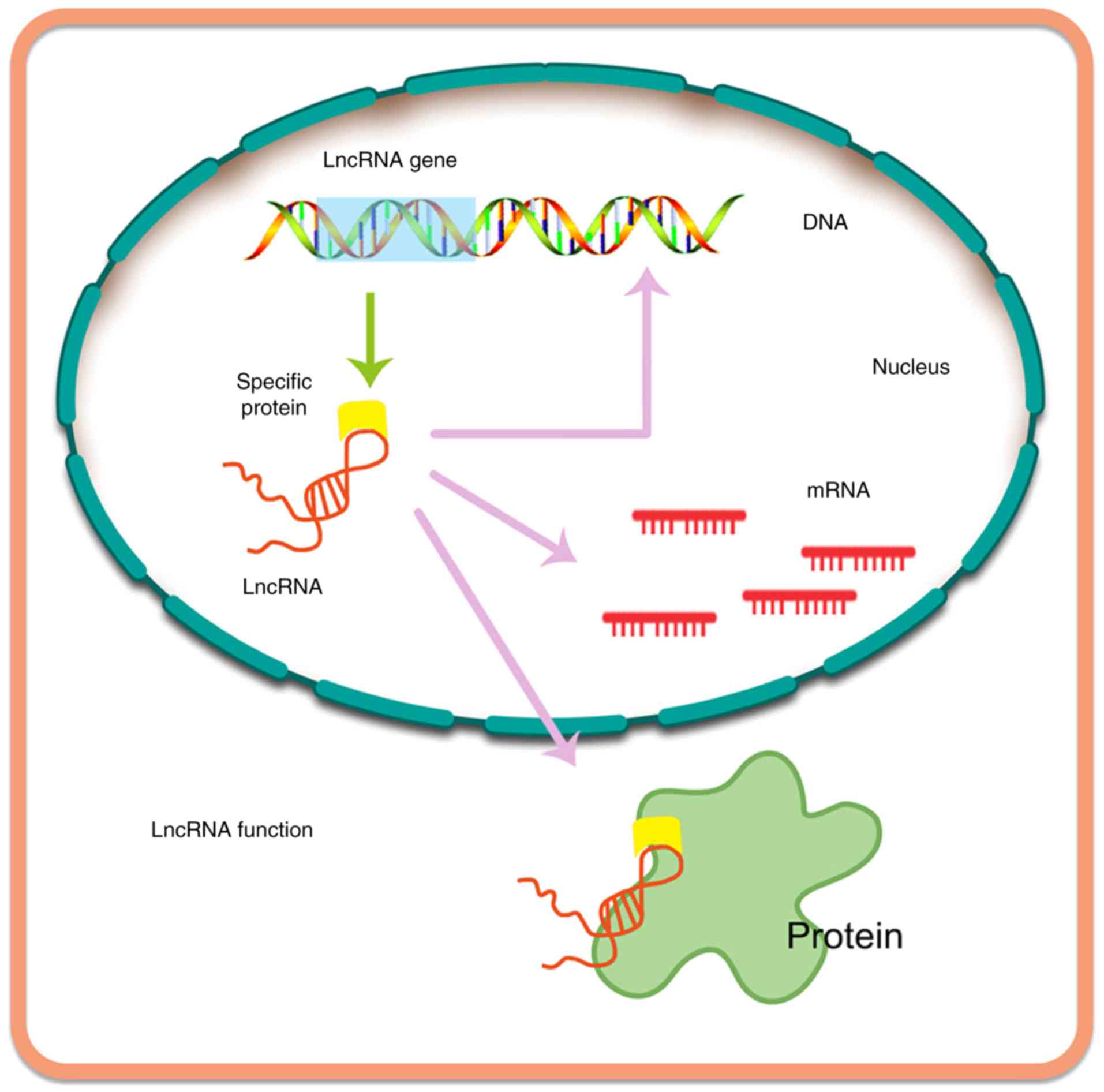
lncRNAs are byproducts of transcription, typically
consisting of >200 bases (105). lncRNAs are primarily transcribed
by the enzymes RNA polymerase II and RNA polymerase III (19,106).
Similarly to miRNAs, lncRNAs are also able to bind to certain
proteins, RNA and nucleating RNA compartments, forming
ribonucleoprotein complexes. Interestingly, lncRNAs can function
immediately after synthesis, acting as flexible scaffolds to
promote dynamic gene control (107). Recently, a number of studies have
reported the potential role of lncRNAs in normal and pathological
processes [e.g., see (20)].
Furthermore, it has been demonstrated that the expression of
thousands of lncRNAs varies according to the types of tumor, and
certain of these have been linked to tumorigenesis (19,108).
Currently, a total of 7,258 sncRNAs and 15,767
annotated lncRNAs have been recorded in the GENCODE database, which
contains the largest known compilation of transcripts (20,109).
lncRNAs are able to modulate multiple levels of gene expression
(Fig. 5), including regulation at
the epigenetic, transcriptional, and post-transcriptional levels,
based on the findings of tissue microarray analyses and
next-generation sequencing assays. Thus, in the future, lncRNAs may
attract interest in various therapeutic areas.
lncRNAs can act as sponges or molecular decoys of
miRNAs, influencing the expression and activities of miRNAs
(65). Furthermore, miRNAs could
directly or indirectly target lncRNAs. Accumulating evidence has
suggested that an active crosstalk between lncRNAs and miRNA exists
via a double-negative feedback circle that is able to manipulate
the levels of gene expression (108).
Curcumin regulates lncRNAs in lung
cancer
lncRNAs participate in epigenetic regulation,
transcriptional regulation, and post-transcriptional processing.
There are two major categories of lncRNAs, which are defined as
oncogene lncRNAs [metastasis-associated lung adenocarcinoma
transcript 1 (MALAT1), HOX transcript antisense RNA (HOTAIR), SOX2
overlapping transcript (SOX2-OT), H19, etc.] and tumor suppressor
lncRNAs [maternally expressed 3 (MEG3), promoter of CDKN1A
antisense DNA damage-activated RNA (PANDAR), growth arrest-specific
5 (GAS5), taurine-upregulated gene 1 (TUG1), etc.], according to
their pathological features (110). Therefore, numerous dysregulated
lncRNAs have been identified in patients with NSCLC as specific
biomarkers for diagnosis. The upregulation of 24 lncRNAs (MALAT1,
HOTAIR, H19, etc.) and downregulation of 9 lncRNAs (GAS5, PANDAR,
MEG3, etc.) have been observed in NSCLC (111).
Furthermore, mechanistic studies have revealed that
curcumin is able to inhibit the growth of A549 cells and the
expression of urothelial cancer associated-1 (UCA1), the
overexpression of which abolished the effect of curcumin on cell
apoptosis. Subsequently, curcumin may inhibit the Wnt and mTOR
pathways by downregulating UCA1, which could provide novel insights
into the treatment of lung cancer (112).
Curcumin regulates lncRNA in CRC
CRC-associated lncRNAs are involved in invasion,
metastasis, chemoresistance and radioresistance by interactions
with different signalling pathways, including the EMT, Wnt, and
transforming growth factor-β (TGF-β) pathways, and also
interactions with miRNAs (113).
Specifically, accumulating evidence has indicated that lncRNAs are
able to directly modulate metastatic pathways in CRC. A total of 28
CRC-associated oncogene lncRNAs (HOTAIR, UCA1, H19, MEG3, etc.)
were identified in a recent study, and 13 tumor suppressor lncRNAs
(MEG3, GAS5, etc.) (114).
Experiments in vitro have indicated that
knockdown of PANDAR lncRNA did not significantly affect
proliferation, senescence, or apoptosis of CRC cells. It has been
reported that curcumin produces senescence of CRC cells without
increasing apoptosis. Furthermore, PANDAR expression was
upregulated in CRC cells incubated with curcumin. The silencing of
PANDAR increased the rate of apoptosis and significantly decreased
the level of senescence, results that are likely to be accounted
for by increasing the levels of p53-upregulated modulator of
apoptosis (PUMA) in CRC cells incubated with curcumin (115).
Curcumin regulates lncRNA in prostate
cancer
Compared with normal tissues, distinct fold changes
of 60–100 for 95% of prostate tumors were detected; therefore,
prostate cancer antigen-3 (PCA3) can be potentially applied as a
specific prostate cancer biomarker, which is undetectable in other
tumor types (116). It was
reported that 17 lncRNAs (HOTAIR, SOCS2-AS1, PVT1, etc.) contribute
to prostate cancer progression. Additionally, low expression levels
of 3 lncRNAs (GAS5, MEG3, and H19) were detected in prostate cancer
(117).
In vitro, HuPCaSCs with overexpression of
miR-145, cell cycle arrest and inhibition of cell proliferation and
invasion were observed following pre-treatment with curcumin.
Luciferase activity assays revealed that Oct4 and lncRNA-ROR are
able to bind to miRNAs competitively via their common binding sites
of miR-145. In general, downregulation of endogenous lncRNA-ROR
increased expression of miR-145 in HuPCaSCs; subsequently, miR-145
inhibited cell proliferation by decreasing Oct4 expression
(67).
Curcumin regulates lncRNA in breast
cancer
Some lncRNAs have particular associations with
specific types of cancer. For example, in the case of breast
cancer, several lncRNAs (HOTAIR, MALAT1, H19, etc.) are associated
with cancer progression (118).
Dendrosomal curcumin treatment in MCF7, MDA-MB231
and SKBR3 cells has been shown to reduce the expression of Tusc7
and GAS5 lncRNAs. GAS5 downregulation leads to an enhancement of
the anticancer effect of dendrosomal curcumin. It appears that a
combination of dendrosomal curcumin and GAS5 overexpression may be
applied as a therapy for drug-resistant breast cancer in the clinic
(119).
Curcumin regulates lncRNA in
pancreatic cancer
Increases in the expression of the lncRNAs ROR, H19,
nuclear-enriched abundant transcript-1 (NEAT1), nuclear transport
factor 2 pseudogene 3 (NUTF2P3), and MIR31HG were identified in
pancreatic cancer via different mechanisms (120). In addition, overexpression of 7
lncRNAs (ROR, H19, NEAT1, etc.) was observed in pancreatic cancer,
whereas that of only one lncRNA (ENST00000480739) was shown to be
decreased in a recently published study (121). Curcumin treatment in BxPC3-GemR
cells promoted a reversal of gemcitabine resistance by inhibiting
the expression of the PRC2 subunit, EZH2, and its associated
lncRNA, PVT1 (122).
Curcumin regulates lncRNA in ovarian
cancer
Various novel methods, including RT-qPCR and
high-throughput techniques, have been applied to build up the
lncRNA expression profile for ovarian cancer. Furthermore, lncRNA
clusters show distinct metastatic potentials with different
expression levels in ovarian cancer cells (123). For example, lncRNA MEG3 has been
shown to be efficacious as a cancer suppressor. Curcumin increases
the expression of MEG3 in ovarian cancer. A mechanistic study
revealed that DNA hypomethylation maybe induced by dendrosomal
curcumin, which resulted in the re-expression of silenced tumor
suppressor genes, such as MEG3 (96).
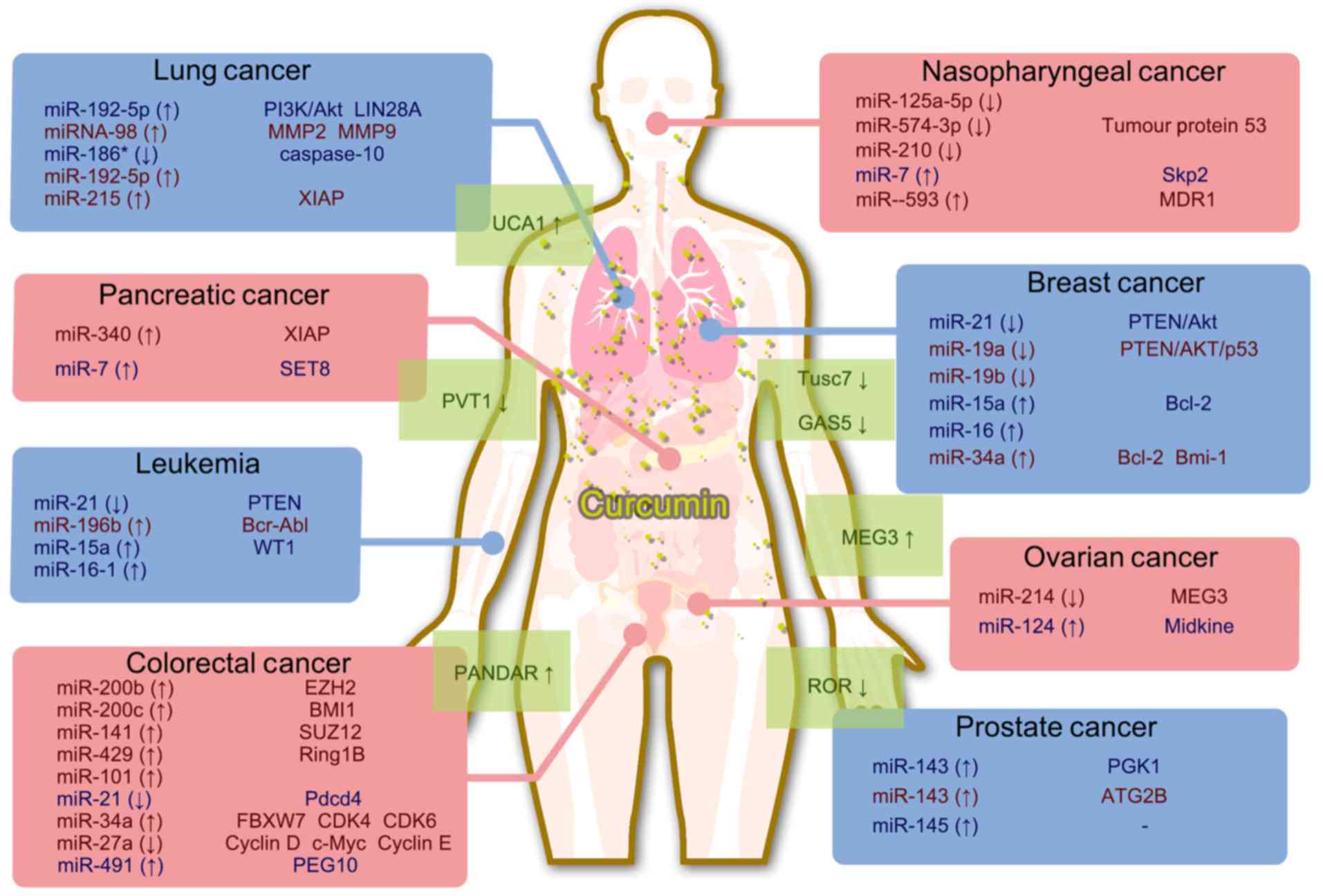
Conclusions and perspectives
Non-coding RNAs have been shown to exert critical
roles in regulating cancer cell biology, and they may serve as
promising targets for novel anti-cancer treatments. The natural
product curcumin has been demonstrated to possess significant
anti-proliferative efficacy in different types of cancer cell. The
known effects of curcumin that have been established with respect
to miRNA/lncRNA expression are summarized in Fig. 6. It is possible that curcumin
inhibits cancer cell proliferation by regulating certain non-coding
RNAs, which are important for cancer cell proliferation. These
findings will help to elucidate the mechanisms that underpin the
efficacy of curcumin, thereby providing valuable information for
the evaluation of novel cancer treatments.
Acknowledgements
We thank Yangmin Chen, who provided editorial
assistance.
Funding
This work was supported by National Natural Science
Foundation of China (grant nos. 81473320 and 81773888), Fund of
Guangdong Science and Technology Department (grant no.
2016A020226024), Fund of Guangzhou Science and Technology Program
(grant no. 201707010048), Fund of Guangdong Education Department
(grant no. 2015KTSCX112), the Fund of Construction of High-Level
Universities in Guangdong (Nanshan Scholars Program and Academic
Backbone Program), and Fund of Xinjiang Science and Technology
Department (grant nos. 2013711020 and 201223111).
Availability of data and materials
Not applicable.
Authors' contributions
JZ and ZC conceived the review; YL, HS, QC, JL, and
CS wrote the review. BM, CRA Jr, ZC, and JZ revised the review. All
authors read and approved the final manuscript.
Ethics approval and consent to
participate
Not applicable.
Patient consent for publication
Not applicable.
Competing interests
The authors declare that they have no competing
interests.
Glossary
Abbreviations
Abbreviations:
|
miRNA
|
microRNA
|
|
lncRNA
|
long non-coding RNA
|
|
mRNA
|
messenger RNA
|
|
oncomiRNA
|
oncogenic miRNA
|
|
CLL
|
chronic lymphocytic leukemia
|
|
pri-miRNA
|
primary miRNA
|
|
RISC
|
RNA-induced silencing complex
|
|
SCLC
|
small cell lung cancer
|
|
NSCLC
|
non-small cell lung cancer
|
|
PI3K
|
phosphoinositide 3-kinase
|
|
MMP
|
matrix metalloproteinase
|
|
EZH2
|
zeste homolog 2
|
|
PTEN
|
phosphatase and tensin homolog
|
|
CRC
|
colorectal cancer
|
|
EMT
|
epithelial-to-mesenchymal
transition
|
|
PRC
|
polycomb repressive complex
|
|
PDCD4
|
programmed cell death protein 4
|
|
AKBA
|
3-acetyl-11-keto-β-boswellic acid
|
|
PEG10
|
paternally expressed gene-10
|
|
Sp
|
specificity protein
|
|
PGK1
|
phosphoglycerate kinase-1
|
|
FOXD3
|
forkhead box D3
|
|
HuPCaSC
|
human prostate cancer stem cell
|
|
NPC
|
nasopharyngeal carcinoma
|
|
TP53
|
tumor protein 53
|
|
Skp2
|
S-phase kinase-associated protein
2
|
|
XIAP
|
X-linked inhibitor of apoptosis
|
|
SET8
|
SET domain-containing lysine
methyltransferase 8
|
|
OCT4
|
octamer-binding transcription factor
4
|
|
SOX-2
|
(sex determining region Y)-box 2
|
|
Nano-CUR
|
nanoparticle formulation of
curcumin
|
|
MALAT1
|
metastasis-associated lung
adenocarcinoma transcript 1
|
|
HOTAIR
|
HOX transcript antisense RNA
|
|
SOX2-OT
|
SOX2 overlapping transcript
|
|
MEG3
|
maternally expressed 3
|
|
PANDAR
|
promoter of CDKN1A antisense DNA
damage-activated RNA
|
|
GAS5
|
growth arrest-specific 5
|
|
UCA1
|
urothelial cancer associated-1
|
|
PCA3
|
prostate cancer antigen-3
|
|
NEAT1
|
nuclear-enriched abundant
transcript-1
|
References
|
1
|
Shi Z, Li Z, Li ZJ, Cheng K, Du Y, Fu H
and Khuri FR: Cables1 controls p21/Cip1 protein stability by
antagonizing proteasome subunit alpha type 3. Oncogene.
34:2538–2545. 2015. View Article : Google Scholar : PubMed/NCBI
|
|
2
|
Jiang QW, Cheng KJ, Mei XL, Qiu JG, Zhang
WJ, Xue YQ, Qin WM, Yang Y, Zheng DW, Chen Y, et al: Synergistic
anticancer effects of triptolide and celastrol, two main compounds
from thunder god vine. Oncotarget. 6:32790–32804. 2015. View Article : Google Scholar : PubMed/NCBI
|
|
3
|
Zhang JY, Wu HY, Xia XK, Liang YJ, Yan YY,
She ZG, Lin YC and Fu LW: Anthracenedione derivative 1403P-3
induces apoptosis in KB and KBv200 cells via reactive oxygen
species-independent mitochondrial pathway and death receptor
pathway. Cancer Biol Ther. 6:1413–1421. 2007. View Article : Google Scholar : PubMed/NCBI
|
|
4
|
Zhang JY, Tao LY, Liang YJ, Yan YY, Dai
CL, Xia XK, She ZG, Lin YC and Fu LW: Secalonic acid D induced
leukemia cell apoptosis and cell cycle arrest of G(1) with
involvement of GSK-3beta/beta-catenin/c-Myc pathway. Cell Cycle.
8:2444–2450. 2009. View Article : Google Scholar : PubMed/NCBI
|
|
5
|
Zhang JY, Huang WJ, Sun HM, Liu Y, Zhao
XQ, Tang SL, Sun MN, Wang S, Li JJ, Zhang LL, et al: Structure
identification and in vitro anticancer activity of
lathyrol-3-phenylacetate-5,15-diacetate. Molecules. 22:e14122017.
View Article : Google Scholar : PubMed/NCBI
|
|
6
|
Zhang J, Lai Z, Huang W, Ling H, Lin M,
Tang S, Liu Y and Tao Y: Apicidin inhibited proliferation and
invasion and induced apoptosis via mitochondrial pathway in
non-small cell lung cancer GLC-82 cells. Anticancer Agents Med
Chem. 17:1374–1382. 2017. View Article : Google Scholar : PubMed/NCBI
|
|
7
|
Shi Z, Peng XX, Kim IW, Shukla S, Si QS,
Robey RW, Bates SE, Shen T, Ashby CR Jr, Fu LW, et al: Erlotinib
(Tarceva, OSI-774) antagonizes ATP-binding cassette subfamily B
member 1 and ATP-binding cassette subfamily G member 2-mediated
drug resistance. Cancer Res. 67:11012–11020. 2007. View Article : Google Scholar : PubMed/NCBI
|
|
8
|
Zhang JY, Mi YJ, Chen SP, Wang F, Liang
YJ, Zheng LS, Shi CJ, Tao LY, Chen LM, Chen HB and Fu LW: Euphorbia
factor L1 reverses ABCB1-mediated multidrug resistance involving
interaction with ABCB1 independent of ABCB1 downregualtion. J Cell
Biochem. 112:1076–1083. 2011. View Article : Google Scholar : PubMed/NCBI
|
|
9
|
Lin M, Bi H, Yan Y, Huang W, Zhang G,
Zhang G, Tang S, Liu Y, Zhang L, Ma J and Zhang J: Parthenolide
suppresses non-small cell lung cancer GLC-82 cells growth via
B-Raf/MAPK/Erk pathway. Oncotarget. 8:23436–23447. 2017.PubMed/NCBI
|
|
10
|
McLoughlin NM, Mueller C and Grossmann TN:
The therapeutic potential of PTEN modulation: Targeting strategies
from gene to protein. Cell Chem Biol. 25:19–29. 2018. View Article : Google Scholar : PubMed/NCBI
|
|
11
|
Zhang JY, Lin MT, Tung HY, Tang SL, Yi T,
Zhang YZ, Tang YN, Zhao ZZ and Chen HB: Bruceine D induces
apoptosis in human chronic myeloid leukemia K562 cells via
mitochondrial pathway. Am J Cancer Res. 6:819–826. 2016.PubMed/NCBI
|
|
12
|
Zhang JY, Tao LY, Liang YJ, Chen LM, Mi
YJ, Zheng LS, Wang F, She ZG, Lin YC, To KK and Fu LW:
Anthracenedione derivatives as anticancer agents isolated from
secondary metabolites of the mangrove endophytic fungi. Mar Drugs.
8:1469–1481. 2010. View Article : Google Scholar : PubMed/NCBI
|
|
13
|
Yang D, Sun Y, Hu L, Zheng H, Ji P, Pecot
CV, Zhao Y, Reynolds S, Cheng H, Rupaimoole R, et al: Integrated
analyses identify a master microRNA regulatory network for the
mesenchymal subtype in serous ovarian cancer. Cancer cell.
23:186–199. 2013. View Article : Google Scholar : PubMed/NCBI
|
|
14
|
Tao YW, Lin YC, She ZG, Lin MT, Chen PX
and Zhang JY: Anticancer activity and mechanism investigation of
beauvericin isolated from secondary metabolites of the mangrove
endophytic fungi. Anticancer Agents Med Chem. 15:258–266. 2015.
View Article : Google Scholar : PubMed/NCBI
|
|
15
|
Zhang JY, Lin MT, Zhou MJ, Yi T, Tang YN,
Tang SL, Yang ZJ, Zhao ZZ and Chen HB: Combinational treatment of
curcumin and quercetin against gastric cancer MGC-803 cells in
vitro. Molecules. 20:11524–11534. 2015. View Article : Google Scholar : PubMed/NCBI
|
|
16
|
Su J, Zhou X, Wang L, Yin X and Wang Z:
Curcumin inhibits cell growth and invasion and induces apoptosis
through down-regulation of Skp2 in pancreatic cancer cells. Am J
Cancer Res. 6:1949–1962. 2016.PubMed/NCBI
|
|
17
|
Sharma V and Pathak K: Effect of hydrogen
bond formation/replacement on solubility characteristics, gastric
permeation and pharmacokinetics of curcumin by application of
powder solution technology. Acta Pharm Sin B. 6:600–613. 2016.
View Article : Google Scholar : PubMed/NCBI
|
|
18
|
Fan Y, Liu Y, Zhang L, Cai F, Zhu L and Xu
J: C0818, a novel curcumin derivative, interacts with Hsp90 and
inhibits Hsp90 ATPase activity. Acta Pharm Sin B. 7:91–96. 2017.
View Article : Google Scholar : PubMed/NCBI
|
|
19
|
Bian EB, Xiong ZG and Li J: New advances
of lncRNAs in liver fibrosis, with specific focus on lncRNA-miRNA
interactions. J Cell Physiol. 234:2194–2203. 2019. View Article : Google Scholar : PubMed/NCBI
|
|
20
|
Castro-Oropeza R, Melendez-Zajgla J,
Maldonado V and Vazquez-Santillan K: The emerging role of lncRNAs
in the regulation of cancer stem cells. Cell Oncol (Dordr).
41:585–603. 2018. View Article : Google Scholar : PubMed/NCBI
|
|
21
|
An X, Sarmiento C, Tan T and Zhu H:
Regulation of multidrug resistance by microRNAs in anti-cancer
therapy. Acta Pharm Sin B. 7:38–51. 2017. View Article : Google Scholar : PubMed/NCBI
|
|
22
|
Ferrajoli A, Ivan C, Ciccone M, Shimizu M,
Kita Y, Ohtsuka M, D'Abundo L, Qiang J, Lerner S, Nouraee N, et al:
Epstein-Barr virus microRNAs are expressed in patients with chronic
lymphocytic leukemia and correlate with overall survival.
EBioMedicine. 2:572–582. 2015. View Article : Google Scholar : PubMed/NCBI
|
|
23
|
Gong C, Tan W, Chen K, You N, Zhu S, Liang
G, Xie X, Li Q, Zeng Y, Ouyang N, et al: Prognostic value of a
BCSC-associated microrna signature in hormone receptor-positive
HER2-negative breast cancer. EBioMedicine. 11:199–209. 2016.
View Article : Google Scholar : PubMed/NCBI
|
|
24
|
Stark MS, Klein K, Weide B, Haydu LE,
Pflugfelder A, Tang YH, Palmer JM, Whiteman DC, Scolyer RA, Mann
GJ, et al: The prognostic and predictive value of melanoma-related
microRNAs using tissue and serum: A microRNA expression analysis.
EBioMedicine. 2:671–680. 2015. View Article : Google Scholar : PubMed/NCBI
|
|
25
|
Montani F and Bianchi F: Circulating
cancer biomarkers: The macro-revolution of the micro-RNA.
EBioMedicine. 5:4–6. 2016. View Article : Google Scholar : PubMed/NCBI
|
|
26
|
Shah MY, Ferrajoli A, Sood AK,
Lopez-Berestein G and Calin GA: microRNA therapeutics in cancer-an
emerging concept. EBioMedicine. 12:34–42. 2016. View Article : Google Scholar : PubMed/NCBI
|
|
27
|
Dou Z, Lin S, Dai C, Lu Y, Tian T, Wang M,
Liu X, Zheng Y, Xu P, Li S, et al: Pooling-analysis for diagnostic
and prognostic value of miRNA-100 in various cancers. Oncotarget.
8:62703–62715. 2017. View Article : Google Scholar : PubMed/NCBI
|
|
28
|
Feng X, Wang Z, Fillmore R and Xi Y:
miR-200, a new star miRNA in human cancer. Cancer Lett.
344:166–173. 2014. View Article : Google Scholar : PubMed/NCBI
|
|
29
|
McGuire A, Brown JA and Kerin MJ:
Metastatic breast cancer: The potential of miRNA for diagnosis and
treatment monitoring. Cancer Metastasis Rev. 34:145–155. 2015.
View Article : Google Scholar : PubMed/NCBI
|
|
30
|
Huang J, Lyu H, Wang J and Liu B: MicroRNA
regulation and therapeutic targeting of survivin in cancer. Am J
Cancer Res. 5:20–31. 2014.PubMed/NCBI
|
|
31
|
Bobbili MR, Mader RM, Grillari J and
Dellago H: OncomiR-17-5p: Alarm signal in cancer? Oncotarget.
8:71206–71222. 2017. View Article : Google Scholar : PubMed/NCBI
|
|
32
|
Yates LA, Norbury CJ and Gilbert RJ: The
long and short of microRNA. Cell. 153:516–519. 2013. View Article : Google Scholar : PubMed/NCBI
|
|
33
|
Mendell JT and Olson EN: MicroRNAs in
stress signaling and human disease. Cell. 148:1172–1187. 2012.
View Article : Google Scholar : PubMed/NCBI
|
|
34
|
Xiao C, Wang L, Zhu L, Zhang C and Zhou J:
Curcumin inhibits oral squamous cell carcinoma SCC-9 cells
proliferation by regulating miR-9 expression. Biochem Biophys Res
Commun. 454:576–580. 2014. View Article : Google Scholar : PubMed/NCBI
|
|
35
|
Rupaimoole R, Calin GA, Lopez-Berestein G
and Sood AK: miRNA deregulation in cancer cells and the tumor
microenvironment. Cancer Discov. 6:235–246. 2016. View Article : Google Scholar : PubMed/NCBI
|
|
36
|
Desgagné V, Guérin R, Guay SP, Corbin F,
Couture P, Lamarche B and Bouchard L: Changes in high-density
lipoprotein-carried miRNA contribution to the plasmatic pool after
consumption of dietary trans fat in healthy men. Epigenomics.
9:669–688. 2017. View Article : Google Scholar : PubMed/NCBI
|
|
37
|
Munoz JL, Bliss SA, Greco SJ, Ramkissoon
SH, Ligon KL and Rameshwar P: Delivery of functional anti-miR-9 by
mesenchymal stem cell-derived exosomes to glioblastoma multiforme
cells conferred chemosensitivity. Mol Ther Nucleic Acids.
2:e1262013. View Article : Google Scholar : PubMed/NCBI
|
|
38
|
Calin GA, Dumitru CD, Shimizu M, Bichi R,
Zupo S, Noch E, Aldler H, Rattan S, Keating M, Rai K, et al:
Frequent deletions and down-regulation of micro-RNA genes miR15 and
miR16 at 13q14 in chronic lymphocytic leukemia. Proc Natl Acad Sci
USA. 99:15524–15529. 2002. View Article : Google Scholar : PubMed/NCBI
|
|
39
|
Mirzaei H, Masoudifar A, Sahebkar A, Zare
N, Sadri Nahand J, Rashidi B, Mehrabian E, Mohammadi M, Mirzaei HR
and Jaafari MR: MicroRNA: A novel target of curcumin in cancer
therapy. J Cell Physiol. 233:3004–3015. 2018. View Article : Google Scholar : PubMed/NCBI
|
|
40
|
Roointan A, Ahmad Mir T, Ibrahim Wani S,
Mati-Ur-Rehman, Hussain KK, Ahmed B, Abrahim S, Savardashtaki A,
Gandomani G, Gandomani M, et al: Early detection of lung cancer
biomarkers through biosensor technology: A review. J Pharm Biomed
Anal. 164:93–103. 2019. View Article : Google Scholar : PubMed/NCBI
|
|
41
|
Lin M, Tang S, Zhang C, Chen H, Huang W,
Liu Y and Zhang J: Euphorbia factor L2 induces apoptosis in A549
cells through the mitochondrial pathway. Acta Pharm Sin B. 7:59–64.
2017. View Article : Google Scholar : PubMed/NCBI
|
|
42
|
Inamura K: Diagnostic and therapeutic
potential of microRNAs in lung cancer. Cancers (Basel). 9:E492017.
View Article : Google Scholar : PubMed/NCBI
|
|
43
|
Mehta A, Dobersch S, Romero-Olmedo AJ and
Barreto G: Epigenetics in lung cancer diagnosis and therapy. Cancer
Metastasis Rev. 34:229–241. 2015. View Article : Google Scholar : PubMed/NCBI
|
|
44
|
MacDonagh L, Gray SG, Finn SP, Cuffe S,
O'Byrne KJ and Barr MP: The emerging role of microRNAs in
resistance to lung cancer treatments. Cancer Treat Rev. 41:160–169.
2015. View Article : Google Scholar : PubMed/NCBI
|
|
45
|
Jin H, Qiao F, Wang Y, Xu Y and Shang Y:
Curcumin inhibits cell proliferation and induces apoptosis of human
non-small cell lung cancer cells through the upregulation of
miR-192-5p and suppression of PI3K/Akt signaling pathway. Oncol
Rep. 34:2782–2789. 2015. View Article : Google Scholar : PubMed/NCBI
|
|
46
|
Liu WL, Chang JM, Chong IW, Hung YL, Chen
YH, Huang WT, Kuo HF, Hsieh CC and Liu PL: Curcumin inhibits
lin-28A through the activation of miRNA-98 in the lung cancer cell
line A549. Molecules. 22:E9292017. View Article : Google Scholar : PubMed/NCBI
|
|
47
|
Zhang J, Zhang T, Ti X, Shi J, Wu C, Ren X
and Yin H: Curcumin promotes apoptosis in A549/DDP
multidrug-resistant human lung adenocarcinoma cells through an
miRNA signaling pathway. Biochem Biophys Res Commun. 399:1–6. 2010.
View Article : Google Scholar : PubMed/NCBI
|
|
48
|
Ye M and Zhang J and Zhang J, Miao Q, Yao
L and Zhang J: Curcumin promotes apoptosis by activating the
p53-miR-192-5p/215-XIAP pathway in non-small cell lung cancer.
Cancer Lett. 357:196–205. 2015. View Article : Google Scholar : PubMed/NCBI
|
|
49
|
Zhan JW, Jiao DM, Wang Y, Song J, Wu JH,
Wu LJ, Chen QY and Ma SL: Integrated microRNA and gene expression
profiling reveals the crucial miRNAs in curcumin anti-lung cancer
cell invasion. Thorac Cancer. 8:461–470. 2017. View Article : Google Scholar : PubMed/NCBI
|
|
50
|
Wu GQ, Chai KQ, Zhu XM, Jiang H, Wang X,
Xue Q, Zheng AH, Zhou HY, Chen Y, Chen XC, et al: Anti-cancer
effects of curcumin on lung cancer through the inhibition of EZH2
and NOTCH1. Oncotarget. 7:26535–26550. 2016.PubMed/NCBI
|
|
51
|
Zhang W, Bai W and Zhang W: miR-21
suppresses the anticancer activities of curcumin by targeting PTEN
gene in human non-small cell lung cancer A549 cells. Clin Transl
Oncol. 16:708–713. 2014. View Article : Google Scholar : PubMed/NCBI
|
|
52
|
Xuan Y, Yang H, Zhao L, Lau WB, Lau B, Ren
N, Hu Y, Yi T, Zhao X, Zhou S and Wei Y: MicroRNAs in colorectal
cancer: Small molecules with big functions. Cancer Lett.
360:89–105. 2015. View Article : Google Scholar : PubMed/NCBI
|
|
53
|
Okugawa Y, Grady WM and Goel A: Epigenetic
alterations in colorectal cancer: Emerging biomarkers.
Gastroenterology. 149:1204–1225. 2015. View Article : Google Scholar : PubMed/NCBI
|
|
54
|
Toden S, Okugawa Y, Jascur T, Wodarz D,
Komarova NL, Buhrmann C, Shakibaei M, Boland CR and Goel A:
Curcumin mediates chemosensitization to 5-fluorouracil through
miRNA-induced suppression of epithelial-to-mesenchymal transition
in chemoresistant colorectal cancer. Carcinogenesis. 36:355–367.
2015. View Article : Google Scholar : PubMed/NCBI
|
|
55
|
Goel A: Utilizing biomarkers in colorectal
cancer: An interview with Ajay Goel. Future Oncol. 13:2511–2514.
2017. View Article : Google Scholar : PubMed/NCBI
|
|
56
|
Mudduluru G, George-William JN, Muppala S,
Asangani IA, Kumarswamy R, Nelson LD and Allgayer H: Curcumin
regulates miR-21 expression and inhibits invasion and metastasis in
colorectal cancer. Biosci Rep. 31:185–197. 2011. View Article : Google Scholar : PubMed/NCBI
|
|
57
|
Riaz Rajoka MS, Jin M, Haobin Z, Li Q,
Shao D, Huang Q and Shi J: Impact of dietary compounds on
cancer-related gut microbiota and microRNA. Appl Microbiol
Biotechnol. 102:4291–4303. 2018. View Article : Google Scholar : PubMed/NCBI
|
|
58
|
Toden S, Okugawa Y, Buhrmann C, Nattamai
D, Anguiano E, Baldwin N, Shakibaei M, Boland CR and Goel A: Novel
evidence for curcumin and boswellic acid-induced chemoprevention
through regulation of miR-34a and miR-27a in colorectal cancer.
Cancer Prev Res (Phila). 8:431–443. 2015. View Article : Google Scholar : PubMed/NCBI
|
|
59
|
Li B, Shi C, Li B, Zhao JM and Wang L: The
effects of Curcumin on HCT-116 cells proliferation and apoptosis
via the miR-491/PEG10 pathway. J Cell Biochem. 4:3091–3098. 2018.
View Article : Google Scholar
|
|
60
|
Gandhy SU, Kim K, Larsen L, Rosengren RJ
and Safe S: Curcumin and synthetic analogs induce reactive oxygen
species and decreases specificity protein (Sp) transcription
factors by targeting microRNAs. BMC Cancer. 12:5642012. View Article : Google Scholar : PubMed/NCBI
|
|
61
|
Dou H, Shen R, Tao J, Huang L, Shi H, Chen
H, Wang Y and Wang T: Curcumin suppresses the colon cancer
proliferation by inhibiting Wnt/β-catenin pathways via miR-130a.
Front Pharmacol. 8:8772017. View Article : Google Scholar : PubMed/NCBI
|
|
62
|
Nascimento-Gonçalves E, Faustino-Rocha AI,
Seixas F, Ginja M, Colaço B, Ferreira R, Fardilha M and Oliveira
PA: Modelling human prostate cancer: Rat models. Life Sci.
203:210–224. 2018. View Article : Google Scholar : PubMed/NCBI
|
|
63
|
Kanwal R, Plaga AR, Liu X, Shukla GC and
Gupta S: MicroRNAs in prostate cancer: Functional role as
biomarkers. Cancer Lett. 407:9–20. 2017. View Article : Google Scholar : PubMed/NCBI
|
|
64
|
Takayama KI, Misawa A and Inoue S:
Significance of microRNAs in androgen signaling and prostate cancer
progression. Cancers (Basel). 9:e1022017. View Article : Google Scholar : PubMed/NCBI
|
|
65
|
Cao H, Yu H, Feng Y, Chen L and Liang F:
Curcumin inhibits prostate cancer by targeting PGK1 in the
FOXD3/miR-143 axis. Cancer Chemother Pharmacol. 79:985–994. 2017.
View Article : Google Scholar : PubMed/NCBI
|
|
66
|
Liu J, Li M, Wang Y and Luo J: Curcumin
sensitizes prostate cancer cells to radiation partly via epigenetic
activation of miR-143 and miR-143 mediated autophagy inhibition. J
Drug Target. 25:645–652. 2017. View Article : Google Scholar : PubMed/NCBI
|
|
67
|
Liu T, Chi H, Chen J, Chen C, Huang Y, Xi
H, Xue J and Si Y: Curcumin suppresses proliferation and in vitro
invasion of human prostate cancer stem cells by ceRNA effect of
miR-145 and lncRNA-ROR. Gene. 631:29–38. 2017. View Article : Google Scholar : PubMed/NCBI
|
|
68
|
Zhang H, Zheng J, Shen H, Huang Y, Liu T,
Xi H and Chen C: Curcumin suppresses in vitro proliferation and
invasion of human prostate cancer stem cells by modulating
DLK1-DIO3 imprinted gene cluster microRNAs. Genet Test Mol
Biomarkers. 22:43–50. 2018. View Article : Google Scholar : PubMed/NCBI
|
|
69
|
Goh JN, Loo SY, Datta A, Siveen KS, Yap
WN, Cai W, Shin EM, Wang C, Kim JE, Chan M, et al: microRNAs in
breast cancer: Regulatory roles governing the hallmarks of cancer.
Biol Rev Camb Philos Soc. 91:409–428. 2016. View Article : Google Scholar : PubMed/NCBI
|
|
70
|
Nassar FJ, Nasr R and Talhouk R: MicroRNAs
as biomarkers for early breast cancer diagnosis, prognosis and
therapy prediction. Pharmacol Ther. 172:34–49. 2017. View Article : Google Scholar : PubMed/NCBI
|
|
71
|
Wang X, Hang Y, Liu J, Hou Y, Wang N and
Wang M: Anticancer effect of curcumin inhibits cell growth through
miR-21/PTEN/Akt pathway in breast cancer cell. Oncol Lett.
13:4825–4831. 2017. View Article : Google Scholar : PubMed/NCBI
|
|
72
|
Li X, Xie W, Xie C, Huang C, Zhu J, Liang
Z, Deng F, Zhu M, Zhu W, Wu R, et al: Curcumin modulates
miR-19/PTEN/AKT/p53 axis to suppress bisphenol A-induced MCF-7
breast cancer cell proliferation. Phytother Res. 28:1553–1560.
2014. View Article : Google Scholar : PubMed/NCBI
|
|
73
|
Yang J, Cao Y, Sun J and Zhang Y: Curcumin
reduces the expression of Bcl-2 by upregulating miR-15a and miR-16
in MCF-7 cells. Med Oncol. 27:1114–1118. 2010. View Article : Google Scholar : PubMed/NCBI
|
|
74
|
Norouzi S, Majeed M, Pirro M, Generali D
and Sahebkar A: Curcumin as an adjunct therapy and microRNA
modulator in breast cancer. Curr Pharm Des. 24:171–177. 2018.
View Article : Google Scholar : PubMed/NCBI
|
|
75
|
Guo J, Li W, Shi H, Xie X, Li L, Tang H,
Wu M, Kong Y, Yang L, Gao J, et al: Synergistic effects of curcumin
with emodin against the proliferation and invasion of breast cancer
cells through upregulation of miR-34a. Mol Cell Biochem.
382:103–111. 2013. View Article : Google Scholar : PubMed/NCBI
|
|
76
|
Kronski E, Fiori ME, Barbieri O, Astigiano
S, Mirisola V, Killian PH, Bruno A, Pagani A, Rovera F, Pfeffer U,
et al: miR181b is induced by the chemopreventive polyphenol
curcumin and inhibits breast cancer metastasis via down-regulation
of the inflammatory cytokines CXCL1 and −2. Mol Oncol. 8:581–595.
2014. View Article : Google Scholar : PubMed/NCBI
|
|
77
|
Spence T, Bruce J, Yip KW and Liu FF:
MicroRNAs in nasopharyngeal carcinoma. Chin Clin Oncol. 5:172016.
View Article : Google Scholar : PubMed/NCBI
|
|
78
|
Lee KT, Tan JK, Lam AK and Gan SY:
MicroRNAs serving as potential biomarkers and therapeutic targets
in nasopharyngeal carcinoma: A critical review. Crit Rev Oncol
Hematol. 103:1–9. 2016. View Article : Google Scholar : PubMed/NCBI
|
|
79
|
Gao W, Chan JY and Wong TS: Curcumin
exerts inhibitory effects on undifferentiated nasopharyngeal
carcinoma by inhibiting the expression of miR-125a-5p. Clin Sci
(Lond). 127:571–579. 2014. View Article : Google Scholar : PubMed/NCBI
|
|
80
|
Feng S, Wang Y, Zhang R, Yang G, Liang Z,
Wang Z and Zhang G: Curcumin exerts its antitumor activity through
regulation of miR-7/Skp2/p21 in nasopharyngeal carcinoma cells.
OncoTargets Ther. 10:2377–2388. 2017. View Article : Google Scholar
|
|
81
|
Fan H, Shao M, Huang S, Liu Y, Liu J, Wang
Z, Diao J, Liu Y, Tong LI and Fan Q: miR-593 mediates
curcumin-induced radiosensitization of nasopharyngeal carcinoma
cells via MDR1. Oncol Lett. 11:3729–3734. 2016. View Article : Google Scholar : PubMed/NCBI
|
|
82
|
Diab M, Muqbil I, Mohammad RM, Azmi AS and
Philip PA: The role of microRNAs in the diagnosis and treatment of
pancreatic adenocarcinoma. J Clin Med. 5:E592016. View Article : Google Scholar : PubMed/NCBI
|
|
83
|
Rachagani S, Macha MA, Heimann N,
Seshacharyulu P, Haridas D, Chugh S and Batra SK: Clinical
implications of miRNAs in the pathogenesis, diagnosis and therapy
of pancreatic cancer. Adv Drug Deliv Rev. 81:16–33. 2015.
View Article : Google Scholar : PubMed/NCBI
|
|
84
|
Chitkara D, Mittal A and Mahato RI: miRNAs
in pancreatic cancer: Therapeutic potential, delivery challenges
and strategies. Adv Drug Deliv Rev. 81:34–52. 2015. View Article : Google Scholar : PubMed/NCBI
|
|
85
|
Yang D, Li Y and Zhao D: Curcumin induces
apoptotic cell death in human pancreatic cancer cells via the
miR-340/XIAP signaling pathway. Oncol Lett. 14:1811–1816. 2017.
View Article : Google Scholar : PubMed/NCBI
|
|
86
|
Ma J, Fang B, Zeng F, Pang H, Zhang J, Shi
Y, Wu X, Cheng L, Ma C, Xia J and Wang Z: Curcumin inhibits cell
growth and invasion through up-regulation of miR-7 in pancreatic
cancer cells. Toxicol Lett. 231:82–91. 2014. View Article : Google Scholar : PubMed/NCBI
|
|
87
|
Sun M, Estrov Z, Ji Y, Coombes KR, Harris
DH and Kurzrock R: Curcumin (diferuloylmethane) alters the
expression profiles of microRNAs in human pancreatic cancer cells.
Mol Cancer Ther. 7:464–473. 2008. View Article : Google Scholar : PubMed/NCBI
|
|
88
|
Siegel RL, Miller KD and Jemal A: Cancer
statistics, 2019. CA Cancer J Clin. 69:7–34. 2019. View Article : Google Scholar : PubMed/NCBI
|
|
89
|
Chen S, Wang Y, Zhou W, Li S, Peng J, Shi
Z, Hu J, Liu YC, Ding H, Lin Y, et al: Identifying novel selective
non-nucleoside DNA methyltransferase 1 inhibitors through
docking-based virtual screening. J Med Chem. 57:9028–9041. 2014.
View Article : Google Scholar : PubMed/NCBI
|
|
90
|
Bertacchini J, Heidari N, Mediani L,
Capitani S, Shahjahani M, Ahmadzadeh A and Saki N: Targeting
PI3K/AKT/mTOR network for treatment of leukemia. Cell Mol Life Sci.
72:2337–2347. 2015. View Article : Google Scholar : PubMed/NCBI
|
|
91
|
Taverna S, Fontana S, Monteleone F, Pucci
M, Saieva L, De Caro V, Cardinale VG, Giallombardo M, Vicario E,
Rolfo C, et al: Curcumin modulates chronic myelogenous leukemia
exosomes composition and affects angiogenic phenotype via exosomal
miR-21. Oncotarget. 7:30420–30439. 2016. View Article : Google Scholar : PubMed/NCBI
|
|
92
|
Taverna S, Giallombardo M, Pucci M, Flugy
A, Manno M, Raccosta S, Rolfo C, De Leo G and Alessandro R:
Curcumin inhibits in vitro and in vivo chronic myelogenous leukemia
cells growth: A possible role for exosomal disposal of miR-21.
Oncotarget. 6:21918–21933. 2015. View Article : Google Scholar : PubMed/NCBI
|
|
93
|
Gao SM, Yang JJ, Chen CQ, Chen JJ, Ye LP,
Wang LY, Wu JB, Xing CY and Yu K: Pure curcumin decreases the
expression of WT1 by upregulation of miR-15a and miR-16-1 in
leukemic cells. J Exp Clin Cancer Res. 31:272012. View Article : Google Scholar : PubMed/NCBI
|
|
94
|
Nakamura K, Sawada K, Yoshimura A, Kinose
Y, Nakatsuka E and Kimura T: Clinical relevance of circulating
cell-free microRNAs in ovarian cancer. Mol Cancer. 15:482016.
View Article : Google Scholar : PubMed/NCBI
|
|
95
|
Li SD, Zhang JR, Wang YQ and Wan XP: The
role of microRNAs in ovarian cancer initiation and progression. J
Cell Mol Med. 14:2240–2249. 2010. View Article : Google Scholar : PubMed/NCBI
|
|
96
|
Zhang J, Liu J, Xu X and Li L: Curcumin
suppresses cisplatin resistance development partly via modulating
extracellular vesicle-mediated transfer of MEG3 and miR-214 in
ovarian cancer. Cancer Chemother Pharmacol. 79:479–487. 2017.
View Article : Google Scholar : PubMed/NCBI
|
|
97
|
Zhao J, Pan Y, Li X, Zhang X, Xue Y, Wang
T, Zhao S and Hou Y: Dihydroartemisinin and curcumin
synergistically induce apoptosis in SKOV3 cells via upregulation of
miR-124 targeting midkine. Cell Physiol Biochem. 43:589–601. 2017.
View Article : Google Scholar : PubMed/NCBI
|
|
98
|
Zhao SF, Zhang X, Zhang XJ, Shi XQ, Yu ZJ
and Kan QC: Induction of microRNA-9 mediates cytotoxicity of
curcumin against SKOV3 ovarian cancer cells. Asian Pac J Cancer
Prev. 15:3363–3368. 2014. View Article : Google Scholar : PubMed/NCBI
|
|
99
|
Tahmasebi Mirgani M, Isacchi B,
Sadeghizadeh M, Marra F, Bilia AR, Mowla SJ, Najafi F and Babaei E:
Dendrosomal curcumin nanoformulation downregulates pluripotency
genes via miR-145 activation in U87MG glioblastoma cells. Int J
Nanomedicine. 9:403–417. 2014.PubMed/NCBI
|
|
100
|
Allegri L, Rosignolo F, Mio C, Filetti S,
Baldan F and Damante G: Effects of nutraceuticals on anaplastic
thyroid cancer cells. J Cancer Res Clin Oncol. 144:285–294. 2018.
View Article : Google Scholar : PubMed/NCBI
|
|
101
|
Saini S, Arora S, Majid S, Shahryari V,
Chen Y, Deng G, Yamamura S, Ueno K and Dahiya R: Curcumin modulates
miRNA-203-mediated regulation of the Src-Akt axis in bladder
cancer. Cancer Prev Res (Phila). 4:1698–1709. 2011. View Article : Google Scholar : PubMed/NCBI
|
|
102
|
Zaman MS, Chauhan N, Yallapu MM, Gara RK,
Maher DM, Kumari S, Sikander M, Khan S, Zafar N, Jaggi M and
Chauhan SC: Curcumin nanoformulation for cervical cancer treatment.
Sci Rep. 6:200512016. View Article : Google Scholar : PubMed/NCBI
|
|
103
|
Chen P, Wang H, Yang F, Chen H, He W and
Wang J: Curcumin promotes osteosarcoma cell death by activating
miR-125a/ERRα signal pathway. J Cell Biochem. 118:74–81. 2017.
View Article : Google Scholar : PubMed/NCBI
|
|
104
|
Mou S, Zhou Z, He Y, Liu F and Gong L:
Curcumin inhibits cell proliferation and promotes apoptosis of
laryngeal cancer cells through Bcl-2 and PI3K/Akt, and by
upregulating miR-15a. Oncol Lett. 14:4937–4942. 2017. View Article : Google Scholar : PubMed/NCBI
|
|
105
|
Zhang L, Peng D, Sood AK, Dang CV and
Zhong X: Shedding light on the dark cancer genomes: Long noncoding
RNAs as novel biomarkers and potential therapeutic targets for
cancer. Mol Cancer Ther. 17:1816–1823. 2018. View Article : Google Scholar : PubMed/NCBI
|
|
106
|
Campos-Parra AD, Lopez-Urrutia E, Orozco
Moreno LT, Lopez-Camarillo C, Meza-Menchaca T, Figueroa Gonzalez G,
Bustamante Montes LP and Perez-Plasencia C: Long non-coding RNAs as
new master regulators of resistance to systemic treatments in
breast cancer. Int J Mol Sci. 19:E27112018. View Article : Google Scholar : PubMed/NCBI
|
|
107
|
Alvarez-Dominguez JR and Lodish HF:
Emerging mechanisms of long noncoding RNA function during normal
and malignant hematopoiesis. Blood. 130:1965–1975. 2017. View Article : Google Scholar : PubMed/NCBI
|
|
108
|
Huang Y: The novel regulatory role of
lncRNA-miRNA-mRNA axis in cardiovascular diseases. J Cell Mol Med.
22:5768–5775. 2018. View Article : Google Scholar : PubMed/NCBI
|
|
109
|
Corrà F, Agnoletto C, Minotti L,
Baldassari F and Volinia S: The network of non-coding RNAs in
cancer drug resistance. Front Oncol. 8:3272018. View Article : Google Scholar : PubMed/NCBI
|
|
110
|
Peng Z, Zhang C and Duan C: Functions and
mechanisms of long noncoding RNAs in lung cancer. Onco Targets
Ther. 9:4411–4424. 2016. View Article : Google Scholar : PubMed/NCBI
|
|
111
|
Wei MM and Zhou GB: Long non-coding RNAs
and their roles in non-small-cell lung cancer. Genomics Proteomics
Bioinformatics. 14:280–288. 2016. View Article : Google Scholar : PubMed/NCBI
|
|
112
|
Wang WH, Chen J, Zhang BR, Lu SJ, Wang F,
Peng L, Dai JH and Sun YZ: Curcumin inhibits proliferation and
enhances apoptosis in A549 cells by downregulating lncRNA UCA1.
Pharmazie. 73:402–407. 2018.PubMed/NCBI
|
|
113
|
Luo J, Qu J, Wu DK, Lu ZL, Sun YS and Qu
Q: Long non coding RNAs: A rising biotarget in colorectal cancer.
Oncotarget. 8:22187–22202. 2017.PubMed/NCBI
|
|
114
|
Li H, Ma SQ, Huang J, Chen XP and Zhou HH:
Roles of long noncoding RNAs in colorectal cancer metastasis.
Oncotarget. 8:39859–39876. 2017.PubMed/NCBI
|
|
115
|
Chen T, Yang P, Wang H and He ZY: Silence
of long noncoding RNA PANDAR switches low-dose curcumin-induced
senescence to apoptosis in colorectal cancer cells. Onco Targets
Ther. 10:483–491. 2017. View Article : Google Scholar : PubMed/NCBI
|
|
116
|
Misawa A, Takayama KI and Inoue S: Long
non-coding RNAs and prostate cancer. Cancer Sci. 108:2107–2114.
2017. View Article : Google Scholar : PubMed/NCBI
|
|
117
|
Mitobe Y, Takayama KI, Horie-Inoue K and
Inoue S: Prostate cancer-associated lncRNAs. Cancer Lett.
418:159–166. 2018. View Article : Google Scholar : PubMed/NCBI
|
|
118
|
Wang J, Ye C, Xiong H, Shen Y, Lu Y, Zhou
J and Wang L: Dysregulation of long non-coding RNA in breast
cancer: An overview of mechanism and clinical implication.
Oncotarget. 8:5508–5522. 2017.PubMed/NCBI
|
|
119
|
Esmatabadi MJD, Motamedrad M and
Sadeghizadeh M: Down-regulation of lncRNA, GAS5 decreases
chemotherapeutic effect of dendrosomal curcumin (DNC) in breast
cancer cells. Phytomedicine. 42:56–65. 2018. View Article : Google Scholar : PubMed/NCBI
|
|
120
|
Duguang L, Jin H, Xiaowei Q, Peng X,
Xiaodong W, Zhennan L, Jianjun Q and Jie Y: The involvement of
lncRNAs in the development and progression of pancreatic cancer.
Cancer Biol Ther. 18:927–936. 2017. View Article : Google Scholar : PubMed/NCBI
|
|
121
|
Huang X, Zhi X, Gao Y, Ta N, Jiang H and
Zheng J: lncRNAs in pancreatic cancer. Oncotarget. 7:57379–57390.
2016.PubMed/NCBI
|
|
122
|
Yoshida K, Toden S, Ravindranathan P, Han
H and Goel A: Curcumin sensitizes pancreatic cancer cells to
gemcitabine by attenuating PRC2 subunit EZH2, and the lncRNA PVT1
expression. Carcinogenesis. 38:1036–1046. 2017. View Article : Google Scholar : PubMed/NCBI
|
|
123
|
Nikpayam E, Tasharrofi B, Sarrafzadeh S
and Ghafouri-Fard S: The role of long non-coding RNAs in ovarian
cancer. Iran Biomed J. 21:3–15. 2017. View Article : Google Scholar : PubMed/NCBI
|