Introduction
Long-term infection of hepatitis B or C virus causes
hepatocellular carcinoma (HCC) in liver cirrhosis. During
metastasis, HCC cells invade connective tissues consisting of
collagen or elastic fiber, since HCC nodules are surrounded by
fibrous septa even at an early stage (1,2).
Invading HCC cells survive with mitotic potential in connective
tissue, which provides low nutrition and growth factors (2,3). It
was revealed that a subclone of Lewis lung carcinoma was resistant
to apoptosis when compared to its parent (4). This same phenomenon may occur during
the invasion of connective tissue by HCC cells. For experiments
involving the cell invasion of connective tissue, serum-free medium
was used as a model of its microenvironment (5).
Sorafenib is a multikinase inhibitor with high
efficacy against tumors (6). It
suppresses cell proliferation and induces apoptosis in HCC cell
lines (7). Sorafenib has been
proven to be effective for HCC and is used in clinical settings
(8). When sorafenib suppresses the
cell proliferation and motility of HCC in invading connective
tissue, it is believed that it can suppress metastasis at an early
stage. However, there are no reports involving the use of sorafenib
to suppress proliferation and motility in a serum-free
condition.
We investigated the possibility that sorafenib
suppresses the proliferation and motility of HCC cells in
serum-free media to search for a molecular therapy for suppressing
the invasion of connective tissue by HCC.
Materials and methods
Cell culture
The HCC cell lines (HLE, HLF, PLC/PRF/5, Huh-7 and
Hep3B) and the hepatoblastoma cell lines (Huh-6 and HepG2) were
purchased from Riken Cell Bank (Tsukuba, Japan) and cultured in
Dulbecco’s minimum essential medium (DMEM) (Sigma, St. Louis, MO,
USA) supplemented with 10% fetal bovine serum (FBS) (Life
Technologies Japan, Tokyo, Japan) in 5% CO2 at 37°C in a
humidified chamber. Cells (104) were spread onto each
well of 6-well plates to count cell numbers. For the wound assay
and analysis of 5-bromo-2′-deoxyuridine (BrdU) labeling, mitosis
and apoptosis, cells were spread onto 4-well chambers (Becton
Dickinson, Franklin Lakes, NJ, USA).
Cell proliferation assay
Cells were trypsinized, harvested and spread onto
96-well flat bottom plates (Asahi Techno Glass, Funabashi, Japan)
at a density of 1,000 cells per well. Following 24 h of culture in
DMEM with 10% FBS, medium was exchanged with DMEM without FBS to
quench the FBS effects. After 24 h of culture in DMEM without FBS,
sorafenib (JS Research Trading e Kfm, Wedel, Germany) was added to
the medium. Seventy-two hours later, the
3-(4,5-dimethylthiazol-2-yl)-5-(3-carboxymethoxyphenyl)-2-(4-sulfophenyl)-2H-tetrazolium
inner salt (MTS) assay was performed according to the
manufacturer’s instructions (Promega Corporation, Tokyo, Japan).
MTS was bio-reduced by the cells into a colored formazan product
that reduces absorbance at 490 nm. The absorbance was analyzed with
a multiple plate reader at a wavelength of 490 nm with a Bio-Rad
Model 550 microplate reader (Bio-Rad, Hercules, CA, USA). Raw data
were normalized against those of 0 μM sorafenib.
BrdU labeling, mitotic cells and
apoptosis
Cells were incubated with BrdU at 10 μM for 2 h and
analyzed immunohistochemically according to the manufacturer’s
instructions (Roche, Tokyo, Japan). The numbers of cells positive
for BrdU were counted for every 100 cells (BrdU labeling index).
The numbers of cells during the mitotic phase were counted for
every 100 cells using H&E-stained slides (mitotic index).
Apoptotic cells were detected by terminal deoxynucleotidyl
transferase-mediated nick end labeling assay (TUNEL) staining,
according to the manufacturer’s instructions (Wako, Osaka, Japan).
The numbers of cells positive for TUNEL were counted for every 100
cells (apoptotic index). For each index, the numbers of cells were
counted for every 100 cells from five different fields.
Wound assay
A cut was made amid the cells with a sterile razor
24 h after plating onto 4-well chamber and cells were stained with
H&E 72 h later (9). For each
experiment, the number of HLF cells migrating >150 μm per 100 μm
cut surface was counted using microscopic images.
Western blot analysis
Protein was isolated from the cells after 72 h of
culture in a serum-free medium. Protein (20 μg) was subjected to
sodium dodecyl sulphate polyacylamide gel electrophoresis and
transferred to a nylon filter. Primary antibodies were rabbit
monoclonal anti-cyclin D1 antibody (Cell Signaling Technology,
Danvers, MA, USA), rabbit monoclonal anti-caspase 3 antibody (Cell
Signaling Technology) and mouse monoclonal anti-tubulin-α antibody
(Lab Vision, Fremont, CA, USA). Secondary antibodies were the
horseradish peroxidase (HRP)-linked anti-rabbit antibody and the
HRP-linked anti-mouse antibody (both from GE Healthcare UK Ltd.,
Buckinghamshire, UK). Dilutions were 1:500 for the primary
antibodies and 1:1,000 for the secondary antibodies. The filter was
reprobed with anti-tubulin-α antibody. The specific
antigen-antibody complexes were visualized by enhanced
chemiluminescence (GE Healthcare UK Ltd.).
Statistical analysis
One-factor analysis of variance was performed with
JMP5.0J (SAS Institute Japan, Tokyo, Japan). Values of P<0.05
were accepted as statistically significant.
Results
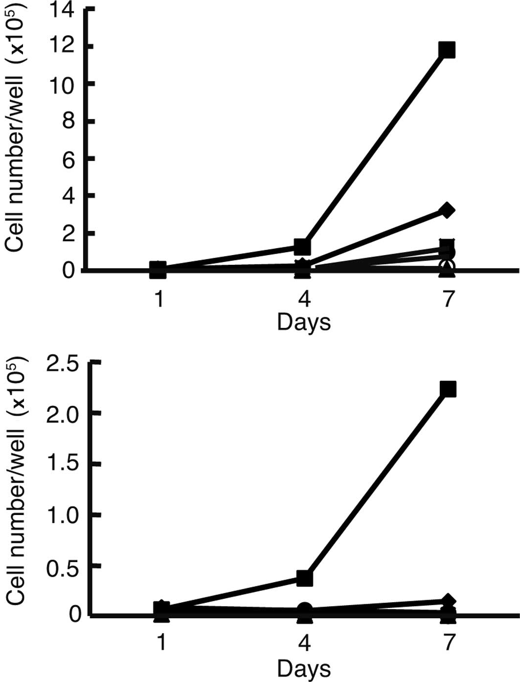
We first analyzed the growth curve of all of the
cell lines in media with or without FBS. In media with FBS, the
cell numbers of all of the cell lines increased (Fig. 1A). The HLF cells exhibited rapid
growth in the media without FBS when compared to growth in the
media with FBS (Fig. 1B). Cell
numbers of the other cell lines decreased compared to the HLE and
HLF cells. Since the HLF cells grew most rapidly compared to the
other cell lines examined in the media without FBS, HFL cells were
used for further analysis.
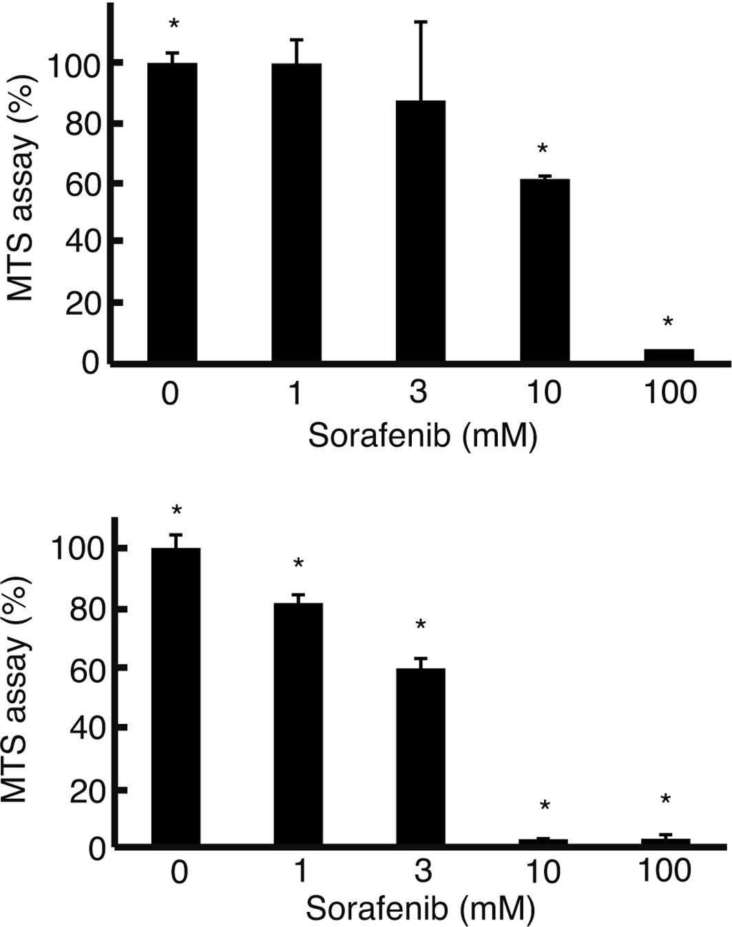
To examine the suppression of cell proliferation by
sorafenib, the MTS assay was performed. In the media with FBS, cell
proliferation of the HLF cells was suppressed at 3 μM sorafenib
(Fig. 2A). In the media without
FBS, suppression of cell proliferation occurred at 1 μM sorafenib
(Fig. 2B). At 10 μM sorafenib, the
MTS assay was 61±1% (mean ± standard deviation) in the media with
FBS, while it was 2.6±0.16% in the media without FBS (P<0.05),
almost the same level as in the media with FBS at 100 μM sorafenib
(2.7±1.3%) (Fig. 2B).
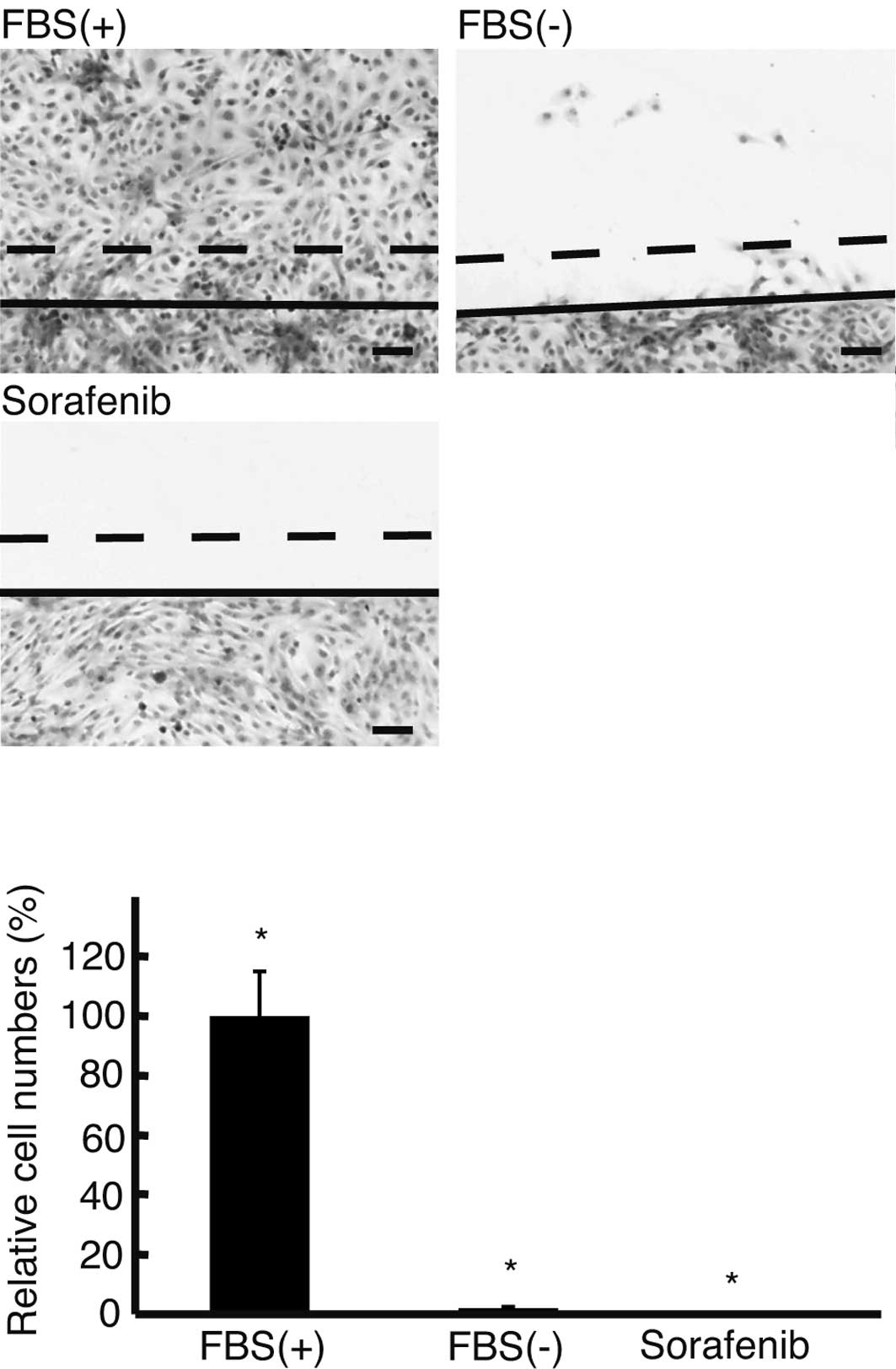
The wound assay was performed to analyze the effect
of sorafenib on cell motility under a serum-free condition
(Fig. 3A). In the media without
FBS, 2±0.5% HLF cells migrated >150 μm, which was significantly
lower than that in the media with FBS (P<0.05) (Fig. 3B). No cells moved >150 μm in the
serum-free media with sorafenib.
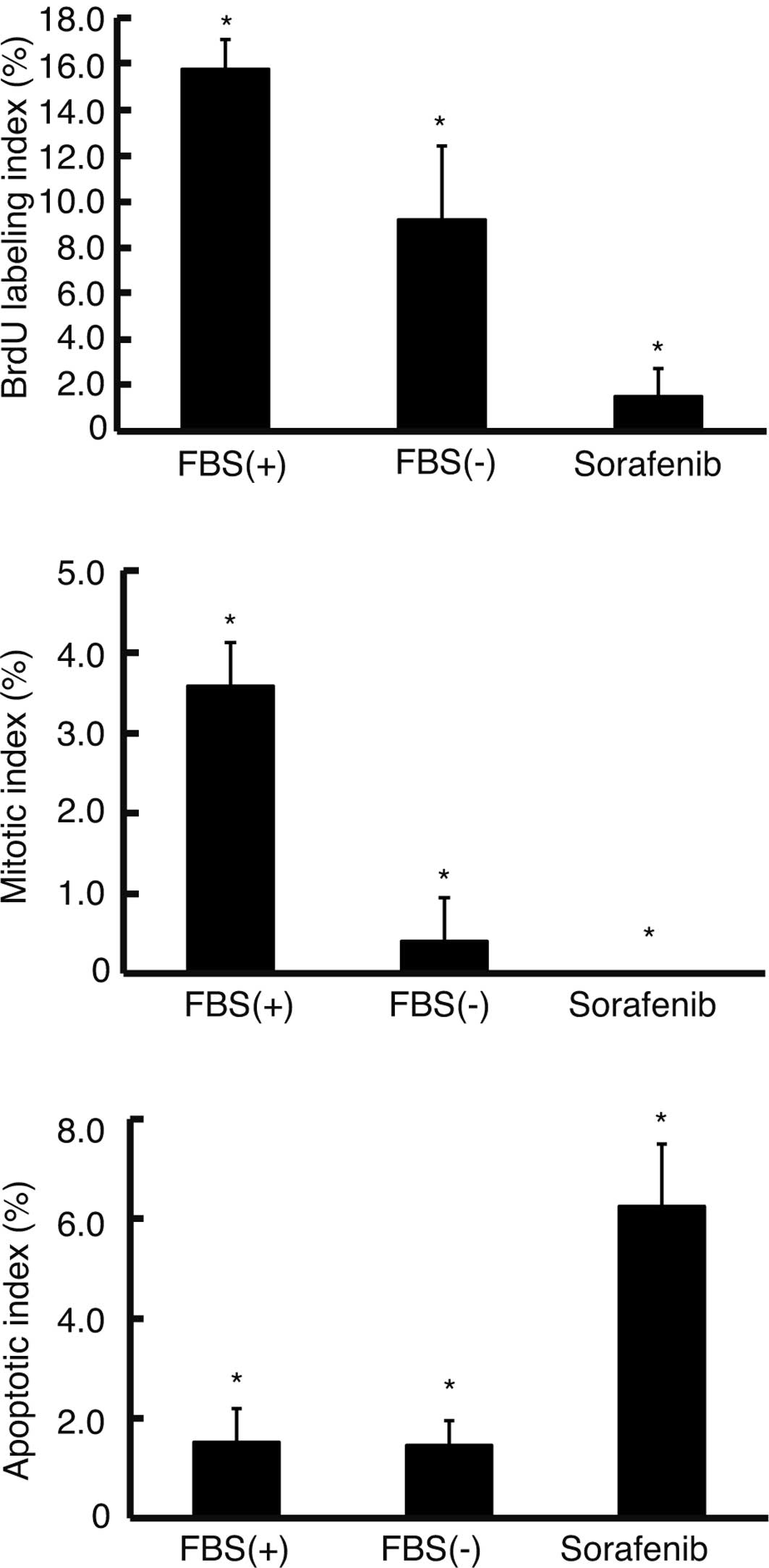
To clarify the effect of sorafenib on the cell
cycle, the BrdU labeling and mitotic indices were analyzed. BrdU
labeling index decreased in the media without FBS (9.2±3.2%) as
compared to the index in the media with FBS (15.7±1.3%) (P<0.05)
(Fig. 4A). Upon the addition of
sorafenib in the media without FBS, the BrdU labeling index
significantly decreased to 1.5±1.2% (P<0.05). The mitotic index
decreased to 0.4±0.55% in the media without FBS, which was
significantly lower than the index in the media with FBS
(3.6±0.55%) (P<0.05) (Fig. 4B).
Upon the addition of sorafenib in the media without FBS, the
mitotic index was 0%. To assess the apoptosis in the serum-free
condition, the TUNEL assay was applied. The ratio of apoptotic
cells did not differ between cells in the media with or without FBS
(1.5±0.6 and 1.4±0.5%, respectively) (Fig. 4C). Sorafenib significantly
increased the ratio of apoptotic cells to 6.3±1.2% in the medium
without FBS.
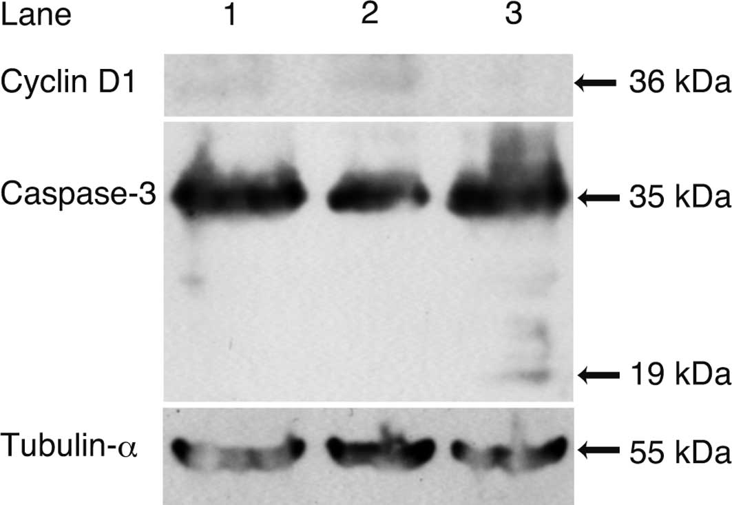
Western blot analysis (Fig. 5) was carried out to ascertain the
mechanism of suppression of the DNA synthesis and stimulation of
apoptosis by sorafenib. The expression of cyclin D1 decreased in
the cells in the media with sorafenib, while 19-kDa caspase-3, a
cleaved form, was noted in the cells in the media with
sorafenib.
Discussion
In the present study, HLF cells significantly
exhibited more rapid proliferation in the media without FBS
compared to the other cell lines examined, while HLE cells
marginally exhibited proliferation. HLE and HLF cells are cloned
from undifferentiated HCC and maintain the morphological features
of poorly differentiated HCC such as a spindle shape (10). HLF cells exhibit the same shape in
media with or without FBS, but their proliferation in a serum-free
condition has not yet been analyzed (11). Our data clearly showed the
significant proliferation of HLF cells in the media without FBS.
Although the mechanism is unclear, HLF cells were found to maintain
proliferation potential under a serum-free condition due to genetic
alteration, such as p53 (11).
Intriguingly, cells invading connective tissues surrounding HCC
exhibit a spindle shape similar to HLF cells (2). In the present study, although the
proliferative potential of the HLF cells in the media without FBS
was weaker than that in the medium with FBS, as shown by the BrdU
labeling and mitotic indices, they grew more rapidly than the other
cell lines studied (Fig. 1B). The
apoptotic index and the level of cleavage of caspase-3 of the HLF
cells in the media without FBS was the same as those in the media
with FBS. HLF cells may survive under a serum-free condition not
causing apoptosis for an unknown reason. A subclone of Lewis lung
carcinoma was found to be resistant to apoptosis with a lower
expression of caspase-3 (4). These
data indicate that the surviving cells were resistant to apoptosis.
HLF cells still marginally maintained the potential of motility.
HLF cells were, thus, further analyzed as a model of the invastion
of connective tissues by HCC cells.
In our study, 10 μM of sorafenib suppressed cell
viability to 61% in the media with FBS, which is higher than that
in a previous report on PLC/PRF5 and HepG2 cells (7). This may be due to the fact that HLF
cells have stronger proliferative potential than PLC/PRL/5 and
HepG2 cells (Fig. 1A). Under a
serum-free condition, suppression of cell viability was noted at 1
μM of sorafenib and declined to 2.6% at 10 μM. These data suggest
that a lower dose of sorafenib can suppress cell viability of HLF
cells in a medium without FBS to a greater exent than that in
medium with FBS. Sorafenib suppressed the cell cycle and stimulated
apoptosis (Fig. 4). Sorafenib was
also found to induce apoptosis in PLC/PRL/5 cells (7). Our data showed that sorafenib induced
apoptosis in a serum-free condition. Notably, sorafenib completely
abolished cell motility, as shown in the scratch assay (Fig. 3). These data indicate that
sorafenib suppresses, not only cell proliferation, but also cell
motility. Sorafenib significantly suppresses invasion of HCC cells
to surrounding connective tissues.
Acknowledgements
This study was, in part, supported by
a Grant-in-Aid for the Encouragement of Scientists from the Japan
Society for the Promotion of Science (JSPS) (grant no.
22931047).
References
|
1.
|
Kondo Y, Kondo F, Wada K and Okabayashi A:
Pathologic features of small hepatocellular carcinoma. Acta Pathol
Jpn. 36:1149–1161. 1986.PubMed/NCBI
|
|
2.
|
Tomizawa M, Kondo F and Kondo Y: Growth
patterns and interstitial invasion of small hepatocellular
carcinoma. Pathol Int. 45:352–358. 1995. View Article : Google Scholar : PubMed/NCBI
|
|
3.
|
Gupta GP and Massague J: Cancer
metastasis: building a framework. Cell. 127:679–695. 2006.
View Article : Google Scholar : PubMed/NCBI
|
|
4.
|
Takasu M, Tada Y, Wang JO, Tagawa M and
Takenaga K: Resistance to apoptosis induced by microenvironmental
stresses is correlated with metastatic potential in Lewis lung
carcinoma. Clin Exp Metastasis. 17:409–416. 1999. View Article : Google Scholar : PubMed/NCBI
|
|
5.
|
Boraldi F, Annovi G, Paolinelli-Devincenzi
C, Tiozzo R and Quaglino D: The effect of serum withdrawal on the
protein profile of quiescent human dermal fibroblasts in primary
cell culture. Proteomics. 8:66–82. 2007. View Article : Google Scholar : PubMed/NCBI
|
|
6.
|
Wilhelm SM, Carter C, Tang L, et al: BAY
43-9006 exhibits broad spectrum oral antitumor activity and targets
the RAF/ MEK/ERK pathway and receptor tyrosine kinases involved in
tumor progression and angiogenesis. Cancer Res. 64:7099–7109. 2004.
View Article : Google Scholar : PubMed/NCBI
|
|
7.
|
Liu L, Cao Y, Chen C, et al: Sorafenib
blocks the RAF/MEK/ ERK pathway, inhibits tumor angiogenesis, and
induces tumor cell apoptosis in hepatocellular carcinoma model
PLC/PRF/5. Cancer Res. 66:11851–11858. 2006. View Article : Google Scholar : PubMed/NCBI
|
|
8.
|
Furuse J, Ishii H, Nakachi K, Suzuki E,
Shimizu S and Nakajima K: Phase I study of sorafenib in Japanese
patients with hepatocellular carcinoma. Cancer Sci. 99:159–165.
2008.PubMed/NCBI
|
|
9.
|
Pennisi PA, Barr V, Nunez NP, Stannard B
and Le Roith D: Reduced expression of insulin-like growth factor I
receptors in MCF-7 breast cancer cells leads to a more metastatic
phenotype. Cancer Res. 62:6529–6537. 2002.
|
|
10.
|
Dor I, Namba M and Sato J: Establishment
and some biological characteristics of human hepatoma cell lines.
Gann. 66:385–392. 1975.PubMed/NCBI
|
|
11.
|
Terai S, Noma T, Kimura T, Nakazawa A,
Kurokawa F and Okita K: Wild-type p53 gene-induced morphological
changes and growth suppression in hepatoma cells. J Gastroenterol.
32:330–337. 1997. View Article : Google Scholar : PubMed/NCBI
|