Introduction
Bamboo salt, as a functional food, originated in
Korea. Approximately 1,000 years ago, Korean doctors and monks
began creating medical salt. The bamboo salt was prepared by
putting sea salt into a case made from young bamboo, which had
grown for just 3 years. The two ends were sealed using natural red
clay and the bamboo case was baked at 1,000–1,500°C (1,2),
using pine as the fuel. In ancient times, the bamboo salt was baked
only 2-3 times. The salt was then used as a special medical
treatment. Eventually, it was identified that bamboo salt acted at
its highest medical efficacy if it was baked at least 9 times. The
9-times-baked bamboo salt was named purple bamboo salt.
Additionally, it was observed that if bamboo salt is completely
melted, the toxic characteristic of the salt disappeared.
Currently, bamboo salt is one of the most well-known
traditional medical treatments, not only in Korea but also in a
number of other Asian countries (3,4).
Bamboo salt contains >70 essential minerals and micronutrients.
Pharmaceutical scientists are researching the special therapeutic
qualities of bamboo salt, including its anticancer and antiviral
effects (5). The researchers noted
that bamboo salt demonstrates anti-inflammatory and antioxidant
effects and that it may also be used as bamboo salt toothpaste for
dental disease and oral hygiene (6,7).
Buccal mucosa cancer is the most common cancer of
the oral cavity (8). In the
present study, the cancer preventive effect of purple bamboo salt
was evaluated using a mouse model of buccal mucosa cancer. The
bamboo salt was shown to enhance anti-cancer activities and the
anti-metastatic effect in mice. As a functional food, purple bamboo
salt demonstrated oral health benefits in mice.
Materials and methods
Preparations of bamboo salt
The purple bamboo salt (9-times-baked bamboo salt)
and sea salt were provided by Taesung Food Company (Gochang,
Jeonbuk, Korea). The salts were dissolved in distilled water.
Cell preparation
TCA8113 human tongue carcinoma cells obtained from
Shanghai Institute of Biochemistry and Cell Biology (Shanghai,
China) and U14 squamous cell carcinoma cells obtained from Chinese
Academy of Medical Sciences (Beijing, China) were used in this
study. The cancer cells were cultured in RPMI-1640 medium (Welgene
Inc., Daegu, Korea) supplemented with 10% fetal bovine serum (FBS)
and 1% penicillin-streptomycin (Gibco-BRL, Grand Island, NY, USA)
at 37°C in a humidified atmosphere with 5% CO2 (incubator model 311
S/N29035; Forma, Waltham, MA, USA). The medium was changed 2 or 3
times a week (5).
In vitro cultured U14 cells
(5×10⁶/mouse) were injected into the abdominal cavity of
7-week-old female Institute of Cancer Research (ICR) mice. After 1
week, the carcinoma ascites were collected and diluted in sterile
saline to a concentration of 1×10⁷/ml.
3-(4,5-Dimethyl-2-thiazolyl)-2,5-diphenyltetrazolium bromide (MTT)
assay
The anticancer effects of purple bamboo salt were
assessed by an MTT assay. The TCA8113 human tongue carcinoma cells
(180 μl) were seeded onto a 96-well plate (2×10⁴
cells/ml/well). The specimen (20 μl) was added to the vessel
to be cultured at 37°C, 5% CO2 for 48 h. MTT solution
(200 μl, 5 mg/ml) was added and the cells were cultured for
a further 4 h under the same conditions. After removing the
supernatant, 150 μl dimethylsulfoxide (DMSO) was added to
each well and mixed for 30 min. Finally, the absorbance of each
well was measured using an enzyme-linked immunosorbent assay
(ELISA) reader (model 680; Bio-Rad, Hercules, CA, USA) at 540 nm
(9).
Nuclear staining with
4,6-diamidino-2-phenylindole (DAPI)
Untreated control and cells treated with purple
bamboo salt were harvested, washed with phosphate-buffered saline
(PBS) and fixed with 3.7% paraformaldehyde (Sigma, St. Louis, MO,
USA) in PBS for 10 min at room temperature. The fixed cells were
washed with PBS and stained with DAPI (1 mg/ml; Sigma) solution for
10 min at room temperature (10).
The cells were washed 2 more times with PBS and examined under a
fluorescence microscope (BX50; Olympus, Tokyo, Japan).
Induction of buccal mucosa cancer
Female ICR mice (n=40, 6 weeks old) were purchased
from the Experimental Animal Center of Chongqing Medical University
(Chongqing, China). They were maintained in a
temperature-controlled (temperature, 25±2°C; relative humidity,
50±5%) facility with a 12 h light/dark cycle and had unlimited
access to a standard mouse chow diet and water.
To investigate the preventive effects of the two
salts against buccal mucosa cancer induced by injecting U14 cells
into the mice, the animals were divided into 4 groups with 10 mice
in each. The experimental design was as follows: the mice in the
two salt sample groups were smeared with the purple bamboo salt and
sea salt solutions (20%) on the buccal mucosa every 12 h for 14
days. The control and salt sample groups were then inoculated with
0.05 ml cancer cell suspension (1×10⁷/ml) on the buccal
mucosa. The salt samples continued to be smeared on the buccal
mucosa of the mice every 12 h. The mice were sacrificed 14 days
later and their tumor volumes and lymph node metastasis rates were
determined (11).
Histological grading of buccal mucosa
cancer
Buccal mucosa and lymph node tissues were removed
and embedded into paraffin for histological analysis with
hematoxylin and eosin (H&E) staining. Buccal mucosa cancer was
graded as follows: i) well-differentiated carcinoma: cells resemble
the adjacent benign squamous epithelium; ii) moderately
differentiated carcinoma: cells form large anastomosing areas in
which keratin pearls are formed, they are not numerous and the main
component consists of cells with pronounced cytonuclear atypia and
iii) poorly differentiated carcinoma: cells have lost the majority
of their squamous epithelial characteristics and architecture
(12).
Reverse transcription-polymerase chain
reaction (RT-PCR) analysis of Bcl-2-associated X protein (Bax), B
cell lymphoma-2 (Bcl-2), inducible nitric oxide synthase (iNOS) and
cyclooxygenase-2 (COX-2) mRNA expression
Total RNA was isolated using TRIzol reagent
(Invitrogen, Carlsbad, CA, USA) according to the manufacturer's
instructions. RNA was digested with RNase-free DNase (Roche, Basel,
Switzerland) for 15 min at 37°C and purified using an RNeasy kit
(Qiagen, Hilden, Germany) according to the manufacturer's
instructions. cDNA was synthesized from 2 μg total RNA by
incubation at 37°C for l h with avian myeloblastosis virus (AMV)
reverse transcriptase (GE Healthcare, Uppsala, Sweden) with random
hexanucleotide, according to the manufacturer's instructions.
Primers used to specifically amplify the genes were as follows:
forward, 5′-AAG CTG AGC GAG TGT CTC CGG CG-3′ and reverse, 5′-CAG
ATG CCG GTT CAG GTA CTC AGT C-3′ for Bax; forward, 5′-CTC GTC GCT
ACC GTC GTG ACT TGG-3′ and reverse, 5′-CAG ATG CCG GTT CAG GTA CTC
AGT C-3′ for Bcl-2; forward, 5′-AGA GAG ATC GGG TTC ACA-3′ and
reverse, 5′-CAC AGA ACT GAG GGT ACA-3′ for iNOS; forward, 5′-TTA
AAA TGA GAT TGT CCG AA-3′ and reverse, 5′-AGA TCA CCT CTG CCT GAG
TA-3′ for COX-2. The internal control gene glyceraldehyde
3-phosphate dehydrogenase (GAPDH) was amplified with the following
primers: forward, 5′-CGG AGT CAA CGG ATT TGG TC-3′ and reverse,
5′-AGC CTT CTC CAT GGT CGT GA-3′. Amplification was performed in a
thermal cycler (Eppendorf, Hamburg, Germany) with 29 Bax cycles, 34
Bcl-2 cycles, 25 iNOS, COX-2 and GAPDH cycles of denaturation. The
amplified PCR products were run in 1.0% agarose gels and visualized
by ethidium bromide (EtBr) staining (13).
Western blot analysis
Total protein was obtained with RIPA buffer as
described by Kim et al(14). Protein concentrations were
determined with a Bio-Rad protein assay kit. For western blot
analysis, aliquots of the lysate containing 30–50 μg protein
were separated by sodium dodecyl sulphate-polyacrylamide gel
electrophoresis (SDS-PAGE) and then electrotransferred onto a
nitrocellulose membrane (Schleicher and Schuell, Keene, NH, USA).
The membranes were subjected to immunoblot analysis and proteins
were visualized by an enhanced chemiluminescence (ECL) method (GE
Healthcare). The cell lysates were separated by 12% SDS-PAGE,
transferred onto a polyvinylidene fluoride membrane (GE
Healthcare), blocked with 5% skimmed milk and hybridized with
primary antibodies (diluted 1:1,000). Antibodies against Bax,
Bcl-2, iNOS and COX-2 were obtained from Santa Cruz Biotechnology,
Inc. (Santa Cruz, CA, USA), then incubated with the horse-radish
peroxidase-conjugated secondary antibody (Santa Cruz Biotechnology,
Inc.) for 1 h at room temperature. Blots were washed 3 times with
PBS/Tween-20 (PBS-T) and then developed by enhanced
chemiluminescence (Amersham Life Science, Arlington Heights, IL,
USA).
Statistical analysis
Data are presented as mean ± standard deviation.
Differences between the mean values for the individual groups were
assessed by a one-way analysis of variance (ANOVA) with Duncan's
multiple range tests. P<0.05 was considered to indicate a
statistically significant difference. The SAS v9.1 statistical
software package (SAS Institute Inc., Cary, NC, USA) was used for
the analysis.
Results
Growth inhibitory effects of salt samples
on TCA8113 cells
The anticancer effects of purple bamboo salt and sea
salt on TCA8113 human tongue carcinoma cells were investigated. The
growth inhibitory rates of TCA8113 cells treated with 0.5% salt
samples were 12 and 35% for sea salt and purple bamboo salt,
respectively. At 1.0%, the growth inhibitory rates of cells treated
with these reagents were 27 and 61%, respectively (P<0.05). It
was clear that the anticancer effect of purple bamboo salt on the
TCA8113 cells was stronger than that of sea salt (Table I).
 | Table I.Inhibition of the growth of TCA8113
human tongue carcinoma cells by salt samples as evaluated by a
3-(4,5-dimethyl-2-thiazolyl)-2,5-diphenyltetrazolium (MTT)
assay. |
Table I.
Inhibition of the growth of TCA8113
human tongue carcinoma cells by salt samples as evaluated by a
3-(4,5-dimethyl-2-thiazolyl)-2,5-diphenyltetrazolium (MTT)
assay.
| Treatment | OD540
(inhibition rate) |
|---|
| Control
(untreated) | 0.815±0.006a |
| Sea salt | |
| 0.5% | 0.717±0.011
(12)b |
| 1.0% | 0.595±0.007
(27)c |
| Purple bamboo
salt | |
| 0.5% | 0.530±0.009
(35)d |
| 1.0% | 0.318±0.009
(61)e |
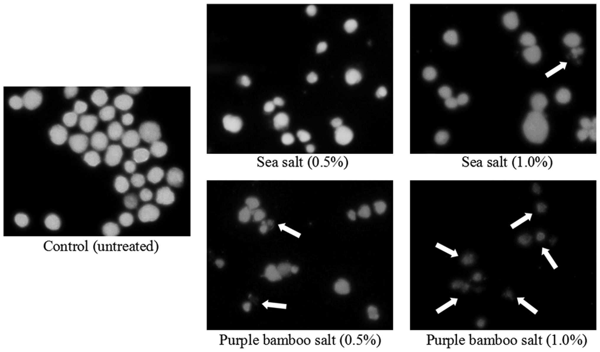
Induction of apoptosis by salt samples on
TCA8113 cells
To further determine whether the growth inhibitory
activity of purple bamboo salt in the TCA8113 cells was related to
the induction of apoptosis, chromatin condensation was analyzed by
fluorescent microscopy using the DNA binding fluorescent dye DAPI
(Fig. 1). In TCA8113 cells, which
normally contain nuclei with homogeneous chromatin distribution,
treatment with salt samples (1.0%) induced chromatin condensation
and nuclear fragmentation, suggesting the presence of apoptotic
cells. Chromatin condensation and formation of apoptotic bodies,
which are characteristic of apoptosis, were observed in the cells
cultured with purple bamboo salt; however, these were not
identified in the cells treated with sea salt. These results
suggest that purple bamboo salt is more effective for inducing the
condensation and formation of apoptotic bodies than sea salt.
Tumor volumes and lymph node metastasis
rates
Buccal mucosa cancer was induced by injecting U14
cells into mice. After 14 days, the mice in all groups presented
carcinogenesis. The tumor volumes of buccal mucosa tissues were
measured. The tumor volumes for the control, sea salt and purple
bamboo salt groups were 12.4, 12.0 and 7.2 mm3,
respectively (Table II). There
were 5 mice demonstrating lymph node metastasis in the control
group, 4 in the sea salt group and 2 in the purple bamboo salt
group. Consequently, the lymph node metastasis rate was 50, 40 and
20%, respectively. These results demonstrate that purple bamboo
salt is more effective than sea salt in impeding carcinogenesis,
proliferation and metastasis.
| Table II.Tumor volumes and lymph node
metastasis rates of mice smeared with salt samples. |
Table II.
Tumor volumes and lymph node
metastasis rates of mice smeared with salt samples.
| Group | Normal | Control | Sea salt | Purple bamboo
salt |
|---|
| Tumor volume
(mm3) | 0 | 12.4±0.6 | 12.0±0.5 | 7.2±0.2 |
| Lymph node metastasis
ratea | 0 | 5/10 (50%) | 4/10 (40%) | 2/10 (20%) |
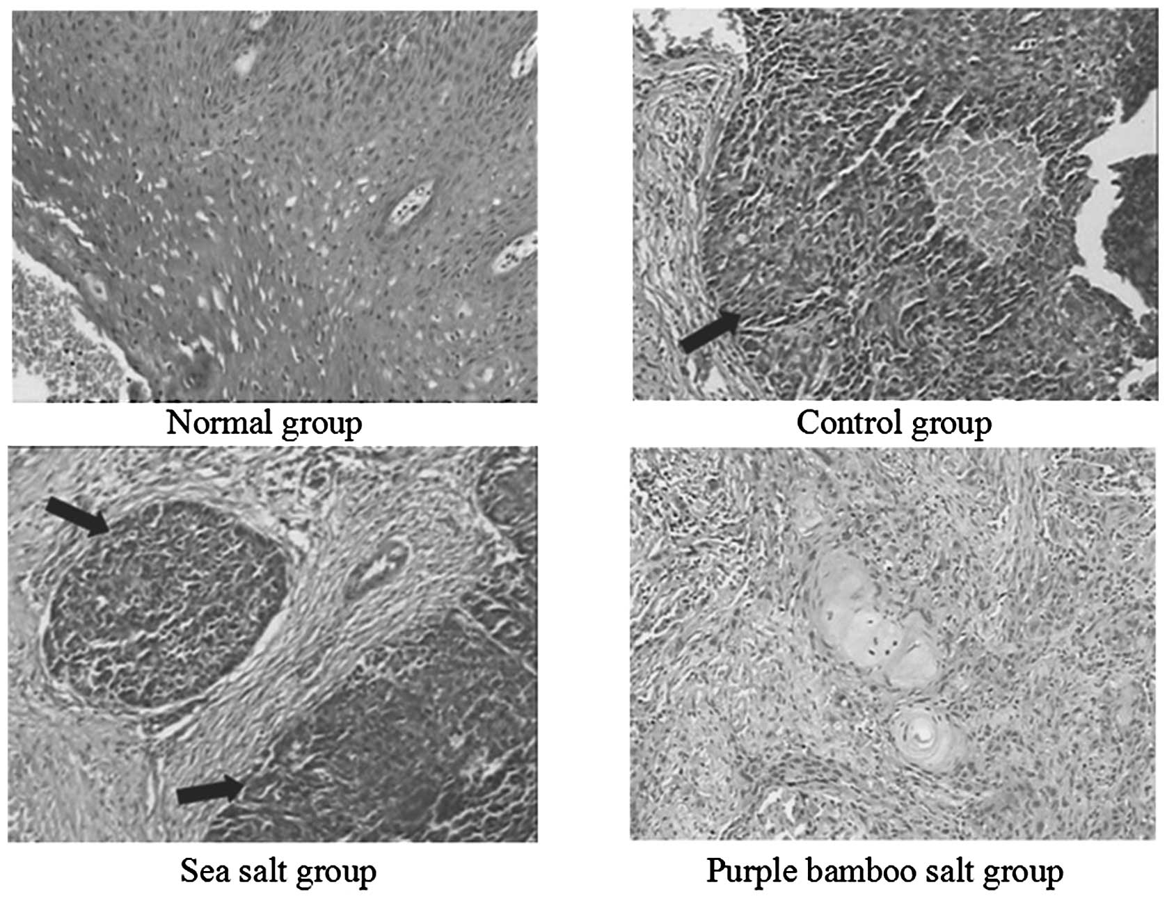
Histopathology of buccal mucosa
tissues
Histological changes in the buccal mucosa of mice
injected with U14 cells were examined by H&E staining. The
histologic tissue sections of mice in the normal group demonstrated
normal histological morphology of squamous epithelium tissue.
Histopathological evaluation revealed indications of buccal mucosa
cancer in both groups receiving U14 cells (Fig. 2). The sections from mice in the
control and sea salt groups showed that the tissue lost its
squamous epithelial characteristics and architecture; however, the
tissues from the sea salt group had some intracellular bridging
between the normal squamous cells. The histopathology sections
indicated that the mice in the control and sea salt groups
developed poorly differentiated carcinoma (grade III) and the
control group experienced more serious carcinogenesis. The tissue
sections of purple bamboo salt group looked less like typical
squamous epithelium. The tumor cells remained in nests and there
were numerous larger, eosinophilic, polygonal cells that were
trying to layer themselves in a squamous-like fashion. However, for
the purple bamboo salt group, the overall resemblance to typical
squamous epithelium was less striking (grade II). From these
sections, purple bamboo salt demonstrated a preventive effect
against buccal mucosa cancer.
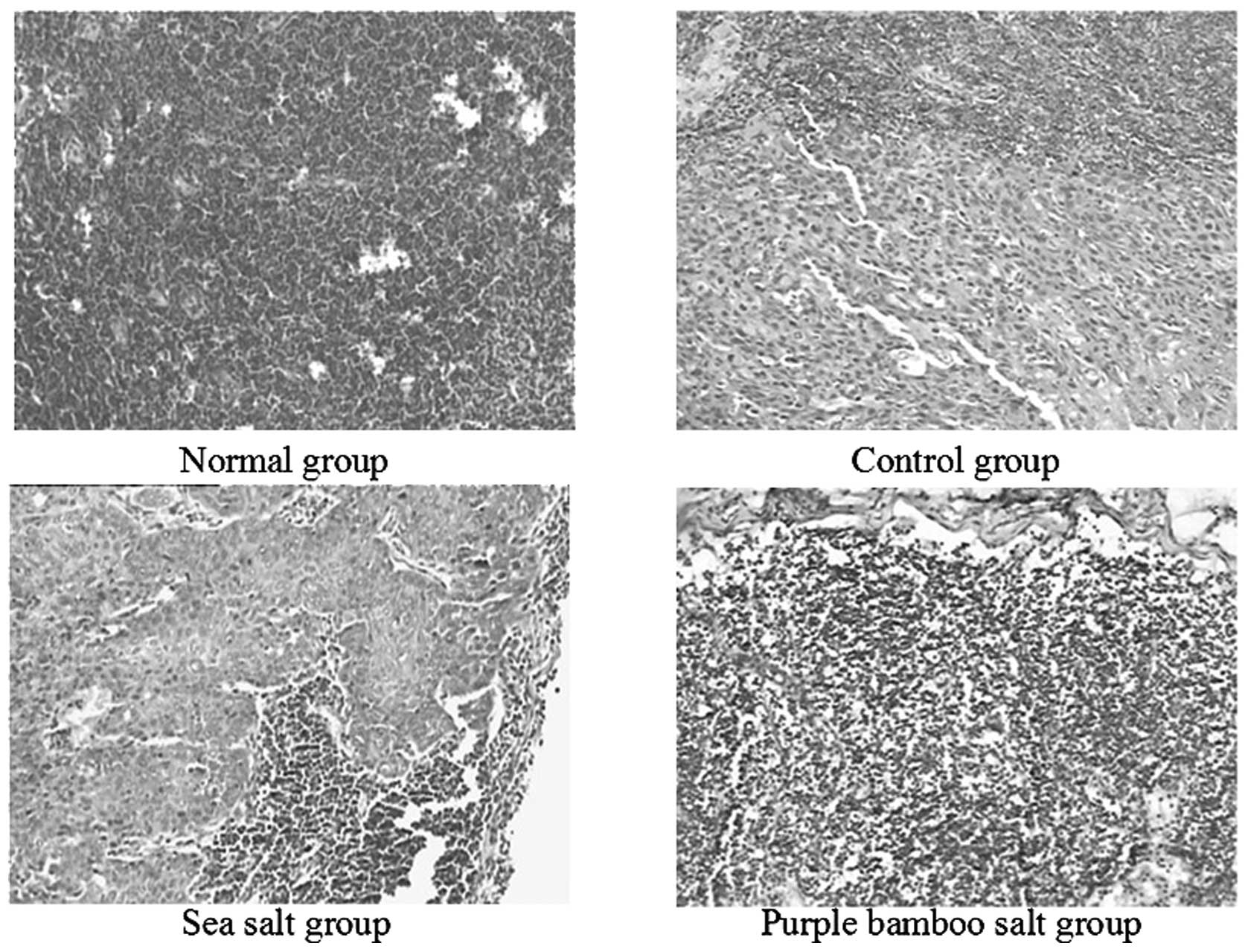
The lymph node sections from the control group
exhibited a large area with liquefaction necrosis of the lymph node
(Fig. 3). Marked liquefaction
necrosis was also identified in the sea salt group, whereas only a
small amount of liquefaction necrosis was observed in the purple
bamboo salt group. These results demonstrate that purple bamboo
salt is more effective than sea salt in preventing lymph node
metastasis.
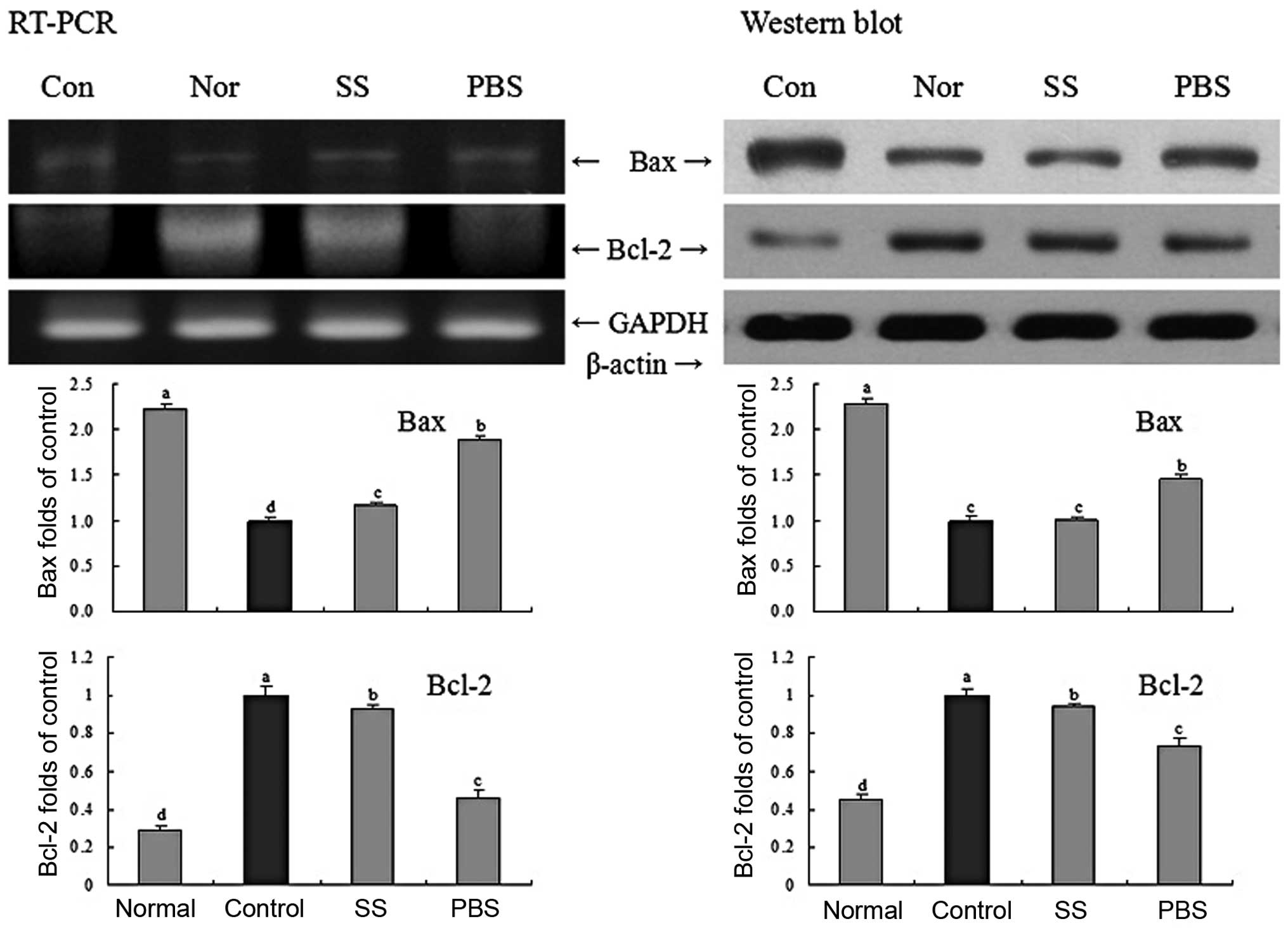
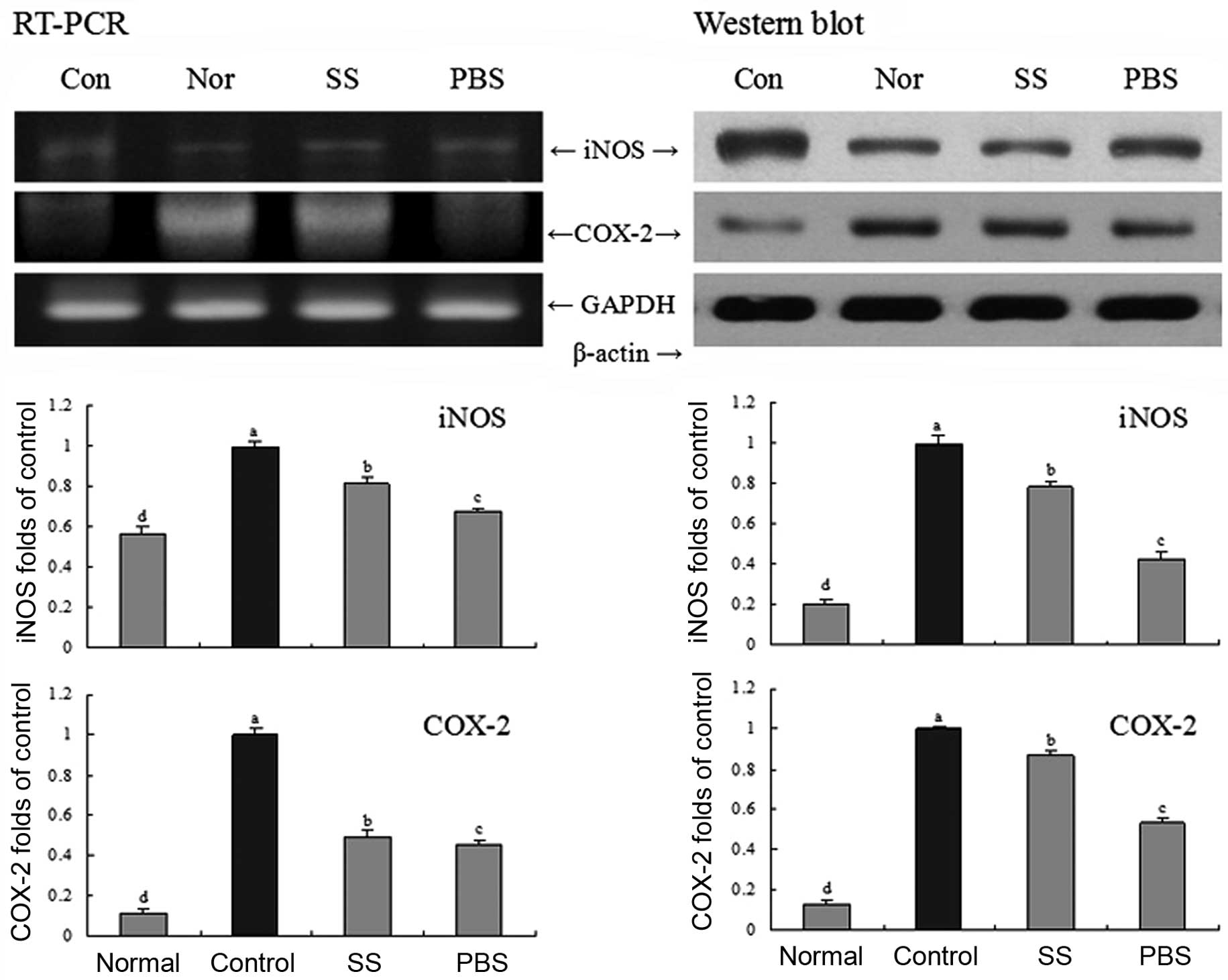
Gene expressions of Bax, Bcl-2, iNOS and
COX-2 in buccal mucosa tissues
To determine the protective mechanisms against
buccal mucosa cancer, the expression levels of Bax and Bcl-2 in
buccal mucosa tissues were determined by RT-PCR and western
blotting. As shown in Fig. 4, in
the group treated with purple bamboo salt, the pro-apoptotic Bax
and the anti-apoptotic Bcl-2 showed significant changes.
Accordingly, the results suggested that the purple bamboo salt
induced apoptosis in buccal mucosa tissues via a Bax- and
Bcl-2-dependent pathway. Thus, apoptosis induction in the purple
bamboo salt group was related to an increase in Bax and a decrease
in Bcl-2 in terms of mRNA and protein expression compared with the
sea salt and control groups. RT-PCR and western blot analysis were
also conducted to investigate whether the inhibitory effect of
salts on inflammation were due to gene regulation of inflammatory
mediators, including iNOS and COX-2. COX-2 and iNOS are important
enzymes that mediate inflammatory processes. As shown in Fig. 5, these inflammatory mediators were
barely detectable in the normal group. However, the control group
demonstrated a sharp increase in mRNA and protein expression
levels. In the purple bamboo salt group, the results showed a
significant decrease in the mRNA and protein expression levels of
COX-2 and iNOS. It was observed that higher decreases in mRNA and
protein expression were related to improved anti-inflammatory
effects.
Discussion
Bamboo salt has been used as a folk medicine;
however, scientific data on the effects of this salt are lacking.
Nine-times-baked bamboo salt, also called purple bamboo salt since
the color changes to purple following the baking process, has high
concentrations of iron, silicon and potassium compared with those
in crude salt (15). This salt has
been previously reported to have various therapeutic effects on
numerous pathological conditions, including inflammation, viral
diseases, diabetes and cancer (16). The bamboo salt is also known to
have in vitro anticancer effects on HT-29 colon cancer cells
(5). We also investigated the
in vitro anticancer effect of purple bamboo salt in the
current study.
Programmed cell death (apoptosis) is a conserved,
natural mechanism for the removal of redundant and unwanted cells
during normal development. Whether apoptosis is the cause or the
consequence of drug-induced cell death remains to be established
(17). The importance of apoptosis
is widely recognized in numerous fields of medicine and is
currently one of the most popular subjects in biomedical research.
Apoptosis is a fundamental cell event and the understanding of its
mechanisms will shed light on its future use in tumor diagnosis and
therapy.
Metastasis is defined as the spread of cancer cells
from one organ or area to another adjacent organ or location
(18,19). It is thought that malignant tumor
cells have the capacity to metastasize. Cancer occurs after cells
in a tissue are genetically damaged in a progressive manner,
resulting in cancer stem cells possessing a malignant phenotype.
After the tumor cells come to rest in another site, they penetrate
the vessel walls, continue to multiply and eventually form another
tumor.
Histopathology is an important tool in anatomical
pathology, since accurate diagnosis of cancer usually requires
histopathological examination of samples. Histopathology is an
important clinical standard for the diagnosis of oral cancer
(20).
In a healthy cell, the outer mitochondrial membrane
expresses the anti-apoptotic protein Bcl-2 on its surface (21). Bcl-2 is bound to a protein named
Apaf-1. Cell internal damage causes Bcl-2 to release Apaf-1
(22) and a Bcl-2-related protein
(Bax) penetrates the mitochondrial membrane, causing cyto-chrome
c to leak out into the cytosol (23). In the current study, purple bamboo
salt demonstrated greater in vivo anticancer activity than
sea salt, as was observed using RT-PCR and western blotting of
buccal mucosa cancer tissues. The anti-cancer mechanisms of purple
bamboo salt include the induction of apoptosis by increasing the
number of apoptotic bodies and by regulating the expression levels
of the apoptosis-related Bax and Bcl-2 mRNA and proteins.
Improper upregulation of COX-2 and/or iNOS has been
associated with the pathophysiology of certain types of human
cancers, as well as inflammatory disorders. Since inflammation is
closely linked to tumor promotion, substances with potent
anti-inflammatory activities are anticipated to exert
chemopreventive effects on carcinogenesis, particularly in the
promotion stage (24).
Accordingly, purple bamboo salt is expected to
contribute to the prevention of buccal mucosa cancer. The
potassium, calcium, magnesium and iron contents in bamboo salts are
higher than those in purified and solar salts. Additionally, bamboo
salt baked for longer periods of time contains more minerals
(25). Increased levels of these
minerals in the salt are important for enhancing the anticancer
effects (26). Bamboo salt also
exhibits a higher reduction potential; this may be due to the fact
that this type of salt contains more hydroxyl (OH) ions than
purified or solar salts (27).
Overall, bamboo salt seems to have more potent anticancer and
anti-inflammatory effects since it contains higher levels of
minerals and more OH ions.
In the current study, we employed in vitro
and in vivo anticancer experimental methods, including MTT
assay, histo-pathology assay and RT-PCR mRNA and western blotting
protein expression assays to determine the anticancer and
anti-inflammatory effects of purple bamboo salt. In addition, we
obtained results demonstrating that the anti-metastatic effect of
purple bamboo salt is greater than that of sea salt. In conclusion,
the increased mineral contents, and other phytochemicals are
important functional compounds that may increase the buccal mucosa
cancer preventive effect of purple bamboo salt.
References
|
1.
|
Hu C, Zhang Y and Kitts DD: Evaluation of
antioxidant and prooxidant activities of bamboo Phyllostachys
nigra var. Henonis leaf extract in vitro. J Agr Food Chem.
48:3170–3176. 2000. View Article : Google Scholar : PubMed/NCBI
|
|
2.
|
Shin HY, Na HJ, Moon PD, Shin TK, Shin TY,
Kim SH, Hong SH and Kim HM: Inhibition of mast cell-dependent
immediate-type hypersensitivity reactions by purple bamboo salt. J
Ethnopharmacol. 91:153–157. 2004. View Article : Google Scholar : PubMed/NCBI
|
|
3.
|
Kim HY, Lee ES, Jeong JY, Choi JH, Choi
YS, Han DJ, Lee MA, Kim SY and Kim CJ: Effect of bamboo salt on the
physico-chemical properties of meat emulsion systems. Meat Sci.
86:960–965. 2010. View Article : Google Scholar : PubMed/NCBI
|
|
4.
|
Shin HY, Lee EH, Kim CY, Shin TY, Kim SD,
Song YS, Lee KN, Hong SH and Kim HM: Anti-inflammatory activity of
Korean folk medicine purple bamboo salt. Immunopharmacol
Immunotoxicol. 25:377–384. 2003. View Article : Google Scholar : PubMed/NCBI
|
|
5.
|
Zhao X, Kim SY and Kun-Young Park: Bamboo
salt has in vitro anti-cancer activity in HCT-116 cells and exerts
anti-metastatic effects in vivo. J Med Food. 1:(In press).
|
|
6.
|
Asada T, Ishihara S, Yamane T, Toba A,
Yamada A and Oikawa K: Science of bamboo charcoal: Study on
carbonizing temperature of bamboo charcoal and removal capability
of harmful gases. J Health Sci. 48:473–479. 2002. View Article : Google Scholar
|
|
7.
|
Li ZH and Kobayashi M: Plantation future
of bamboo in China. J For Res. 15:233–242. 2004. View Article : Google Scholar
|
|
8.
|
Kolanjiappan K, Ramachandran CR and
Manoharan S: Biochemical changes in tumor tissues of oral cancer
patients. Clin Biochem. 36:61–65. 2003. View Article : Google Scholar : PubMed/NCBI
|
|
9.
|
Skehan P, Storeng R, Scudiero D, Monks SA,
McMahon J, Vistica D, Warren JT, Bokesch H, Kenney S and Boyd MR:
New colorimetric cytotoxicity assay for anticancer-drug screening.
J Natl Cancer Inst. 82:1107–1112. 1990. View Article : Google Scholar : PubMed/NCBI
|
|
10.
|
Choi YH, Baek JH, Yoo M, Chung H, Kim ND
and Kim KW: Induction of apoptosis by ursolic acid through
activation of caspases and downregulation of c-IAPs in human
prostate epithelial cells. Int J Oncol. 17:565–571. 2000.PubMed/NCBI
|
|
11.
|
Pang L, Qiu LH, Gao Z, Li P, Xu P and Luo
DP: Experimental study on contrast-enhanced ultrasound imaging of
metastatic lymph modes of cheek carcinoma. J Ultrasound Clin Med.
13:581–583. 2011.(In Chinese).
|
|
12.
|
Schrader M and Laberke HG: Differential
diagnosis of verrucous carcinoma in the oral cavity and larynx. J
Laryngol Otol. 102:700–703. 1988. View Article : Google Scholar : PubMed/NCBI
|
|
13.
|
Bak SS, Kong CS, Rhee SH, Rho CW, Kim NK,
Choi KL and Park KY: Effect of sulfur enriched young radish kimchi
on the induction of apoptosis in AGS human gastric adenocarcinoma
cells. J Food Science Nutr. 12:79–83. 2007. View Article : Google Scholar
|
|
14.
|
Kim YA, Rhee SH, Park KY and Choi YH:
Antiproliferative effect of resveratrol in human prostate carcinoma
cells. J Med Food. 6:273–280. 2003. View Article : Google Scholar : PubMed/NCBI
|
|
15.
|
Woodhouse EC, Chuaqui RF and Liotta LA:
General mechanisms of metastasis. Cancer. 80(Suppl 8): S1529–S1537.
1997. View Article : Google Scholar
|
|
16.
|
Shin HY, Na HJ, Moon PD, Seo SW, Shin TY,
Hong SH and Lee KN: Biological activity of bamboo salt. Food Ind
Nutr. 9:36–45. 2004.
|
|
17.
|
Smets LA: Programmed cell death
(apoptosis) and response to anti-cancer drugs. Anticancer Drugs.
5:3–9. 1994. View Article : Google Scholar : PubMed/NCBI
|
|
18.
|
Chiang AC and Massagué J: Molecular basis
of metastasis. New Engl J Med. 359:2814–2823. 2008. View Article : Google Scholar
|
|
19.
|
Klein CA: Cancer: The metastasis cascade.
Science. 321:1785–1787. 2008. View Article : Google Scholar : PubMed/NCBI
|
|
20.
|
Sankaranarayanan R, Ramadas K, Thomas G,
Muwonge R, Thara S, Mathew B and Rajan B: Effect of screening on
oral cancer mortality in Kerala, India: a cluster-randomised
controlled trial. Lancet. 365:1927–1933. 2005. View Article : Google Scholar : PubMed/NCBI
|
|
21.
|
Cao Y and Cao R: Angiogenesis inhibited by
drinking tea. Nature. 398:3811999. View
Article : Google Scholar : PubMed/NCBI
|
|
22.
|
Song ZW and Steller H: Death by design:
mechanism and control of apoptosis. Trends Cell Biol. 9:M49–M52.
1999. View Article : Google Scholar : PubMed/NCBI
|
|
23.
|
Yang J, Liu XS, Bhalla K, Kim CN, Ibrado
AM, Cai JY, Peng TI, Jones DP and Wang XD: Prevention of apoptosis
by Bcl-2: release of cytochrome c from mitochondria blocked.
Science. 275:1129–1132. 1997. View Article : Google Scholar : PubMed/NCBI
|
|
24.
|
Surh YJ, Chun KS, Cha HH, Han SS, Keum YS,
Park KK and Lee SS: Molecular mechanisms underlying chemopreventive
activities of anti-inflammatory phytochemicals: down-regulation of
COX-2 and iNOS through suppression of NF-kappa B activation. Mutat
Res. 480–481:243–268. 2001.
|
|
25.
|
Zhao X: Anticancer and antiinflammatory
effects of bamboo salt and Rubus coreanus Miquel bamboo salt
(unpublished PhD thesis). Pusan National University; Busan, Korea:
2011
|
|
26.
|
Zhao X, Jung OS and Park KY: Alkaline and
antioxidant effects of bamboo salt. J Korean Soc Food Sci Nutr. (In
press).
|
|
27.
|
Welch AA, Fransen H, Jenab M,
Boutron-ruault MC, Tumino R, Agnoli C, Ericson U, Johansson I,
Ferrari P, Engeset D, Lund E, Lentjes M, Key T, Touvier M, Niravong
M, Larrañaga N, Rodríguez L, Ockém C, Peeters PH, Tjønneland A,
Bjerregaard L, Vasilopoulou E, Dilis V, Linseisen J, Nöthlings U,
Riboli E, Slimani N and Bingham S: Variation in intakes of calcium,
phosphorus, magnesium, iron and potassium in 10 countries in the
European prospective investigation into cancer and nutrition study.
Eur J Clin Nutr. 63(Suppl 4): 101–121. 2009. View Article : Google Scholar : PubMed/NCBI
|