Introduction
Reflux esophagitis (RE) is an inflammation of the
lower esophagus due to the regurgitation of gastric acid,
characterized by a burning pain in the chest (so-called heartburn)
and nausea following eating. RE is usually a result of a
malfunction of the lower esophageal sphincter (1). In addition, it is associated with an
increase in gastric acid secretion and a westernization of
lifestyle and diet (i.e. a high-fat diet), as well as a low
prevalence of Helicobacter pylori (H. pylori) infection.
H. pylori is generally accepted as the most
important type of bacteria in gastrointestinal disease. However,
certain studies have suggested that the increased prevalence of RE
following H. pylori eradication may be due to the protective
role of H. pylori infection in patients with RE (2–4).
In humans, prolonged gastroesophageal reflux,
including acidic gastric fluid, leads to esophageal mucosal injury,
such as bleeding, erythema, erosions and ulcers (5). Medication used to treat RE include
antacids, acid blockers, gastric motility agents and surgery. Acid
blockers, which comprise histamine type 2 (H2) antagonists and
proton pump inhibitors (PPIs), are the most commonly used
treatments for RE. H2 antagonists, such as ranitidine and
cimetidine, reduce acid production in the stomach, and PPIs, such
as omeprazole and esomeprazole, also arrest the production of
stomach acid (6). Usually, PPIs
are more effective than H2 antagonists (7,8).
However, in spite of the marked therapeutic effect, a number of
patients have suffered from incidences of relapse and shown
incomplete mucosal healing, continued symptoms and complications
(9,10). Even with an adequate administration
of H2 antagonists and PPIs, 40–60% of patients have suffered from
stricture of the esophagus or cancer, instead of recovering from
the RE (8).
Previous studies have revealed a number of serious
and unusual side-effects resulting from the long-term use of PPIs,
such as hypomagnesemia, bowel symptoms and small intestinal
bacterial overgrowths (11,12).
As a consequence of this fact, there are, at present, safety
concerns regarding the long-term use of PPIs, making it necessary
to search for effective and safe alternatives (13). Our previous study was carried out
to evaluate the potential therapeutic effect of Curculiginis
Rhizoma in RE by the suppression of proinflammatory cytokines
(14). The reduction of factors
that are associated with inflammation is important in the
alleviation of RE.
The present study was performed to evaluate the
effect of berberine (BB) in an acute model of RE in rats. RE was
induced in the rats by pylorus and forestomach ligation, a
technique that is considered to create a valuable simple animal
model to mimic human RE.
BB, a major natural constituent of the Chinese herb
Coptidis Rhizoma, has been shown to exert potent antitumor,
anti-inflammatory, antidiarrhea and antidiabetic effects (14–16).
BB has been demonstrated to suppress proinflammatory responses
through AMP-activated protein kinase (AMPK) activation (17–19)
and to inhibit inflammatory cytokines, such as tumor necrosis
factor (TNF)-α, interleukin (IL)-1β and IL-6, and inflammatory
mediators, such as nitric oxide [NO; produced by inducible nitric
oxide synthase (iNOS)] and prostaglandin E2 [PGE2; produced by
cyclooxygenase (COX)-2] (20–25).
The anti-inflammatory effects of BB in a rat model
of acute RE were investigated by the analysis of gastric
secretions, a histological assay of esophageal tissue, an
enzyme-linked immunosorbent assay (ELISA) and the analysis of gene
expression by quantitative polymerase chain reaction (qPCR). In
vitro effects in RAW 264.7 cells were also evaluated.
Materials and methods
Materials and animals
BB chloride was obtained from Waco Pure Chemical
Industries, Ltd (cat. no. 022-05501, lot no. STL2430; Osaka, Japan)
and was dissolved in distilled water. Omeprazole was purchased from
Sigma-Aldrich (St. Louis, MO, USA) and dissolved in polyethylene
glycol (Sigma-Aldrich) at a concentration of 0.1%.
Five-week-old male Sprague-Dawley rats (Central Lab.
Animal Inc., Seoul, Korea), weighing 160–180 g, were housed under
normal laboratory conditions at 25±1ºC with a controlled 12-h
light-dark cycle and maintained on standard rodent chow and tap
water. The experimental protocols were performed in accordance with
the internationally accepted principles for the use and care of
laboratory animals, as stated in the US guidelines (26). When necessary, the rats were
deprived of food, although access to water was maintained, 18 h
prior to the experiments. All animals were kept in raised
mesh-bottom cages to prevent coprophagy. Nine rats were used in
each group. The study was approved by the Institutional Review
Board (number DHU2012-23).
Cell culture and chemical treatment
The RAW 264.7 cells were obtained from the American
Tissue Culture Collection (Manassas, VA, USA) and were cultured in
Dulbecco's modified Eagle’s medium (DMEM) containing 10% fetal
bovine serum (FBS) in an atmosphere containing 5% CO2.
The cells were treated with BB diluted in DMEM with 5% FBS for 24
h, depending on the experimental designs.
Cell viability assay
The effect of BB on the viability of the cells was
estimated using the Cell Counting kit (CCK)-8 (Dojindo Molecular
Technologies, Inc., Rockville, MD, USA), in accordance with the
manufacturer's instructions. Cells were seeded in a 96-well plate
and then incubated with various concentrations (10, 20 and 40 μM)
of BB for 24 h. Absorbance was measured with a microplate reader
(Thermo Fisher Scientific Inc., Waltham, MA, USA) at 450 nm.
NO production
RAW 264.7 cells (2.5×10⁴ cells/ml in a
96-well plate) were treated with lipopolysaccharide (LPS; 1 μg/ml)
alone or with BB (10, 20 or 40 μM) for 24 h. The culture
supernatants were mixed with an equal volume of Griess reagent
(Promega Corporation, Madison, WI, USA) and incubated at room
temperature for 10 min. Absorbance was measured at 540 nm with a
microplate reader. Nitrite levels in the samples were determined by
comparisons against a sodium nitrite curve.
PGE2 and IL-1β production
RAW 264.7 cells were seeded in a 96-well plate at a
density of 2.5×10⁵ cells/ml and then treated with LPS (1
μg/ml) alone or with BB (10, 20 or 40 μM) for 24 h. The PGE2
concentration in the culture supernatants was quantified using a
competitive enzyme immunoassay kit (R&D Systems Inc.,
Minneapolis, MN, USA) in accordance with the manufacturer's
instructions. IL-1β levels were determined using a commercially
available ELISA kit (R&D Systems Inc.), in accordance with the
manufacturer's instructions. The ELISA was performed in 96-well
polystyrene microplates with a specific monoclonal antibody
coating. Absorbance was measured at 540 nm in a microplate
reader.
Acute RE induction
All 54 rats were starved of food for 18 h prior to
the RE induction surgery; however, free access to water was
maintained. The rats were anesthetized with an intraperitoneal
injection of 0.75 mg/kg Zoletil® (Virbac S.A., Carros,
France). A midline laparotomy was performed to expose the stomach,
prior to the pylorus and the transitional junction between the
forestomach and the corpus being exposed and subsequently ligated
with 2-0 silk thread (only sham control rats were not ligated). The
vagus nerves were left intact. Following surgery, the 54 rats were
divided into six groups of nine rats each. In the intact control
group and the RE control group, no further treatment was performed
in addition to the previously mentioned surgical procedure.
However, the rats in the omeprazole group were additionally treated
with 20 mg/kg omeprazole 1 h prior to surgery and the rats in the
BB groups were treated with 20, 40 and 60 mg/kg BB, respectively, 1
h prior to abdominal surgery.
Analysis of gastric secretions
After sacrifice, each rat stomach was washed with 1
ml phosphate-buffered saline (PBS) with a 1,000 μl micropipette and
the gastric contents were collected. In addition, the volume of
gastric juice was examined. The pH of the collected gastric juice
was measured using a pH meter (EcoMet; iSTEK Co., Seoul, South
Korea).
Effect of BB on cytokine levels in the
serum
In order to study the effect of BB on cytokine
levels in the serum, immediately following the termination of the
experiment, venous blood samples were drawn from the abdominal
vein, placed into vials and used for the determination of plasma
TNF-α, IL-1β, IL-6 and monocyte chemoattractant protein (MCP)-1
levels. Blood samples were collected and centrifuged at 1,800 × g
for 15 min at a temperature of 15ºC, prior to the plasma being
collected using a micropipette and stored at −80ºC until the ELISA
was performed.
The serum levels of the proinflammatory cytokines,
TNF-α, IL-1β and IL-6, and the chemokine, MCP-1, were evaluated
with a Multi-Analyte ELISArray® kit (Millipore
(Rockford, IL, USA), in accordance with the manufacturer's
instructions. The color intensity of the reaction was estimated
using a Luminex luminometer (Awareness Technology Inc., Palm City,
FL, USA) at 490 nm.
Effect of BB on TNF-α, IL-1β, IL-6 and
plasminogen activator inhibitor (PAI)-1 mRNA transcript expression
level by qPCR
Total RNA was extracted from the intestinal graft
from the esophagus using TRIzol reagent (Invitrogen Life
Technologies, Inc., Grand Island, NY, USA), in accordance with the
manufacturer's instructions. RNA content was measured by 260/280 UV
spectrophotometry. qPCR analysis, using a SYBR-Green PCR kit, was
conducted using glyceraldehyde 3-phosphate dehydrogenase (GAPDH),
IL-6, TNF-α, IL-1β and PAI-1 primers (Table I), as described previously
(1). The expression of all
transcripts was normalized to GAPDH levels. qPCR analysis was
conducted using an ABI PRISM® 7000 Sequence Detection
System (Applied Biosystems, Foster City, CA, USA), in accordance
with the manufacturer's instructions. The thermal cycling
conditions were: 10 min at 95ºC to activate the Amplitaq
Gold® DNA polymerase, followed by 40 cycles of 95ºC for
15 sec and 60ºC for 1 min with the ABI PRISM® 7000
Sequence Detection System (Applied Biosystems). Using the
manufacturer's software, qPCR data were plotted as the fluorescence
signal versus the cycle number. The cycle threshold was defined as
the cycle number at which the fluorescence signal crossed the
threshold.
 | Table ISequences of primers. |
Table I
Sequences of primers.
| Gene | Primer
direction | Sequence |
|---|
| IL-1β | Forward | 5′-CAC CTC TCA AGC
AGA GCA CAG-3′ |
| Reverse | 5′-GGG TTC CAT GGT
GAA GTC AAC-3′ |
| TNF-α | Forward | 5′-CCA GGA GAA AGT
CAG CCT CCT-3′ |
| Reverse | 5′-TCA TAC CAG GGC
TTG AGC TCA-3′ |
| IL-6 | Forward |
5′-CGAAAGTCAACTCCATCTGCC-3′ |
| Reverse |
5′-GGCAACTGGCTGGAAGTCTCT-′3 |
| PAI-1 | Forward |
5′-CCGATGGGCTCGAGTATGA-3′ |
| Reverse |
5′-TTGTCTGATGAGTTCAGCATCCA-3′ |
| GAPDH | Forward |
5′-ATGGCACAGTCAAGGCTGAGA-3′ |
| Reverse |
5′-CGCTCCTGGAAGATGGTGAT-3′ |
The expression of each gene was normalized to GAPDH
mRNA content and calculated relative to the control using the
comparative cycle threshold method.
Determination of gross and microscopic
esophageal mucosal damage
Rats were sacrificed and the entire esophagus was
removed, prior to a lengthwise incision being made in the esophagus
using scissors. Following this, the esophagus was gently rinsed
with 0.9% NaCl and an image was captured using a digital camera
(Sony, Tokyo, Japan). Examination of gross mucosal injury was
conducted using the i-Solution Lite software program (Innerview
Co., Sungnam, South Korea) and a gross lesion index was applied as
follows: 0, appeared as normal glistening mucosa; 1, edematous
mucosa with focal hemorrhage spots; 2, multiple erosions with
hematins attached; 3, linear ulcerations with yellowish exudates
and 4, coalesced ulcerations. The gross esophageal mucosa
protecting ratio was calculated as follows: Esophageal mucosa
protecting ratio (%)=[total area of esophagus
(mm2)-width of area with esophageal mucosal injury
(mm2)]/width of total area of esophagus (mm2)
×100.
For microscopic evaluation, the opened esophagus was
cut to isolate the middle segment. This segment was embedded in
paraffin, cut into 2-μm sections and stained using hematoxylin and
eosin (H&E) for microscopic evaluation. The stained slices were
subsequently observed under an optical microscope, analyzed using
the i-Solution Lite software program (Innerview Co.) and assessed
using the three-part histological activity index as follows: i)
extent of esophageal ulcers: 0, none; 1, erosion; 2, multiple
erosions; 3, ulceration and 4, large, excavated ulcer; ii) degree
of inflammation: 0, none; 1, mild; 2, moderate; 3, severe and 4,
absent; iii) damage to the mucosa: 0, 0–10%; 1, 10–30%; 2, 30–60%
and 3, 60–100%.
Statistical analysis
Results are expressed as the mean ± standard
deviation. Statistical analysis was performed using analysis of
variance (ANOVA) and two-way ANOVA tests with a Tukey post hoc test
where appropriate. Data are expressed as the mean ± standard
deviation of each group. A Student's t-test was also used for
statistical analysis. P<0.05, P<0.01 and P<0.001 were
considered to indicate statistically significant differences.
Results
Cytotoxicity and anti-inflammatory
activity of BB in vitro
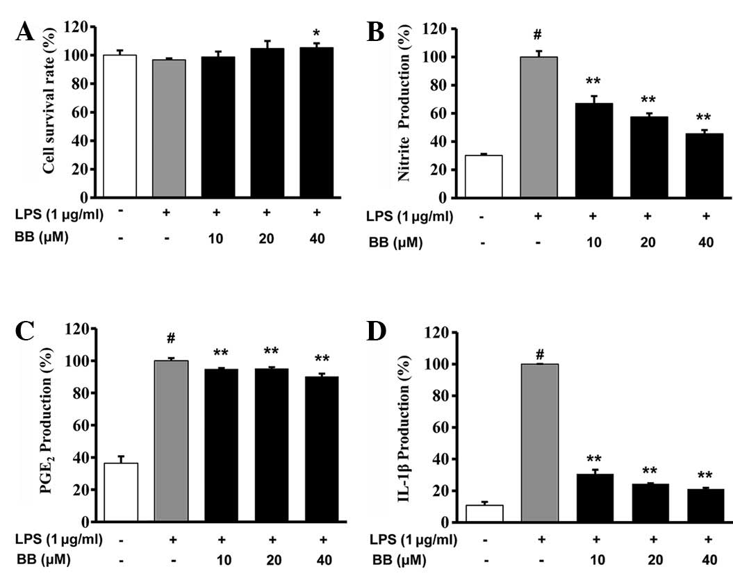
To examine the effect of BB on cell growth in RAW
264.7 cell, the cells were treated with various concentrations (10,
20 and 40 μM) of BB for 24 h. It was observed that BB did not
affect normal cell growth (Fig.
1A). Thus, in the following experiments, the effects of BB at
all concentrations were studied on cells with a normal growth
status. Inflammatory leukocytes are stimulated by LPS, leading to
the release of inflammatory mediators of NO and PGE2. In the
evaluation of the anti-inflammatory effect of BB on RAW 264.7
cells, it was observed that BB inhibited the LPS-induced elevation
of NO production; at concentrations of 10, 20 and 40 μM, BB
inhibited the production of NO by RAW 264.7 cells in a
concentration-dependent manner. Furthermore, the amount of PGE2
released by the BB-treated cells was reduced in comparison with
that by the LPS-treated control cells (Fig. 1C). The level of IL-1β production by
the activated RAW 264.7 cells was significantly increased compared
with that of the normal control cells (P<0.05); however, the
IL-1β production of the cells treated with each concentration of BB
was significantly reduced compared with that of the LPS-treated
control group (P<0.01, Fig.
1D).
Effects on gross mucosal damage
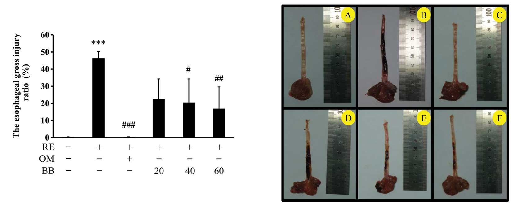
In the normal intact group, no lesion damage or
mucosal injury, such as hyperemia, multiple erosions with hematins
attached, coalesced ulcerations or serosanginous exudates were
observed (Fig. 2A). However, a
severe longitudinal lesion with hyperemia, multiple erosions with
hematins attached, coalesced ulcerations and serosanginous exudates
were observed in the esophagi of the RE control group (Fig. 2B). The pathological area of the
esophagi of the RE control group was grossly increased compared
with that in the intact group. However, the positive control group
treated with 20 mg/kg omeprazole had less damage than the RE
control group (Fig. 2C). The
gastric injury of each BB-treated group comprised only scattered
erosions or mild hemorrhagic spots with whitish exudates scattered
along the esophagus (Fig.
2D–F).
The gross injury index of the RE control was
46.4±3.9%, whereas the index of the positive control treated with
omeprazole was 0.5±0%, and the indices of the groups treated with
20, 40 and 60 mg/kg BB were 22.6±11.7, 20.5±13.8 and 16.9±12.6%,
respectively. Therefore, the gross injury indices of the BB groups
were significantly dose-dependently decreased compared with that of
the RE control (Fig. 2).
The macroscopic observations of the rats with RE
with and without pretreatment with BB are shown in Fig. 3. Mucosal ulceration was markedly
suppressed in the rats pretreated with BB (Fig. 3).
Effect on gastric volume and gastric
juice pH
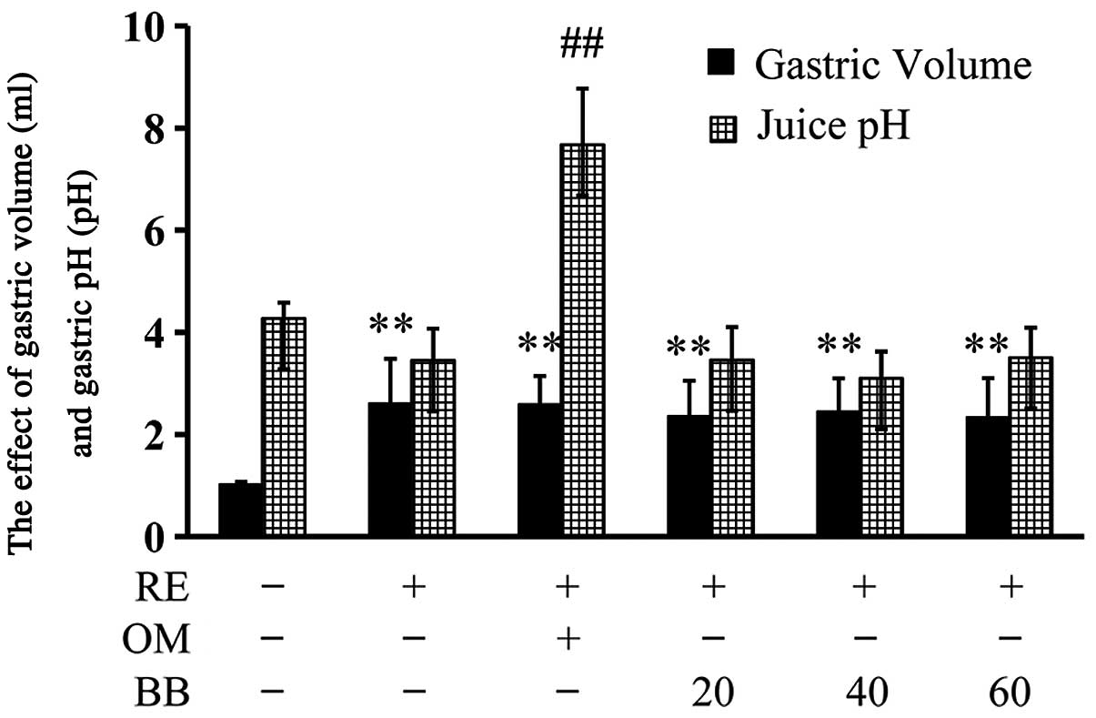
The gastric volume of the normal intact group was
1.03±0.04 ml (containing 1 ml PBS). However, the gastric volume of
the RE control group was significantly increased compared with that
of the intact group (2.61±0.8 ml; P<0.01). The gastric volumes
of the positive control group treated with 20 mg/kg omeprazole
(2.6±0.8 ml) and the RE groups pretreated with 20, 40 and 60 mg/kg
BB (2.4±0.7, 2.45±0.6 and 2.3±0.8 ml, respectively) were not
significantly different compared with that of the RE control group;
however, they were significantly higher than that of the intact
control group (P<0.01 for all; Fig.
4).
The administration of omeprazole (20 mg/kg)
significantly increased the gastric acid pH in the RE rats
(P<0.01). However, the gastric acid pH values of the groups
treated with BB were not significantly different from those of the
RE group (Fig. 4).
Effects on serum TNF-α, IL-1β, IL-6 and
MCP-1 levels
To examine the effects of BB on proinflammatory
cytokines (TNF-α, IL-1β and IL-6) and a chemokine (MCP-1) in
surgically induced acute RE, cytokine levels in the serum were
analyzed.
The serum level of TNF-α was 17.3±3.5 pg/ml in
intact control rats. However, in the RE rats there was an increase
in TNF-α production (27.6±1.0 pg/ml). Treatment of the RE rats with
omeprazole (21.9±3.3 pg/ml, P<0.05), 40 mg/kg BB (20.9±1.4
pg/ml, P<0.05) and 60 mg/kg BB (18.9±1.6 pg/ml, P<0.01)
significantly inhibited this increase (Fig. 5A).
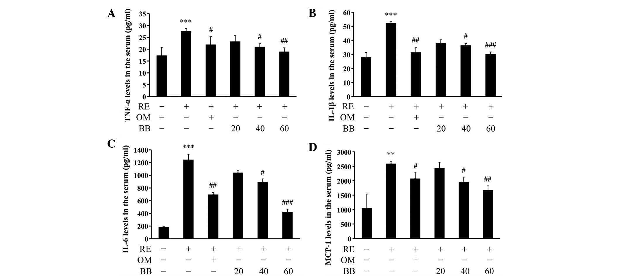 | Figure 5Effect of berberine (BB) on serum
levels of tumor necrosis factor (TNF)-α, interleukin (IL)-1β, IL-6
and monocyte chemoattractant protein (MCP)-1. Whole blood was
collected from the abdominal vein with a 5 ml syringe, at the time
of sacrifice. Collected blood was centrifuged at 1,800 × g for 15
min to collect the serum. Serum levels of the proinflammatory
biomarkers (A) TNF-α, (B) IL-1β, (C) IL-6 and (D) MCP-1 were
evaluated with the Multi-Analyte ELISArray® Kit (Millipore,
Rockford, IL, USA) in accordance with the manufacturer's
instructions. Color intensity of the reaction was estimated using
the Luminex luminometer(Awareness Technology Inc., Palm City, FL,
USA) at 490 nm. Values are expressed as the mean ± standard
deviation. ***P<0.001 compared with the intact rat
group; #P<0.05, ##P<0.01 and
###P<0.001 compared with the reflux esophagitis (RE)
control rat group. RE, control rat with pylorus and forestomach
ligation treated with distilled water; OM, positive control rat
with pylorus and forestomach ligation treated with omeprazole (20
mg/kg). BB, rat with pylorus and forestomach ligation treated with
BB (20, 40 and 60 mg/kg, respectively). |
The serum IL-1β level was 52.1±6.4 pg/ml in the RE
control rats, which was significantly higher than the levels in the
rats treated with omeprazole (31.3±8.2 pg/ml, P<0.01), 40 mg/kg
BB (36.2±9.6 pg/ml, P<0.05) and 60 mg/kg BB (30±11 pg/ml,
P<0.001), indicating that these treatments significantly
inhibited the increase in IL-1β level following RE induction
(Fig. 5B).
The serum IL-6 level of the normal intact group was
178.7±11.5 pg/ml. However, surgically inducing acute RE in the rats
resulted in an increase in IL-6 production (1,243±89.5 pg/ml,
P<0.001) compared with the control group. Rats treated with 20
mg/kg omeprazole (694.4±35.4 pg/ml), 40 mg/kg BB and 60 mg/kg BB
demonstrated significantly lower serum IL-6 levels than the RE
control group (P<0.01, P<0.05, and P<0.001, respectively;
Fig. 5C).
In the normal intact group, the MCP-1 level was
1052.2±488 pg/ml. However, RE induction in the rats resulted in an
increase in the level of MCP-1 production (2,583.5±71.8 pg/ml). The
rats treated with 20 mg/kg omeprazole (2,066.7±232.1 pg/ml), 40
mg/kg BB (1,950.4±175.4 pg/ml) and 60 mg/kg BB (1,669.4±150.1
pg/ml) had significantly decreased MCP-1 levels compared with those
of the RE control group (P<0.05 for omeprazole and 40 mg/kg BB
and P<0.01 for 60 mg/kg BB, Fig.
5D).
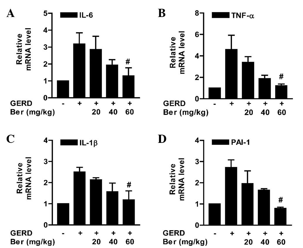
Effects on TNF-α, IL-1β, IL-6 and PAI-1
mRNA expression, analyzed using qPCR
As shown in Fig. 6,
the expression levels of TNF-α, IL-1β, IL-6 and PAI-1 mRNA were low
in the intact esophageal mucosa. However, mRNA expression levels in
the RE control were significantly increased, due to the
inflammatory reaction in the esophagus. The expression levels of
TNF-α, IL-1β, IL-6 and PAI-1 mRNA in the rats pretreated with a
concentration of 60 mg/kg BB were significantly decreased compared
with those of the RE control group (P<0.05, Fig. 6).
Histological analysis of esophageal
mucosa
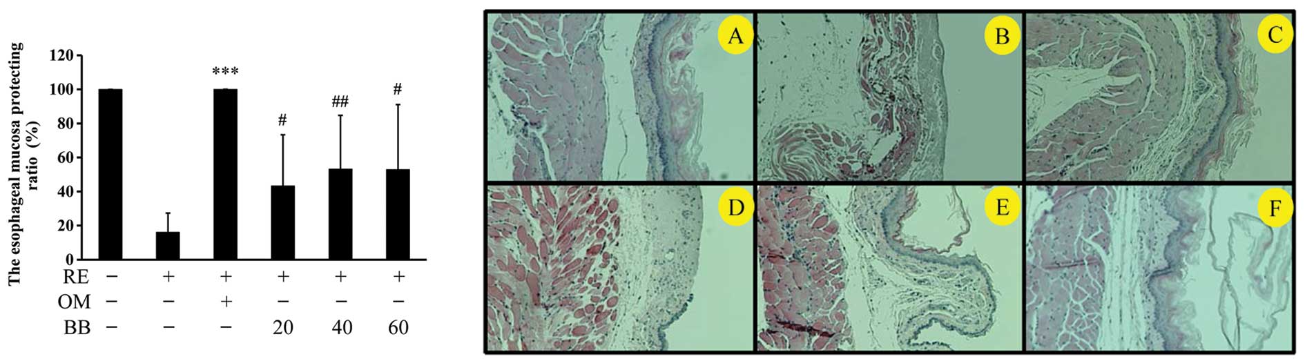
Esophageal tissue stained with H&E revealed no
microscopic mucosal changes in the intact rat (Fig. 3A). The normal esophagus exhibited a
thin epithelial layer with squamous cells and inflammatory cells
were not observed in the submucosal layers. By contrast, 6 h
subsequent to the induction of RE, the RE group rats developed
large coalesced longitudinal ulcers in the lower and middle
sections of the esophagus. Mucosal damage and hyperemia of the
epithelial layers and edema and hemorrhage in the mucosa and
submucosa were observed in the RE control animals (Fig. 3B). Furthermore, the mucosal layers
were damaged by gastric acid. By contrast, esophageal damage,
edema, neutrophil infiltration and gastric hemorrhage were not
observed in the rats treated with omeprazole (Fig. 3C) and the RE rats treated with 40
and 60 mg/kg BB (Fig. 3E and F,
respectively) also showed less severe pathological changes.
The histological activity indices were significantly
reduced in the rats pretreated with omeprazole and BB (Table II).
| Table IIHistological activity index in
esophageal tissue assessed by hematoxylin and eosin staining. |
Table II
Histological activity index in
esophageal tissue assessed by hematoxylin and eosin staining.
| Score | |
|---|
| |
|---|
| Group | Extent of
esophageal ulcers | Degree of
inflammation | Damage to
mucosa |
|---|
| Intact | 0 | 0 | 0 |
| Reflux
esophagitis | 3.33±0.71a | 2.78±0.67a | 3a |
| Omeprazole | 0.11±0.33d | 0d | 0d |
| Berberine 20
mg/kg | 2.83±0.75 | 2.33±0.51 | 2.16±1.1 |
| Berberine 40
mg/kg | 2.50±0.54c | 1.33±0.81b | 2.00±1.3b |
| Berberine 60
mg/kg | 1.70±0.51d | 1.00±0.63d | 1.10±1.0c |
Discussion
The present study demonstrated that the
administration of BB significantly inhibited gastric acid-induced
esophageal damage. A number of herbal therapies have been proposed
for the treatment of RE; however, the efficacy of BB as a treatment
for RE has not, to the best of our knowledge, been
investigated.
The pathogenesis of RE is associated with oxidative
stress, inflammation and apoptosis. A number of studies have
demonstrated the suppressive effect of BB on inflammation (27–29).
The proinflammatory cytokines, TNF-α, IL-1β and IL-6, are important
in the response to microbial infection or tissue damage. BB
treatment has been reported to significantly downregulate the
expression of proinflammatory genes, such as TNF-α, IL-1β, IL-6 and
MCP-1 (30).
In the present study, we investigated whether BB
inhibited the inflammation associated with RE. The suppressive
effects of BB on the production of inflammatory mediators in
LPS-stimulated RAW 264.7 cells and rat serum were observed.
Two of the major cytokines are TNF-α and IL-1. TNF-α
induces a number of physiological effects, including septic shock,
inflammation and cytotoxicity. TNF-α is also known as cachectin,
due to the fact that it mediates fever and cachexia, and is
responsible for the numerous detrimental effects associated with
bacterial sepsis, rheumatoid arthritis and Crohn's disease. TNF-α
is released by monocytes and macrophages in response to various
stimuli including bacterial LPS, which is a principal mediator of
the deleterious effects of endotoxin (31). IL-1 is an inflammatory cytokine
that is released in response to infection or cell injury by cells
of the innate immune system, such as macrophages. Keratinocytes
store and release IL-1 following wounding of the skin, rapidly
signaling to the surrounding cells that the external barrier has
been damaged. IL-1β is important for the initiation and enhancement
of the inflammatory response (32,33).
IL-6 is produced in various cells, such as
fibroblasts, monocytes, T cells, B cells, microglia, endothelial
cells, neurons and astrocytes (34). IL-6, originally identified as a B
cell differentiation factor, and synthesized in response to IL-1β,
has a critical role in the host reaction to inflammation, inducing
the synthesis of acute inflammatory proteins (35). IL-1β and IL-6 are involved in the
acute response phase of the immune response (36). IL-6 is a key regulator of cell
growth, survival and differentiation, and, as such, is involved in
a variety of biological responses, including the immune response,
inflammation, hematopoiesis and oncogenesis.
MCP-1 is a member of the C-C chemokine family, and
possesses inflammatory properties (37). MCP-1 is important in the
recruitment and activation of leukocytes during acute inflammation
(38). MCP-1 upregulation is
associated with macrophage recruitment, angiogenesis and survival
in human breast cancer.
Elizabeth et al(39) demonstrated that PGE2 regulated the
production of PAI-1 in primary cultures of rat calvarial
osteoblasts. PAI-1 production is increased by TNF-α (40) and IL-1 (41). It has been revealed that the
induction and transcriptional regulation of the PAI-1 gene may be
mediated by cytokines and inflammatory mediators, such as
endotoxins, IL-1, transforming growth factor (TGF)-β, insulin,
TNF-α, hepatocyte growth factor (HGF) and phorbol 12-myristate
13-acetate (42).
BB is known to inhibit inflammatory cytokines, such
as TNF-α, IL-1β and IL-6, and inflammatory mediators, such as NO
(produced by iNOS) and PGE2 (produced by COX-2) (20–25,27–29).
In the present study, the gastric volume and the pH of the gastric
juice in the RE rats were not significantly altered following
treatment with BB, so the BB-treated rats were stimulated by
gastric acid to the same extent as the RE control rats. However,
the gross esophageal lesions and histological indications of
mucosal damage in the BB-treated rats were significantly reduced
compared with those in the control RE rats, with regard to the
extent of esophageal ulcers, the degree of inflammation, the damage
to the mucosa and the survival ratio of the mucosal layer.
In conclusion, the present results indicate that BB
suppresses inflammation of the esophagus. In support of our
hypothesis, NO, PGE2 and IL-1β production levels were significantly
diminished in vitro in RAW 264.7 cells that had been
stimulated with LPS. Furthermore, serum levels of the inflammatory
biomarkers, TNF-α, IL-1β, IL-6 and MCP-1, were significantly
reduced in vivo in rats treated with BB, compared with the
levels in untreated RE rats. TNF-α, IL-1β, IL-6 and PAI-1 mRNA
expression levels in esophageal tissue was also significantly
reduced compared with the levels in the untreated RE control
rats.
Acknowledgements
This study was supported by the National Research
Foundation of Korea (NRF) and grant funded by the Korea
government(MSIP; No. 2012-0009400).
References
|
1
|
Mahattanadul S, Ridtitid W, Nima S,
Phdoongsombut N, Ratanasuwon P and Kasiwong S: Effects of
Morinda citrifolia aqueous fruit extract and its biomarker
scopoletin on reflux esophagitis and gastric ulcer in rats. J
Ethnopharmacol. 134:243–250. 2011.
|
|
2
|
Fujiwara Y, Higuchi K, Hamaguchi M,
Takashima T, Watanabe T, Tominaga K, Oshitani N, Matsumoto T and
Arakawa T: Increased expression of transforming growth factor-alpha
and epidermalgrowthfactor receptors in rat chronic reflux
esophagitis. J Gastroenterol Hepatol. 19:521–517. 2004. View Article : Google Scholar : PubMed/NCBI
|
|
3
|
Yoshida N: Inflammation and oxidative
stress in gastroesophageal reflux disease. J Clin Biochem Nutr.
40:13–23. 2007. View Article : Google Scholar : PubMed/NCBI
|
|
4
|
Labenz J, Blum AL, Bayendörffer E, Meining
A, Stolte M and Börsch G: Curing Helicobacter pylori
infection in patients with duodenal ulcer disease may provoke
reflux esophagitis. Gastroenterology. 112:1442–1447. 1997.
|
|
5
|
Varanasi RV, Fantry GT and Wilson KT:
Decreased prevalence of Helicobacter pylori infection in
gastroesophageal reflux disease. Helicobacter. 3:188–194. 1998.
|
|
6
|
Haruma K, Hamada H, Mihara M, Kamada T,
Yoshihara M, Sumii K, Kajiyama G and Kawanishi M: Negative
associated between Helicobacter pylori infection and reflux
esophagitis in older patients: case-control study in Japan.
Helicobacter. 5:24–29. 2000.
|
|
7
|
Shin JM and Kim N: Pharmacokinetics and
pharmacodynamics of the proton pump inhibitors. J
Neurogastroenterol Motil. 19:25–35. 2013. View Article : Google Scholar : PubMed/NCBI
|
|
8
|
Carling L, Axelsson CK, Forssell H,
Stubberöd A, Kraglund K, Bonnevie O and Ekström P: Lansoprazole and
omeprazole in the prevention of relapse of reflux oesophagitis: a
long-term comparative study. Aliment Pharmacol Ther. 12:985–990.
1998. View Article : Google Scholar : PubMed/NCBI
|
|
9
|
Youssef SS, Iskandar SB, Scruggs J and Roy
TM: Acute pancreatitis associated with omeprazole. Int J Clin
Pharmacol Ther. 43:558–561. 2005. View
Article : Google Scholar : PubMed/NCBI
|
|
10
|
Katz PO: Optimizing medical therapy for
gastroesophageal reflux disease: state of the art. Rev
Gastroenterol Disord. 3:59–69. 2003.PubMed/NCBI
|
|
11
|
Lahiri S, Singh P, Singh S, Rasheed N,
Palit G and Pant KK: Melatonin protects against experimental reflux
esophagitis. J Pineal Res. 46:207–213. 2009. View Article : Google Scholar : PubMed/NCBI
|
|
12
|
Colin-Jones DG: Histamine-2-receptor
antagonists in gastro- oesophageal reflux. Gut. 30:1305–1308. 1989.
View Article : Google Scholar : PubMed/NCBI
|
|
13
|
Doornebal J, Bijlsma R and Brouwer RM: An
unknown but potentially serious side effect of proton pump
inhibitors: hypomagnesaemia. Ned Tijdschr Geneeskd.
153:A7112009.(In Dutch).
|
|
14
|
Ku SK, Kim JS, Seo YB, et al: Effect of
Curculigo orchioides on reflux esophagitis by suppressing
proinflammatory cytokines. Am J Chin Med. 40:1241–1255. 2012.
|
|
15
|
Compare D, Pica L, Rocco A, De Giorgi F,
Cuomo R, Sarnelli G, Romano M and Nardone G: Effects of long-term
PPI treatment on producing bowel symptoms and SIBO. Eur J Clin
Invest. 41:380–386. 2011. View Article : Google Scholar : PubMed/NCBI
|
|
16
|
Allescher HD and Wagner H: STW
5/Iberogast: multi-target-action for treatment of functional
dyspepsia and irritable bowel syndrome. Wien Med Wochenschr.
157:301–307. 2007.(In German).
|
|
17
|
Wang XH, Jiang SM and Sun QW: Effects of
berberine on human rheumatoid arthritis fibroblast-like
synoviocytes. Exp Biol Med (Maywood). 236:859–866. 2011. View Article : Google Scholar : PubMed/NCBI
|
|
18
|
Lou T, Zhang Z, Xi Z, Liu K, Li L, Liu B
and Huang F: Berberine inhibits inflammatory response and
ameliorates insulin resistance in hepatocytes. Inflammation.
34:659–667. 2011. View Article : Google Scholar : PubMed/NCBI
|
|
19
|
Kuo CL, Chi CW and Liu TY: The
anti-inflammatory potential of berberine in vitro and in vivo.
Cancer Lett. 203:127–137. 2004. View Article : Google Scholar : PubMed/NCBI
|
|
20
|
Jeong HW, Hsu KC, Lee JW, Ham M, Huh JY,
Shin HJ, Kim WS and Kim JB: Berberine suppresses proinflammatory
responses through AMPK activation in macrophages. Am J Physiol
Endocrinol Metab. 296:E955–E964. 2009. View Article : Google Scholar : PubMed/NCBI
|
|
21
|
Lu DY, Tang CH, Chen YH and Wei IH:
Berberine suppresses neuroinflammatory responses through
AMP-activated protein kinase activation in BV-2 microglia. J Cell
Biochem. 110:697–705. 2010. View Article : Google Scholar : PubMed/NCBI
|
|
22
|
Wang Q, Zhang M, Liang B, Shirwany N, Zhu
Y and Zou MH: Activation of AMP-activated protein kinase is
required for berberine-induced reduction of atherosclerosis in
mice: the role of uncoupling protein 2. PLoS One. 6:e254362011.
View Article : Google Scholar : PubMed/NCBI
|
|
23
|
Shang W, Liu J, Yu X and Zhao J: Effects
of berberine on serum levels of inflammatory factors and
inflammatory signaling pathway in obese mice induced by high fat
diet. Zhongguo Zhong Yao Za Zhi. 35:1474–1477. 2010.(In
Chinese).
|
|
24
|
Shen YB, Piao XS, Kim SW, Wang L and Liu
P: The effects of berberine on the magnitude of the acute
inflammatory response induced by Escherichia coli
lipopolysaccharide in broiler chickens. Poult Sci. 89:13–19. 2010.
View Article : Google Scholar : PubMed/NCBI
|
|
25
|
Singh P, Singh N and Palit G: Analysing
the role of COX-2 in acute oesophagitis and in melatonin-exerted
protection against experimental reflux oesophagitis in rats. J
Pharm Pharmacol. 63:1572–1580. 2011. View Article : Google Scholar : PubMed/NCBI
|
|
26
|
US Environmental Protection Agency. Health
Effects Test Guidelines OPPTS 870.100. Washington: US EPA; 2012
|
|
27
|
McAdam E, Haboubi HN, Forrester G, Eltahir
Z, Spencer-Harty S, Davies C, Griffiths AP, Baxter JN and Jenkins
GJ: Inducible nitric oxide synthase (iNOS) and nitric oxide (NO)
are important mediators of reflux-induced cell signalling in
esophageal cells. Carcinogenesis. 33:2035–2043. 2012. View Article : Google Scholar : PubMed/NCBI
|
|
28
|
Lee CH, Chen JC, Hsiang CY, Wu SL, Wu HC
and Ho TY: Berberine suppresses inflammatory agents-induced
interleukin-1beta and tumor necrosis factor-alpha productions via
the inhibition of IkappaB degradation in human lung cells.
Pharmacol Res. 56:193–201. 2007. View Article : Google Scholar
|
|
29
|
Choi BH, Ahn IS, Kim YH, Park JW, Lee SY,
Hyun CK and Do MS: Berberine reduces the expression of adipogenic
enzymes and inflammatory molecules of 3T3-L1 adipocyte. Exp Mol
Med. 38:599–605. 2006. View Article : Google Scholar : PubMed/NCBI
|
|
30
|
Xiao HB, Sun ZL, Zhang HB and Zhang DS:
Berberine inhibits dyslipidemia in C57BL/6 mice with
lipopolysaccharide induced inflammation. Pharmacol Rep. 64:889–895.
2012. View Article : Google Scholar : PubMed/NCBI
|
|
31
|
Jia L, Liu J, Song Z, Pan X, Chen L, Cui X
and Wang M: Berberine suppresses amyloid-beta-induced inflammatory
response in microglia by inhibiting nuclear factor-kappaB and
mitogen-activated protein kinase signalling pathways. J Pharm
Pharmacol. 64:1510–1521. 2012.
|
|
32
|
Yan F, Wang L, Shi Y, et al: Berberine
promotes recovery of colitis and inhibits inflammatory responses in
colonic macrophages and epithelial cells in DSS-treated mice. Am J
Physiol Gastrointest Liver Physiol. 302:G504–G514. 2012. View Article : Google Scholar : PubMed/NCBI
|
|
33
|
Jeong HW, Hsu KC, Lee JW, Ham M, Huh JY,
Shin HJ, Kim WS and Kim JB: Berberine suppresses proinflammatory
responses through AMPK activation in macrophages. Am J Physiol
Endocrinol Metab. 296:E955–E964. 2009. View Article : Google Scholar : PubMed/NCBI
|
|
34
|
Yoon HD, Jeong EJ, Choi JW, Lee MS, Park
MA, Yoon NY, Kim YK, Cho DM, Kim JI and Kim HR: Anti-inflammatory
effects of ethanolic extracts from Codium fragile on
LPS-stimulated RAW 264.7 macrophages via nuclear factor kappaB
inactivation. Fish Aquat Sci. 14:267–274. 2011.
|
|
35
|
Rodríguez-Hernández H, Simental-Mendía LE,
Rodríguez-Ramírez G and Reyes-Romero MA: Obesity and inflammation:
epidemiology, risk factors, and markers of inflammation. Int J
Endocrinol. 2013:6781592013.PubMed/NCBI
|
|
36
|
Rieder F, Biancani P, Harnett K, Yerian L
and Falk GW: Inflammatory mediators in gastroesophageal reflux
disease: impact on esophageal motility, fibrosis, and
carcinogenesis. Am J Physiol Gastrointest Liver Physiol.
298:G571–G581. 2010. View Article : Google Scholar : PubMed/NCBI
|
|
37
|
Gruol DL and Nelson TE: Physiological and
pathological roles of interleukin-6 in the central nervous system.
Mol Neurobiol. 15:3307–3339. 1997. View Article : Google Scholar
|
|
38
|
Heinrich PC, Castell JV and Andus T:
Interleukin-6 and acute phase response. Biochem J. 265:621–636.
1990.PubMed/NCBI
|
|
39
|
Allan EH and Martin TJ: Prostaglandin E2
regulates production of plasminogen activator isoenzymes, urokinase
receptor, and plasminogen activator inhibitor-1 in primary cultures
of rat calvarial osteoblasts. J Cell Physiol. 165:521–529. 1995.
View Article : Google Scholar
|
|
40
|
Ortiz-Muñoz G, Martin-Ventura JL,
Hernandez-Vargas P, et al: Suppressors of cytokine signaling
modulate JAK/STAT-mediated cell responses during atherosclerosis.
Arterioscler Thromb Vasc Biol. 29:525–531. 2009.PubMed/NCBI
|
|
41
|
Wu AC, Morrison NA, Kelly WL and Forwood
MR: MCP-1 expression is specifically regulated during activation of
skeletal repair and remodeling. Calcif Tissue Int. 92:566–575.
2013. View Article : Google Scholar : PubMed/NCBI
|
|
42
|
Dawson SJ, Wiman B, Hamsten A, Green F,
Humphries S and Henney AM: The two allele sequences of a common
polymorphism in the promoter of the plasminogen activator
inhibitor-1 (PAI-1) gene respond differently to interleukin-1 in
HepG2 cells. J Biol Chem. 268:10739–10745. 1993.PubMed/NCBI
|