Introduction
Urolithiasis is a frequently occurring global
disease. The formation of stones in the urinary tract affects 5–10%
of the population in Europe and the United States (1,2), and
the annual incidence of stone formation in the industrialized world
is generally considered to be 1,500–2,000 cases per million
(3). Furthermore, the prevalence
of nephrolithiasis appears to have increased in the last quarter of
the twentieth century (4). The
prevalence of stone disease shows a geographical variability, with
China being an area with a high prevalence (5). At present, renal stones are the most
common form of urolithiasis in China (6). Although there is currently little
epidemiological information available regarding urinary stones in
China, clinical data has shown an increasing trend in the incidence
of urinary stones (6).
At present, extracorporal shock wave lithotripsy
(ESWL) and percutaneous nephrolithotomy (PCNL) are the primary
treatments for renal stones. Circular stone increments have been
frequently observed during PCNL procedures in our clinical
practice. Similar observations have been apparent in the ear stones
of fish, known as otoliths. Otolith micro-structure investigative
techniques may be applicable to the examination of the age and
growth of the juvenile chum salmon Oncorhynchus keta, which
inhabits coastal waters (7).
There are, at present, few techniques that may be
used to study stone growth, particularly in the evaluation of drug
efficacy in medical expulsive therapy. Approximately 80% of kidney
stones contain calcium, and the majority of calcium stones consist
primarily of calcium oxalate (8,9). In
the present study, stone circular increments were labeled using
calcium-tracing fluorochromes in a rat calcium oxalate
xeno-plantation model.
Materials and methods
Establishment of the rat calcium oxalate
xenoplantation model
Stone particles were extracted by PCNL from one male
patient with renal stones. Informed consent from the patient was
obtained prior to the study. One stone particle was sent for
analysis by infrared spectroscopy. The result showed that it was
predominantly composed of calcium oxalate. Following this, other
particles were selected, cut with a blunt instrument into sections
with a diameter of 2–3 mm, weighed and maintained in a sterile
environment, prior to use.
Eight-week-old male Wistar rats (Vital River
Laboratory Animal Technology Co., Ltd., Beijing, China), weighing
250–300 g, were housed in a specific pathogen-free (SPF)
environment. All animals had free access to drinking water and
regular chow every day, and were kept under a controlled 12-h
light/dark cycle at 22±2°C. All animal experimentation was
performed in accordance with the Chinese Home Office-approved
guidelines, and was approved by the Animal Care Committee of Peking
University People’s Hospital (Beijing, China).
The rats were anesthetized by intraperitoneal
injection of sodium pentobarbital [50 mg/kg body weight (bw)] and
the bladder was exposed by a suprapubic incision. Following this, a
4–5 mm incision was made at the top of bladder and one prepared
human stone particle was inserted. The bladder and suprapubic
incision were closed respectively. Ethylene glycol (EG) was
supplied in the drinking water at a final concentration of 1% from
the second day (day 1) postoperatively for 4 weeks.
Fluorochrome application
The method of fluorochrome administration was
modified from previous studies (10). Briefly, three fluorescent
chromophores, calcein, alizarin complexone and xylenol orange, were
administered by intraperitoneal injection from day 15,
postoperatively. The dosages used were 15, 30 and 90 mg/kg bw,
respectively (10). Two protocols
were used in the fluorochrome application. Protocol 1 entailed each
fluorescent chromophore being intraperitoneally injected twice a
week for 2 weeks (continuous labeling), while protocol 2 entailed
the sequential injection of calcein, alizarin complexone and
xylenol on each consecutive day for 1 week (sequential labeling).
All fluorochromes were purchased from the Beijing Chemical Reagent
Company (Beijing, China) and were sterilized by filtration, prior
to use.
Four weeks after EG was supplied in the drinking
water, the rats were sacrificed with an intraperitoneal injection
of an overdose of sodium pentobarbital. The kidneys and urinary
bladder were dissected and the kidneys were dehydrated in a graded
ethanol series and embedded in paraffin.
The bladder stones were harvested, weighed and
maintained in 75% ethanol for 24 h, prior to the stones being
embedded in autopolymerizing resin and sectioned transversely with
a diamond wire saw in order to select the best section plane.
Sectioned blocks were then fixed to a glass slide with a
thermoplastic glue and polished successively using a 1,200 grit
sandpaper and a mix of alumina polishing compounds (3, 1 and 0.3
μm) with a small volume of water, until it was possible to
observe the core clearly under a transmitted light microscope.
Thermoplastic glue, which softens when heated, enabled the block to
be turned over so that was possible to polish the other side and
for the core to be approached cautiously.
Detection of renal stone formation
Renal stone formation was assessed using von Kossa
histochemical staining. Briefly, 5-μm-thick cross sections
of the paraffin-embedded rat kidneys were deparaffinized and placed
in distilled water, prior to being exposed to 2% silver nitrate for
60 min. Subsequent to being washed in 5% sodium thiosulfate for 5
min, the sections were counterstained with neutral red and examined
under a light microscope. The 5-μm cross sections were also
observed under a fluorescence microscope.
Bladder stone analysis
Bladder stone analysis was undertaken using a
confocal laser scanning microscope (Leica TCS-SP2; Leica
Microsystems, Wetzlar, Germany) fitted with spectrophotometers for
emission band wavelength selection. The calcein and alizarin
complexone were excited with the 476 and 530 nm laser lines from an
argon laser, with the laser intensity set at 9% of the available
power. For the visualization of calcein and alizarin complexone,
the emission windows were set at 496–505 and 530–580 nm,
respectively. Any areas of interest that were well-focused and
exhibited sufficient reflection or fluorescence were captured using
the Leica Confocal software.
Results
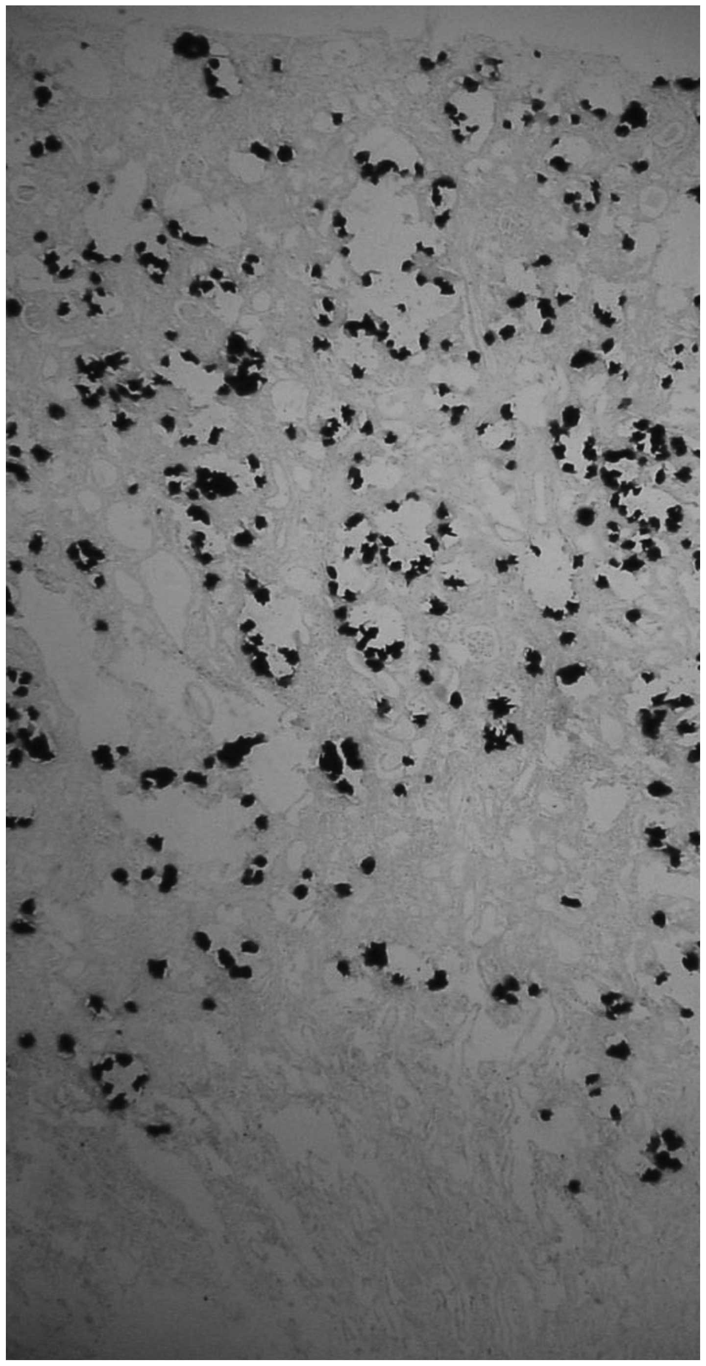
Renal stone confirmation
Kidney stone formation was confirmed by von Kossa
histochemical staining in all seven rats. The stones were
predominantly formed in the renal tubules located at the border
between the renal cortex and the medulla (Fig. 1).
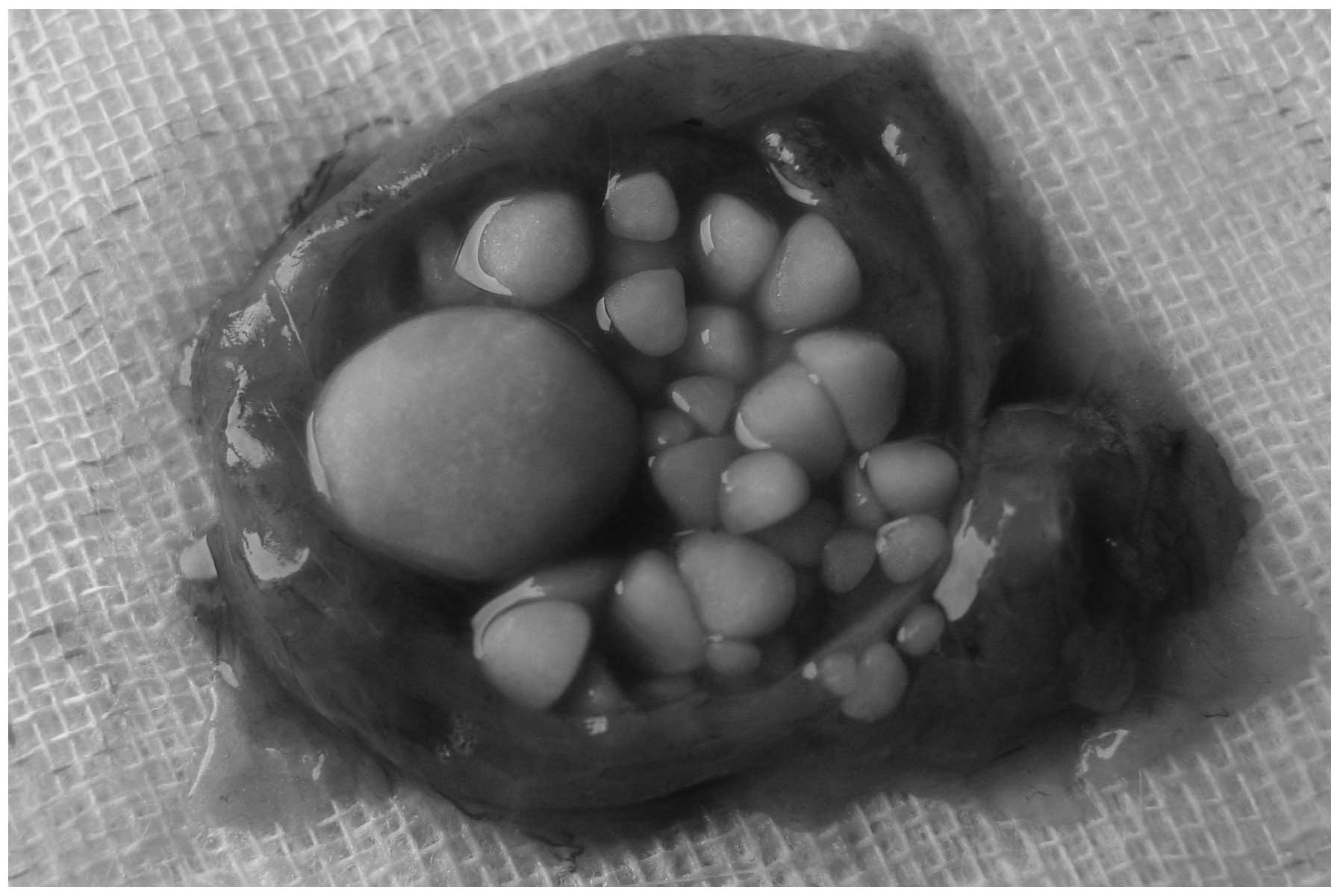
Bladder stone regrowth
The bladder stones showed a large variation in the
size following regrowth in the rats (Fig. 2). The average stone weight was 0.14
g (range, 0.01–0.61 g).
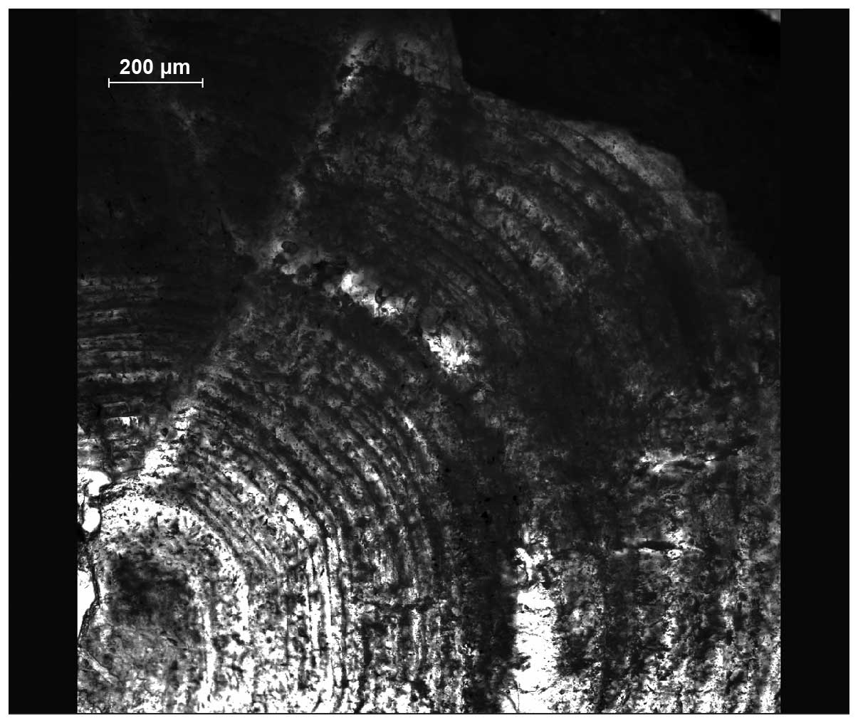
Stone microscopic analysis
The circular bladder stone increments were observed
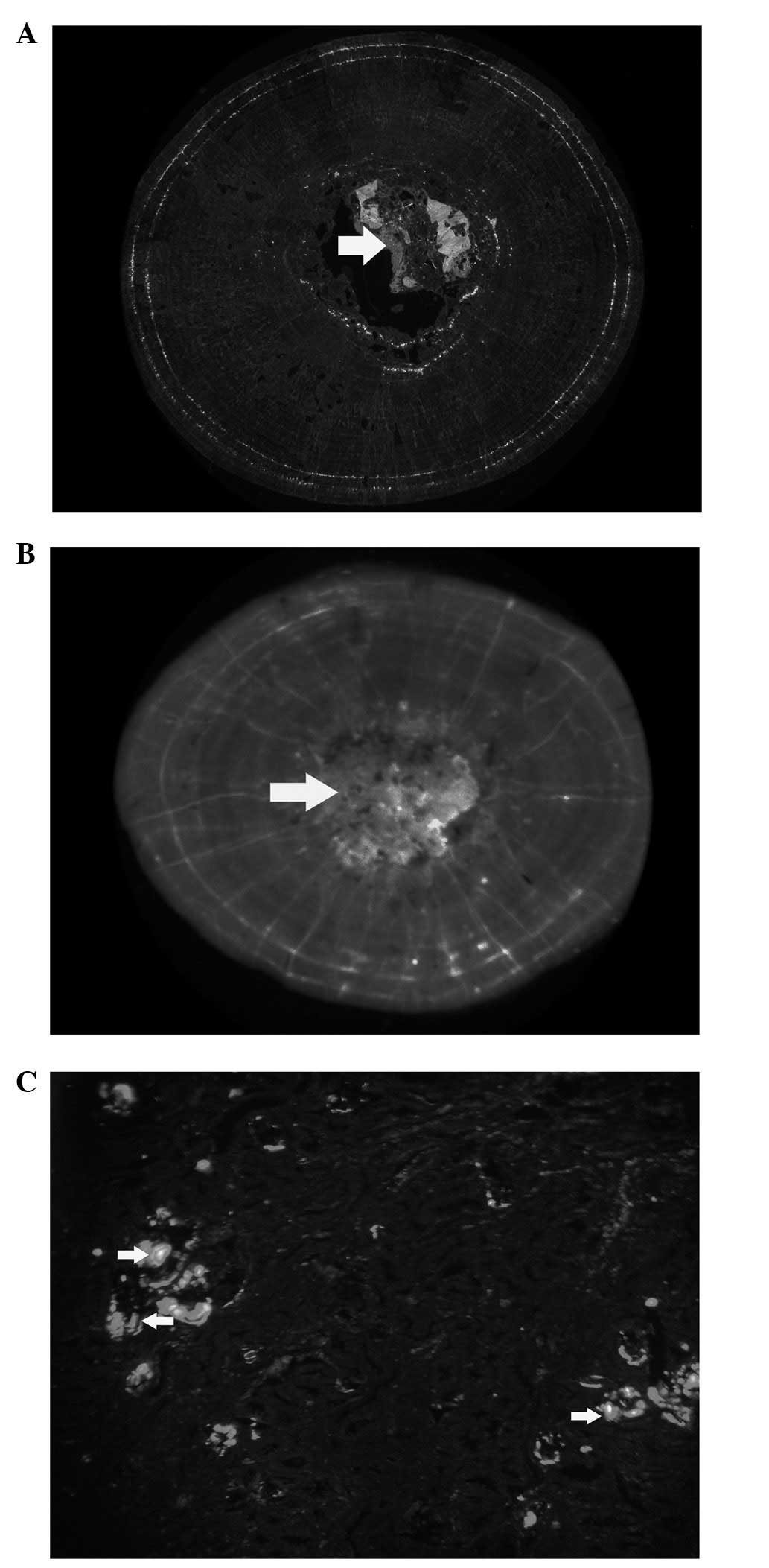
under the laser scanning confocal microscope (Fig. 3). Under fluorescence emission,
clear colored marks were apparent in the sections that had been
prepared by continuous labeling with calcein and alizarin
complexone (Fig. 4A and B);
however xylenol orange did not produce the same effect. Green and
red labeling was also observed in the renal sections prepared by
sequential labeling (Fig. 4C).
Discussion
Nephrolithiasis is a particularly common clinical
condition in the industrialized world. Up to 15% of Caucasian males
and 6% of all females are likely to have at least one stone during
their lifetime, and half of these individuals will experience
recurrent episodes (11,12). During recent decades, the incidence
of the disease has appeared to increase, although the exact cause
of the disease has yet to be elucidated. Among the various types of
renal stones, those composed of calcium oxalate are by far the most
prevalent, accounting for 75% of all stones (13).
With regard to calcium oxalate crystallization,
there are numerous complexities. Crystal nucleation, growth,
aggregation and retention are considered to be the fundamental
steps in stone formation. The final step of the process is poorly
understood; however, the assembly of crystalline material is a
necessary factor in stone formation. Circular increments have been
observed in the majority of renal stones during PCNL procedures.
Similar stone increments were apparent in the rat calcium oxalate
xenoplantation model in the present study. This phenomenon has been
described in detail in previous studies (14,15).
To define the time-course of stone growth in the
clinic is difficult, due to the challenge presented in marking the
stone at a specific time-point. An identical problem is encountered
in studies with animal models. Enabling the effective labeling of
the stone at specific time-points is likely to be beneficial for
clinical and animal model studies. Calcium-tracing fluorochromes
are widely used in investigations concerning the process of
mineralization, including otoliths, tooth growth and bone
remodeling (10,16–18).
The fluorochromes that are frequently used are alizarin complexone,
calcein, xylenol orange and oxytetracycline. The sites labeled by
these fluorochromes are revealed as different colors under the
fluorescence microscope. In the present study, clear circular
labeling was observed with intraperitoneal injections of alizarin
complexone and calcein, although not with xylenol orange. By
labeling the stone at a specific time, alizarin complexone and
calcein may be used to evaluate stone growth. In a previous study,
Figueiredo et al applied the calcium-binding fluorescence
probes, Osteosense 680 and Osteosense 750, in the identification of
calculi in the urinary tract. In the study, the Osteosense 680 and
Osteosense 750 probes demonstrated a variety of binding affinities
for different types of stones. It was concluded that the improved
visualization of these stones was likely to reduce the difficulties
encountered in endoscopic procedures, decrease the risk of
complications and increase the chance of rendering the patient
stone-free (19).
Animal models are frequently used in the study of
urolithiasis, and, as such, there are numerous animal models of
urolithiasis (20). The rat
calcium oxalate model, induced by ethylene glycol, is frequently
used. Ethylene glycol administration has been demonstrated to
result in an increase in urinary oxalate levels and calcium oxalate
supersaturation, and to induce calcium oxalate crystalluria with a
decreased calcium level (14,20).
Since human urolithogenesis is different to that in the rat calcium
oxalate model, it is not certain whether calcium tracers are likely
to lead to the desired results in human urolithiasis labeling.
Furthermore, the size of the bladder stones demonstrated a large
variance in the present study, which makes this xenoplantation
model unsuitable for urinary stone growth studies.
In conclusion, calcein and alizarin complexone
effectively labeled the stone increments in the present rat calcium
oxalate xenoplantation model. This technique may potentially be
used to evaluate the efficacy of medical expulsive therapy by
marking the stone increments at specific time-points and monitoring
the rate of stone growth between two specific time-points.
References
|
1.
|
Ljunghall S: Renal stone disease. Studies
of epidemiology and calcium metabolism. Scand J Urol Nephrol. 1–96.
1977.PubMed/NCBI
|
|
2.
|
Pak CY, Resnick MI and Preminger GM:
Ethnic and geographic diversity of stone disease. Urology.
50:504–507. 1997. View Article : Google Scholar : PubMed/NCBI
|
|
3.
|
Tiselius HG: Metabolic evaluation and
therapy. Curr Opin Urol. 10:545–549. 2000. View Article : Google Scholar
|
|
4.
|
Stamatelou KK, Francis ME, Jones CA,
Nyberg LM and Curhan GC: Time trends in reported prevalence of
kidney stones in the United States: 1976–1994. Kidney Int.
63:1817–1823. 2003.PubMed/NCBI
|
|
5.
|
Finlayson B: Symposium on renal lithiasis.
Renal lithiasis in review. Urol Clin North Am. 1:181–212.
1974.PubMed/NCBI
|
|
6.
|
Sun C: Epidemiology of urinary stone. Wu
Jieping’s Urology. Wu J: Shandong Science & Technology Press;
Jinan: pp. 745–747. 2005, (In Chinese).
|
|
7.
|
Saito T, Kaga T, Seki J and Otake T:
Otolith microstructure of chum salmon Oncorhynchus keta:
formation of sea entry check and daily depositon of otolith
increments in seawater conditions. Fish Sci. 73:27–37. 2007.
|
|
8.
|
Coe FL, Parks JH and Asplin JR: The
pathogenesis and treatment of kidney stones. N Engl J Med.
327:1141–1152. 1992. View Article : Google Scholar : PubMed/NCBI
|
|
9.
|
Levy FL, Adams-Huet B and Pak CY:
Ambulatory evaluation of nephrolithiasis: an update of a 1980
protocol. Am J Med. 98:50–59. 1995. View Article : Google Scholar
|
|
10.
|
Pautke C, Tischer T, Vogt S, et al: New
advances in fluorochrome sequential labelling of teeth using seven
different fluorochromes and spectral image analysis. J Anat.
210:117–121. 2007. View Article : Google Scholar
|
|
11.
|
Barbas C, García A, Saavedra L and Muros
M: Urinary analysis of nephrolithiasis markers. J Chromatogr B
Analyt Technol Biomed Life Sci. 781:433–455. 2002. View Article : Google Scholar
|
|
12.
|
Bihl G and Meyers A: Recurrent renal stone
disease - advances in pathogenesis and clinical management. Lancet.
358:651–656. 2001. View Article : Google Scholar : PubMed/NCBI
|
|
13.
|
Ramello A, Vitale C and Marangella M:
Epidemiology of nephrolithiasis. J Nephrol. 13(Suppl 3): S45–S50.
2000.
|
|
14.
|
Khan SR and Hackett RL: Urolithogenesis of
mixed foreign body stones. J Urol. 138:1321–1328. 1987.
|
|
15.
|
Murphy BT and Pyrah LN: The composition,
structure, and mechanisms of the formation of urinary calculi. Br J
Urol. 34:129–159. 1962. View Article : Google Scholar
|
|
16.
|
Lee TC, Mohsin S and Taylor D: Detecting
microdamage in bone. J Anat. 2003:161–172. 2003.
|
|
17.
|
Nkenke E, Kloss F, Wiltfang J, et al:
Histomorphometric and fluorescence microscopic analysis of bone
remodelling after installation of implants using an osteotome
technique. Clin Oral Implants Res. 13:595–602. 2002. View Article : Google Scholar : PubMed/NCBI
|
|
18.
|
O’Brien FJ, Taylor D and Lee TC: An
improved labelling technique for monitoring microcrack growth in
compact bone. J Biomech. 35:523–526. 2002.PubMed/NCBI
|
|
19.
|
Figueiredo JL, Passerotti CC, Sponholtz T,
Nguyen HT and Weissleder R: A novel method of imaging calcium
urolithiasis using fluorescence. J Urol. 179:1610–1614. 2008.
View Article : Google Scholar : PubMed/NCBI
|
|
20.
|
Khan SR: Animal models of kidney stone
formation: an analysis. World J Urol. 15:236–243. 1997. View Article : Google Scholar : PubMed/NCBI
|