Introduction
Acute organophosphorus pesticide poisoning (AOPP) is
a global threat to human health. According to a report by the World
Health Organization (WHO) (1),
~3,000,000 people worldwide are affected by pesticide poisoning
each year, with AOPP being the most common type. Although an
effective antidote for AOPP may be administered, severe cases are
likely to lead to the development of multiple organ dysfunction
syndromes (MODS). It has been found that the inhibition of
cholinesterase (ChE) activity by organic phosphorus in patients
with AOPP induces the accumulation of large amounts of
acetylcholine within the body, causing cholinergic system
dysfunction, hypoxia, inadequate tissue perfusion, microcirculation
dysfunction, disseminated intravascular coagulation (DIC) and,
ultimately, MODS (2). Human
leukocyte antigen (HLA)-DR, the most important effector molecule in
antigen presentation in the monocyte-macrophage system, is crucial
during specific T lymphocyte immune responses. CD4+ T
lymphocytes are capable of combining with peptide-loaded HLA-DR
molecules on the surface of monocytes or macrophages, initiating
T-cell activation and proliferation. HLA-DR expression on the
surface of mononuclear cells (MNCs) is closely associated with
immune function (3). In addition,
previous studies have indicated that HLA-DR expression is
associated with the immune state (4,5).
However, the role of HLA-DR antigen expression in patients with
AOPP and MODS has yet to be elucidated. To date, there have not
been any studies indicating that HLA-DR is involved in the
occurrence and development of AOPP and MODS. The aim of this study
was to explore the correlations between HLA-DR expression and
AOPP-associated parameters in order to evaluate their roles in
clinical applications.
Materials and methods
Patient data
From January 2003 to August 2009, 75 patients were
admitted to the emergency room, nephropathy department and
intensive care unit of Affiliated Hospital of North Sichuan Medical
College (Nanchong, China), having ingested 10–380 ml
organophosphorus pesticides. These 75 patients met the diagnosis of
AOPP. There were 28 cases of methamidophos poisoning, 15 cases of
dichlorvos poisoning, 13 cases of omethoate poisoning, 11 cases of
phorate poisoning, five cases of parathion poisoning and three
cases of rogor poisoning. There was a time-period of <6 h from
poisoning to treatment in all cases. None of the patients had
complications from diseases of the heart, brain, liver, lungs or
kidneys, or from diabetes, hypertension, malignant tumors or
connective tissue diseases. In accordance with the diagnostic
criteria for MODS (6) proposed by
the American College of Chest Physicians and Critical Care Medicine
(ACCP/SCCM) in 1991, the patients were divided into a MODS group
(39 cases) and a non-MODS group (36 cases). Patients were scored
according to their disease severity by means of the acute
physiology and chronic health evaluation II (APACHE II) (7), with the lowest score being 13 points,
the highest score being 38 points and the average score being 23.5
points. The study was approved by the hospital Medical Ethics
Committee of Affiliated Hospital of North Sichuan Medical College
and informed consent was signed by all patients. There were 30
healthy individuals in the control group, which included nine males
and 21 females with an age range of 22–65 years and a mean age of
33.5 years.
Flow cytometry
Peripheral blood samples were collected on admission
to the hospital using ethylenediamine tetraacetic acid (EDTA)
anticoagulant tubes. Fluorescein isothiocyanate (FITC)-conjugated
anti-human HLA-DR monoclonal antibodies (Becton-Dickinson Company,
Franklin Lakes, NJ, USA) were used to detect HLA-DR expression
according to the manufacturer’s instructions. The mean fluorescence
channel number (MCF) was used to measure the expression of
HLA-DR.
Biochemical analysis
Liver function, renal function and creatine kinase
levels were examined using a Beckman CX 7 Automatic analyzer
(Beckman Coulter Inc., Brea, CA, USA). Cardiac troponin I (cTnI)
levels were assessed using a chemiluminescent microparticle
immunoassay (CMIA) with a Beckman ACCESS autoimmune luminescence
analyzer (Beckman Coulter Inc.). Creatine kinase isoenzyme (CK-MB)
levels were examined using a Nissan 7170S automatic chemical
analyzer (Nissan, Tokyo, Japan). Serum ChE activity was assessed
using the dibutyryl thiocholine method, with reagents provided by
Biological Engineering Co., Ltd. of Zhejiang Eastern Europe
(Zhejiang, China).
Statistical analysis
SPSS 13.0 statistical software (SPSS, Inc., Chicago,
IL, USA) was used for data analysis and processing. Measurement
data are expressed as the mean ± standard deviation. Comparisons
between the two groups were conducted using a t-test, and
correlation tests were performed using a linear correlation
analysis. P<0.05 was considered to indicate a statistically
significant difference.
Results
Clinical data
Of the 75 patients with AOPP, there were eight
deaths from complications of MODS. The mortality rate was 10.7%.
All 36 patients in the non-MODS group recovered (Table I).
 | Table IClinical data and HLA-DR antigen
expression level in different groups. |
Table I
Clinical data and HLA-DR antigen
expression level in different groups.
| | Gender (n) | | | |
|---|
| |
| | | |
|---|
| Group | Cases (n) | Male | Female | Age (years) | HLA-DR antigen
expression (MFI) | T-value |
|---|
| Healthy | 30 | 9 | 21 | 33.5±11.6 | 27.85±4.86 | 5.549a |
| AOPP | 75 | 28 | 47 | 34.5±11.3 | 21.59±5.36 | |
| Non-MODS | 36 | 13 | 23 | 33.5±10.2 | 25.15±6.15 | 5.764a |
| MODS | 39 | 15 | 24 | 35.5±13.6 | 18.17±4.23 | |
| Surviving | 67 | 25 | 42 | 34.1±10.5 | 22.34±2.76 | 6.510a |
| Deceased | 8 | 3 | 5 | 37.5±12.2 | 15.29±3.97 | |
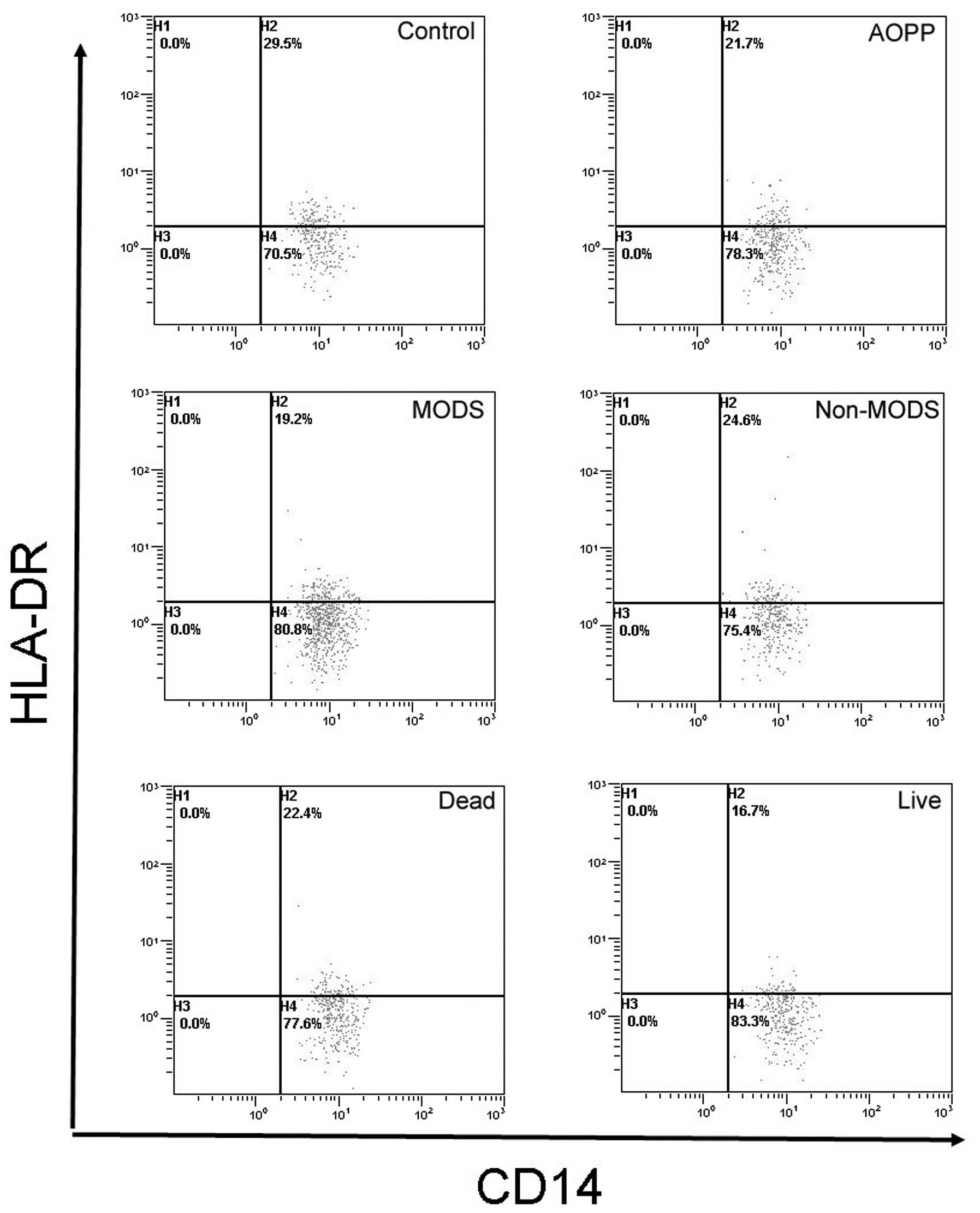
Comparisons of HLA-DR antigen expression
levels among the groups
HLA-DR antigen expression levels in the AOPP group
were lower than those in the normal control group (P<0.001). The
levels in the MODS group were lower than those in the non-MODS
group (P<0.001). There were no significant differences between
the non-MODS and the normal control groups (P>0.05) (Fig. 1).
Correlation of HLA-DR antigen expression
levels with APACHE II score
The APACHE II score (data not shown) of the patients
who died in the AOPP group (30.2±7.7) was significantly higher than
that of those who survived (22.7±9.7) (P<0.05). HLA-DR antigen
expression levels in the patients who died in the AOPP group were
significantly lower than the levels in those who survived
(P<0.001).
Correlation of HLA-DR antigen expression
levels with clinical indicators
The HLA-DR antigen expression level was positively
correlated with serum ChE (r=0.52, P<0.01) and negatively
correlated with the APACHE II score, CK-MB, cTnI, lactate
dehydrogenase (LDH), alanine aminotransferase (ALT), aspartate
aminotransferase (AST), blood urea nitrogen (BUN) and serum
creatinine (Scr) (r=−0.61~−0.29, P<0.01 or P<0.05) (Table II).
| Table IICorrelations of HLA-DR antigen with
other indicators in patients with AOPP. |
Table II
Correlations of HLA-DR antigen with
other indicators in patients with AOPP.
| Indicators | r | Indicators | r | Indicators | r |
|---|
| APACHE II score | −0.61b | CK-MB | −0.51b | AST | −0.45b |
| ChE | 0.52b | LDH | −0.46b | BUN | −0.29a |
| cTnI | −0.49b | ALT | −0.41b | Scr | −0.35b |
Discussion
HLA-DR antigen is one of the class II major
histocompatibility (MHC) antigens, mainly expressed in B
lymphocytes, monocytes, macrophages, dendritic cells, vascular
endothelial cells and activated T cells. It is also expressed in
gastrointestinal epithelial cells (8). HLA-DR is crucial for immune system
function. In normal circumstances, macrophages, monocytes and other
antigen-presenting cells (APCs) engulf and process foreign
microbial or exogenous protein into peptides presented on MHC-II
molecules, which is capable of initiating an immune response
through binding with T helper cells. If HLA-DR antigen expression
levels decrease, or its antigen-presenting role is hindered, an
effective immune response is not able to be produced (9,10).
Thus, the body is not able to effectively remove pathogens and
inflammatory mediators. Liao et al evaluated the HLA-DR
antigen expression of peripheral blood monocytes in patients with
severe multiple trauma, demonstrating significant correlations
between antigen expression and trauma severity and prognosis. Wang
et al observed the HLA-DR expression of monocytes in 32
patients with trauma, and found that HLA-DR expression decreased
after one day, reached its lowest level on day four, and then
gradually recovered. HLA-DR expression in patients with trauma was
significantly negatively correlated with the APACHE II score. A
sustained decrease in HLA-DR expression in monocytes was associated
with a poor prognosis in patients with sepsis, which was one
crucial reason for post-injury complications following severe
trauma. Tschoeke and Ertel (11)
and Cheron et al(12)
reported that HLA-DR antigen expression decreased among patients
with severe pancreatitis. HLA-DR expression gradually returned to
normal levels in surviving patients, and progressively decreased in
patients who ultimately died; expression levels in these patients
were closely associated with mortality (13). The present study showed that the
HLA-DR antigen expression of peripheral blood MNCs in the AOPP
group was lower than that in the normal control group (P<0.001).
HLA-DR antigen expression in patients with AOPP and MODS was lower
than that in patients in the non-MODS group. HLA-DR antigen
expression was positively correlated with serum ChE, reflecting the
degree of AOPP, and was negatively correlated with the APACHE II
score (P<0.01), indicating that monocyte HLA-DR antigen
expression may be involved in the pathogenesis of AOPP, and may be
used as a clinical indicator reflecting AOPP severity. HLA-DR
antigen expression levels in patients with MODS were lower than
those in patients in the non-MODS group (P<0.01), and were
negatively correlated with CK-MB, cTnI, LDH, ALT, AST, BUN and Scr
(P<0.01 or P<0.05). This demonstrated that HLA-DR antigen
expression of monocytes was closely associated with MODS subsequent
to AOPP, which may be an important supplementary mechanism of MODS
caused by AOPP. It has been shown that organophosphorus pesticides
stimulate the release of cytokines, such as interleukin-1,
interleukin-6 and tumor necrosis factor (14), which may increase HLA-DR
expression. Endotoxin has been shown to inhibit
γ-interferon-induced HLA-DR antigen expression (15). Among patients with AOPP, tumor
necrosis factor and endorphin levels may be significantly increased
(14), both of which are involved
in negative regulation of monocyte HLA-DR antigen expression
(16). As HLA-DR antigen
expression is reduced, the antigen presenting function becomes
impaired (17–20). APACHE II score is positively
correlated with disease severity, and is extensively used in
monitoring disease conditions and estimating prognosis. In a
postoperative study of 30 critically ill patients, Lekkou et
al(20) observed that monocyte
HLA-DR expression was lower in these patients than that in the
normal control group. The present study showed that the APACHE II
score among those who died in the AOPP group was significantly
higher than that among those who survived (P<0.05), and that the
monocyte HLA-DR antigen expression level among those who died in
the AOPP group was significantly lower than that among those who
survived (P<0.01). The APACHE II score of patients with AOPP was
significantly negatively correlated with the HLA-DR antigen
expression level (P<0.01), indicating that immune function
inhibition becomes more apparent with increasing severity of the
disease (16). The dynamic
observation of monocyte HLA-DR antigen expression levels at
different stages may be beneficial in predicting AOPP severity and
prognosis. However, in a study of patients with multiple trauma and
systemic inflammatory response syndrome by Ploder et
al(21), HLA-DR antigen
expression of monocytes was decreased in all patients compared with
the expression levels in the healthy controls, and there was no
difference between the group of patients that died and the group
that survived. The reasons for the difference between the studies
may be associated with different underlying diseases, sample size,
genetic background and examination methods. Studies of HLA-DR in
AOPP are relatively rare, and the pathogenesis of AOPP and MODS
requires further discussion.
Acknowledgements
The authors would like to thank Professor Mei Wang,
(Department of Nephropathy, People’s Hospital of Peking University,
Beijing, China) who provided guidance and help in writing this
study.
References
|
1
|
Cavaliere MJ, Puga FR, Calore EE, et al:
Protective effect of pralidoxime on muscle fiber necrosis induced
by organophosphate compounds. J Toxicol Clin Toxicol. 36:295–300.
1998. View Article : Google Scholar : PubMed/NCBI
|
|
2
|
Mizuno Y, Ohama E, Hirato J, et al: Nestin
immunoreactivity of Purkinje cells in Creutzfeldt-Jakob disease. J
Neurol Sci. 246:131–137. 2006. View Article : Google Scholar : PubMed/NCBI
|
|
3
|
Lukaszewicz AC, Faivre V and Payen D: Is
monocyte HLA-DR expression monitoring a useful tool to predict the
risk of secondary infection? Minerva Anestesiol. 76:737–743.
2010.PubMed/NCBI
|
|
4
|
Itakura Sumi Y, Ogura H, Tanaka H, et al:
Paradoxical cytoskeleton and microparticle formation changes in
monocytes and polymorphonuclear leukocytes in severe systemic
inflammatory response syndrome patients. J Trauma. 55:1125–1132.
2003.
|
|
5
|
Ono S, Tsujimoto H, Matsumoto A, Ikuta S,
Kinoshita M and Mochizuki H: Modulation of human leukocyte
antigen-DR on monocytes and CD16 on granulocytes in patients with
septic shock using hemoperfusion with polymyxin B-immobilized
fiber. Am J Surg. 188:150–156. 2004. View Article : Google Scholar : PubMed/NCBI
|
|
6
|
Bone RC, Balk RA, Cerra FB, et al:
Definitions for sepsis and organ failure and guidelines for the use
of innovative therapies in sepsis. The ACCP/SCCM Consensus
Conference Committee American College of Chest Physicians/Society
of Critical Care Medicine. Chest. 101:1644–1655. 1992. View Article : Google Scholar
|
|
7
|
Knaus WA, Draper EA, Wagner DP and
Zimmerman JE: APACHE II: a severity of disease classification
system. Crit Care Med. 13:818–829. 1985. View Article : Google Scholar : PubMed/NCBI
|
|
8
|
Chiba M, Ishii N, Ishioka T, et al:
Topographic study of Helicobacter pylori and HLA-DR antigen
expression on gastric epithelium. J Gastroenterol. 30:149–155.
1995. View Article : Google Scholar : PubMed/NCBI
|
|
9
|
Wakefield CH, Carey PD, Foulds S, Monson
JR and Guillou PJ: Changes in major histocompatibility complex
class II expression in monocytes and T cells of patients developing
infection after surgery. Br J Surg. 80:205–209. 1993. View Article : Google Scholar
|
|
10
|
Klava A, Windsor A, Boylston AW, Reynolds
JV, Ramsden CW and Guillou PJ: Monocyte activation after open and
laparoscopic surgery. Br J Surg. 84:1152–1156. 1997. View Article : Google Scholar : PubMed/NCBI
|
|
11
|
Tschoeke SK and Ertel W: Immunoparalysis
after multiple trauma. Injury. 38:1346–1357. 2007. View Article : Google Scholar : PubMed/NCBI
|
|
12
|
Cheron A, Floccard B, Allaouchiche B, et
al: Lack of recovery in monocyte human leukocyte antigen-DR
expression is independently associated with the development of
sepsis after major trauma. Crit Care. 14:R2082010. View Article : Google Scholar : PubMed/NCBI
|
|
13
|
Ho YP, Sheen IS, Chiu CT, Wu CS and Lin
CY: A strong association between down-regulation of HLA-DR
expression and the late mortality in patients with severe acute
pancreatitis. Am J Gastroenterol. 101:1117–1124. 2006. View Article : Google Scholar : PubMed/NCBI
|
|
14
|
Chadban SJ, Tesch GH, Foti R, Lan HY,
Atkins RC and Nikolic-Paterson DJ: Interleukin-10 differentially
modulates MHC class II expression by mesangial cells and
macrophages in vitro and in vivo. Immunology. 94:72–78. 1998.
View Article : Google Scholar : PubMed/NCBI
|
|
15
|
Otsuka A, Hanafusa T, Kono N and Tarui S:
Lipopolysaccharide augments HLA-A,B,C molecule expression but
inhibits interferon-gamma-induced HLA-DR molecule expression on
cultured human endothelial cells. Immunology. 73:428–432. 1991.
|
|
16
|
Hershman MJ, Cheadle WG, Wellhausen SR,
Davidson PF and Polk HC Jr: Monocyte HLA-DR antigen expression
characterizes clinical outcome in the trauma patient. Br J Surg.
77:204–207. 1990. View Article : Google Scholar : PubMed/NCBI
|
|
17
|
Melhus O, Koerner TJ and Adams DO: Effects
of TNF alpha on the expression of class II MHC molecules in
macrophages induced by IFN gamma: evidence for suppression at the
level of transcription. J Leukoc Biol. 49:21–28. 1991.PubMed/NCBI
|
|
18
|
Bone RC, Grodzin CJ and Balk RA: Sepsis: a
new hypothesis for pathogenesis of the disease process. Chest.
112:235–243. 1997. View Article : Google Scholar : PubMed/NCBI
|
|
19
|
Volk HD, Reinke P and Döcke WD: Clinical
aspects: from systemic inflammation to ‘immunoparalysis’. Chem
Immunol. 74:162–177. 2000.
|
|
20
|
Lekkou A, Karakantza M, Mouzaki A,
Kalfarentzos F and Gogos CA: Cytokine production and monocyte
HLA-DR expression as predictors of outcome for patients with
community-acquired severe infections. Clin Diagn Lab Immunol.
11:161–167. 2004.PubMed/NCBI
|
|
21
|
Ploder M, Pelinka L, Schmuckenschlager C,
et al: Lipopolysaccharide-induced tumor necrosis factor alpha
production and not monocyte human leukocyte antigen-DR expression
is correlated with survival in septic trauma patients. Shock.
25:129–134. 2006.
|