Introduction
Heat stroke (HS) is a type of nerve damage caused by
thermoregulatory dysfunction and excessive accumulation of body
heat due to high temperature. The clinical symptoms mainly include
high fever, no sweat and central nervous system disorders (1,2). HS
is the most severe form of heat stress, with extensive damage to
the body, and may lead to functional and morphological changes of
numerous organs and systems. Once HS occurs, the mortality rate is
as high as 63%, unless timely and proper treatment is received
(3).
The pathophysiological process of HS is similar to
that of severe sepsis. Cytokines may mediate the systemic
inflammatory response, and play key roles in the process of HS
(4). The uncontrolled systemic
inflammatory response causes a cascade resulting in multiple organ
dysfunction syndrome (MODS). Bouchama and Knochel (1) consider that the characteristic
pathological and clinical manifestations of HS are the interaction
results of complex physiological and biochemical mechanisms prior
to body collapse, including thermoregulatory imbalance, enlargement
of the acute-phase response and the expression of heat shock
protein (HSP).
The traditional Chinese medicine preparation
Xuebijing injection (XBJ) is produced and applied clinically. XBJ
is able to antagonize bacterial toxins, reduce endotoxin levels,
regulate immune and inflammatory mediators, improve
microcirculation and protect vascular endothelial cells. It has
been demonstrated that XBJ can significantly increase the survival
rate of mice with sepsis (5).
However, to the best of our knowledge, the protective effects of
XBJ on HS have not been reported.
In the present study, the vital signs and survival
times of rats with HS were observed, and the plasma levels of
certain cytokines, biochemical indicators and coagulation
indicators were detected. The mechanism by which XBJ protects
against HS in rats was examined.
Materials and methods
Animals and main reagents
A total of 56 healthy adult male Sprague Dawley rats
(clean grade; weight, 331–410 g; average weight, 376±23.6 g;
provided by the Experimental Animal Center of the Chinese People’s
Liberation Army General Hospital, Beijing, China) were included in
this study. The rats were raised in cages (22±1°C, free access to
food and water, 12-h light/dark cycle). XBJ was purchased from
Tianjin Hongri Pharmaceutical Stock Co., Ltd. (Tianjin, China).
[125I]-labeled tumor necrosis factor-α (TNF-α),
interleukin (IL)-1β and IL-6 radioimmunoassay kits were provided by
Beijing North Institute of Biological Technology (Beijing, China).
This study was carried out in strict accordance with the
recommendations in the Guide for the Care and Use of Laboratory
Animals of the National Institutes of Health (4th edition, 2008).
The animal use protocol was reviewed and approved by the
Institutional Animal Care and Use Committee of the Chinese People’s
Liberation Army General Hospital.
Animal treatment
Eight rats were randomly selected for the
establishment of animal models of HS in a preliminary experiment.
The rats were exposed to a high temperature environment
(42.5–43.5°C). The reduction of the mean arterial pressure (MAP)
from the peak by 25 mmHg indicated the occurrence of HS (6). The time taken for HS to occur
(tHS), the rectal temperature (Tr) and the heart rate
(HR) were recorded.
Following anesthesia by intraperitoneal injection of
3% sodium pentobarbital (1 ml/kg), 24 rats were randomly divided
into the normal control (NC), normal saline-treated HS (NS-HS) and
XBJ-treated HS (XBJ-HS) groups, with eight rats in each group. In
the NC group, the rats were placed in a 26°C environment and the Tr
was maintained at 34°C, without any treatment. In the NS-HS group,
prior to heat exposure, normal saline was injected into the femoral
vein (4 ml/kg), followed by treatment with heat stress at 43°C for
47 min (in the preliminary experiment, the average tHS
was 46.88±1.25 min). Subsequently, the heat stress was removed and
the rats were placed in a 26°C environment. In the XBJ-HS group,
prior to heat exposure, XBJ was injected into the femoral vein (4
ml/kg), followed by treatment with heat stress at 43°C for 47 min.
Subsequently, the heat stress was removed and the rats were placed
in a 26°C environment. The MAP, Tr and HR were consecutively
recorded and the changes in survival time (tS, from HS
occurrence to death) were observed.
XBJ effects on blood indicators and liver
damage in the HS rats
Arterial blood (2.3 ml) was drawn at 0, 47 and 57
min after the initiation of the heat stress. The detection indices
were as follows: i) After centrifugation at 4°C and 1,610 × g for
10 min, the serum was separated, and the concentrations of cytokine
IL-1β, IL-6 and TNF-α were determined by radioimmunoassay (reagents
were provided by Beijing North Institute of Biological Technology,
Beijing, China); ii) the plasma levels of creatinine (Cr), blood
urea nitrogen (BUN), aspartate aminotransferase (AST), alanine
aminotransferase (ALT) and alkaline phosphatase (ALP) were
determined by spectrophotometry (HITACHI7600; Hitachi
High-Technologies, Tokyo, Japan); and iii) 0.9 ml arterial blood
was combined with 0.1 ml 3.8% sodium citrate, was followed by
centrifugation at 4°C and 716 × g for 7 min. The serum was
separated, and the levels of activated partial thromboplastin time
(APTT), prothrombin time (PT), fibrinogen degradation products
(FDP) and D-dimer (D-D) were measured using CA-1500 automated
coagulation instrument (SYSMEX Corporation, Kobe, Japan). Following
the last blood drawing, the rats were sacrificed and three small
sections of liver tissue (0.5×0.5×0.3 cm) were obtained. After
fixation with neutral formalin, paraffin sections were prepared,
followed by hematoxylin and eosin staining and observation under a
XSP-10C light microscope (Shanghai Optical Instrument Factory,
Shanghai, China).
Statistical analysis
Data are expressed as the mean ± standard deviation.
Statistical analysis was performed using SPSS statistical software,
version 12.0 (SPSS, Inc., Chicago, IL, USA). The F-test was
performed for analyzing the measurement data and the SNK-q test was
used for multiple comparisons. P<0.05 was considered to indicate
a statistically significant difference.
Results
Vital sign changes and tHS of
the HS rats
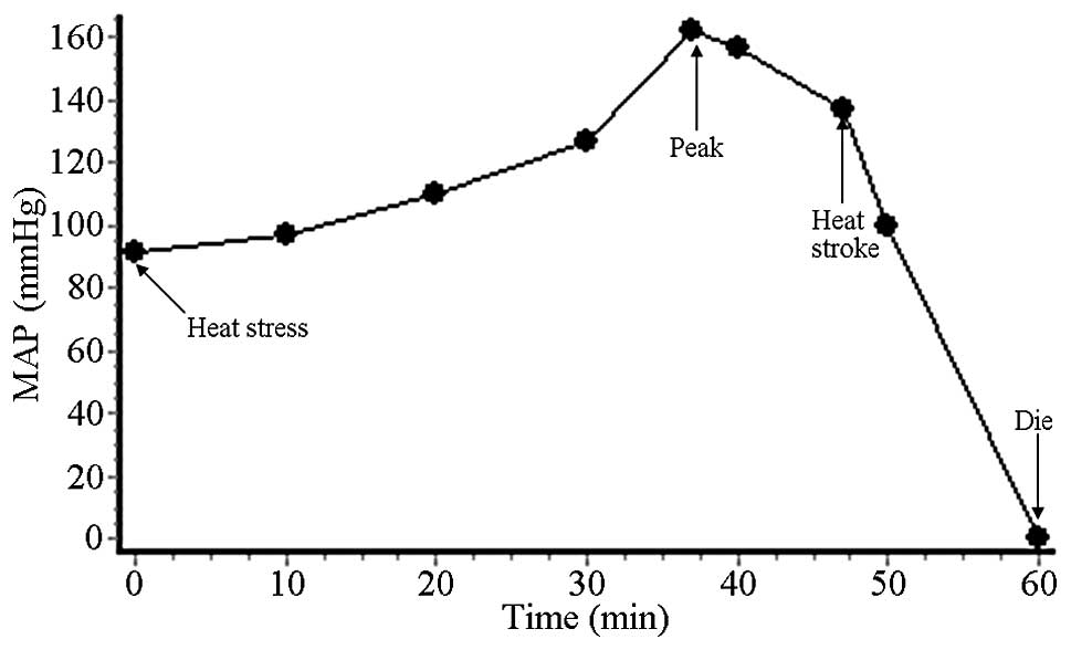
Changes in the MAP under the high temperature
environment (42.5–43.5°C) are shown in Fig. 1 and Table I. In the first 10 min of heat
stress, the MAP did not significantly increase. In the subsequent
20 min, the rate of ascent of the MAP began to increase. From 30 to
37 min, the MAP quickly rose to a peak of ~160 mmHg. In the
subsequent 10 min it declined to 136 mmHg, with a descent of 24
mmHg from the peak which indicated the occurrence of HS. The MAP at
this moment was slightly higher than the basic level. In the final
13 min, the MAP quickly dropped to 0 mmHg and the rats died.
 | Table ITime taken for HS to occur and vital
signs at the time of HS occurrence. |
Table I
Time taken for HS to occur and vital
signs at the time of HS occurrence.
| Rat | tHS
(min) | MAPHS
(mmHg) | TrHS
(°C) | HRHS
(bpm) |
|---|
| 1 | 46 | 141 | 42.8 | 605 |
| 2 | 48 | 131 | 43.4 | 580 |
| 3 | 47 | 129 | 43.0 | 595 |
| 4 | 45 | 141 | 42.8 | 606 |
| 5 | 47 | 143 | 43.6 | 616 |
| 6 | 49 | 134 | 42.9 | 578 |
| 7 | 47 | 137 | 42.4 | 591 |
| 8 | 46 | 138 | 43.0 | 599 |
| Mean ± SD | 46.875±1.246 | 136.75±5.036 | 42.987±0.372 | 596.25±13.069 |
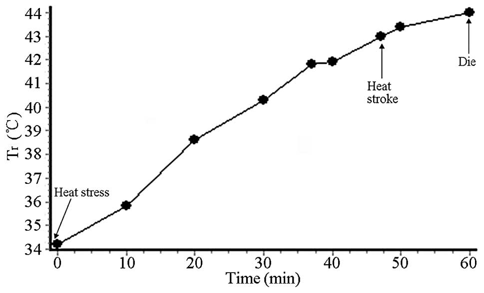
Fig. 2 shows the Tr
changes in the HS rats. Following the initiation of the heat
stress, the Tr presented a continuous, linear and rapid ascent. At
the time of HS occurrence, the Tr reached 43.2°C. When the rats
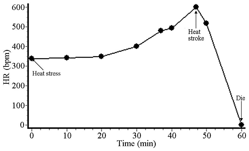
died, it was as high as 44°C. As shown in Fig. 3, in the first 20 min after the
beginning of the heat stress, the HR was basically stable. In the
subsequent 27 min, the HR rapidly rose and reached a peak [600
beats per minute (bpm)], with occurrence of HS. In the subsequent
13 min, heart arrhythmia appeared until the HR reached 0 bpm (the
rats died). Under high-temperature heat stress in the preliminary
experiment, the average tHS was 46.875±1.246 min.
Therefore, the tHS in the following experiments was set
as 47 min.
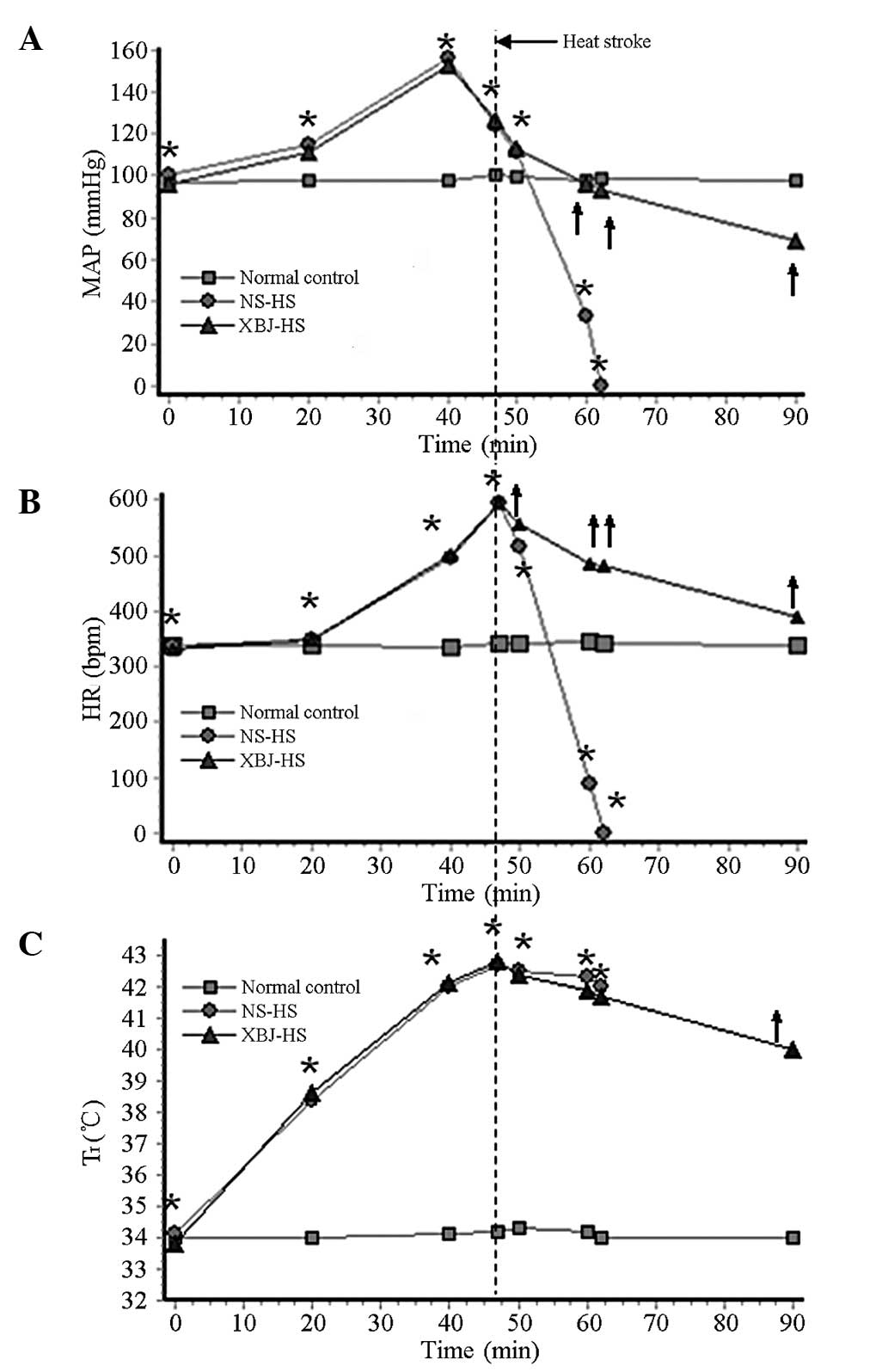
Effects of XBJ on the vital signs and
tS of the HS rats
Prior to the removal of heat stress, the trends of
the changes of the vital signs in the NS-HS group were roughly the
same as those in the XBJ-HS group. The MAP increased slowly in the
first 20 min, then rapidly rose and reached a peak at 40 min,
followed by a rapid reduction. The HR rose slowly in the first 20
min and then rapidly rose. At the HS time point, the HR reached a
peak (600 bpm). The Tr gradually increased with the increase of
heat stress time and reached 43°C at the HS time point. Following
the removal of the heat stress, significant differences were
identified in MAP and HR between the NS-HS and XBJ-HS groups. The
MAP and HR in the NS-HS group decreased rapidly, and the rats died
at 15 min after HS. In the XBJ-HS group, the MAP and HR decreased
slowly. Following the removal of heat stress, the Tr did not
markedly decrease in the NS-HS and XBJ-HS groups, and no
significant differences were observed between the two groups
(Fig. 4).
The effect of the heat stress on the tS
in the HS rats is shown in Table
II. In the NC group, the rats were sacrificed by the
intraperitoneal injection of a double dose of sodium pentobarbital
at 480 min. The mean tS in the XBJ-HS group was
74.625±4.627 min, which was significantly longer than that of the
NS-HS group (15±2.07 min) (P<0.05).
| Table IIEffects of XBJ on the survival time of
HS rats. |
Table II
Effects of XBJ on the survival time of
HS rats.
| Rat | NS-HS | XBJ-HS | NC |
|---|
| 1 | 11 | 75 | 480 |
| 2 | 15 | 68 | 480 |
| 3 | 13 | 80 | 480 |
| 4 | 17 | 73 | 480 |
| 5 | 16 | 69 | 480 |
| 6 | 16 | 75 | 480 |
| 7 | 15 | 76 | 480 |
| 8 | 17 | 81 | 480 |
| Mean ± SD |
15±2.07* | 74.625±4.627a,b | 480 |
Effects of XBJ on blood indicators and
liver damage in the HS rats
Tables III,
IV and V show that at 47 min (the termination of
heat stress) and 57 min, the levels of certain cytokines (IL-1β,
IL-6 and TNF-α), coagulation indicators (APTT, PT, FDP and D-D) and
biochemical indicators (Cr, BUN, AST, ALT and ALP) in the NS-HS
group were significantly higher than those in the NC group
(P<0.05). In the NS-HS group, the levels of the aforementioned
indices were significantly reduced compared with those of the NC
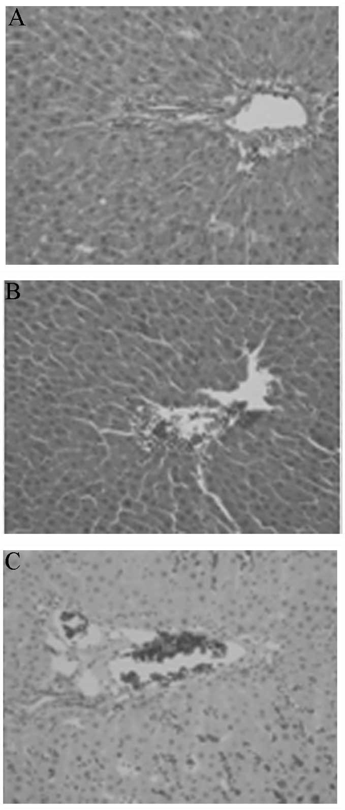
and NS-HS groups (P<0.05). Representative images of the liver
tissue pathology results of each group are shown in Fig. 5. In the NS-HS group, serious liver
cell congestion, nuclear swelling and central vein dilation were
visible, along with the appearance of bubbles. In the XBJ-HS group,
only a small number of congestive liver cells were identified, with
occasional nuclear swelling but no bubbles, which was similar to
that appearance of the NC group.
| Table IIIEffects of XBJ on certain cytokines in
HS rats. |
Table III
Effects of XBJ on certain cytokines in
HS rats.
| Group | Heat stress time
(min) | IL-1β (ng/ml) | IL-6 (pg/ml) | TNF-α (ng/ml) |
|---|
| NC | 0 | 0.315±0.040 | 89.054±6.061 | 0.991±0.133 |
| 47 | 0.305±0.042 | 93.685±3.841 | 0.940±0.076 |
| 57 | 0.314±0.036 | 94.633±4.697 | 0.966±0.065 |
| NS-HS | 0 | 0.288±0.024 | 90.850±4.227 | 1.011±0.096 |
| 47 | 1.246±0.090a |
389.482±18.904a | 5.620±0.321a |
| 57 | 2.547±0.146a |
494.754±18.249a | 5.867±0.212a |
| XBJ-HS | 0 | 0.305±0.042 | 91.164±4.339 | 0.979±0.085 |
| 47 | 0.686±0.069a,b |
192.679±11.568a,b | 2.620±0.321a,b |
| 57 | 0.710±0.046a,b |
209.912±9.779a,b | 2.503±0.261a,b |
| Table IVEffects of XBJ on certain coagulation
indicators in HS rats. |
Table IV
Effects of XBJ on certain coagulation
indicators in HS rats.
| Group | Heat stress time
(min) | APTT (sec) | FDP (mg/l) | D-D (μg/ml) | PT (sec) |
|---|
| NC | 0 | 23.887±1.476 | 172.375±7.009 | 45.563±1.868 | 15.800±0.338 |
| 47 | 24.225±1.419 | 169.500±7.211 | 45.250±2.112 | 15.950±0.434 |
| 57 | 24.075±1.492 | 173.750±8.172 | 45.425±2.218 | 15.850±0.302 |
| NS-HS | 0 | 23.100±1.533 | 167.250±7.760 | 46.375±2.000 | 15.762±0.233 |
| 47 |
69.362±7.751a |
251.375±8.501a |
85.850±3.113a |
20.213±0.340a |
| 57 |
89.975±7.674a |
281.875±8.593a |
112.85±3.275a |
23.575±0.212a |
| XBJ-HS | 0 | 23.938±1.814 | 168.625±8.280 | 45.588±2.149 | 15.830±0.282 |
| 47 |
45.962±5.756a,b |
199.750±5.994a,b |
78.100±3.106a,b |
16.988±0.368a,b |
| 57 |
48.725±4.642a,b |
215.750±6.840a,b |
81.775±3.397a,b |
17.587±0.300a,b |
| Table VEffects of XBJ on certain biochemical
indicators in HS rats. |
Table V
Effects of XBJ on certain biochemical
indicators in HS rats.
| Group | Heat stress time
(min) | Cr (mg/dl) | BUN (mg/dl) | AST (U/l) | ALT (U/l) | ALP (U/l) |
|---|
| NC | 0 | 18.825±1.930 | 6.031±1.014 | 101.263±11.696 | 45.475±5.393 | 196.388±20.508 |
| 47 | 20.125±1.807 | 6.106±0.588 | 100.962±11.924 | 43.213±3.991 | 196.688±14.283 |
| 57 | 20.900±1.373 | 6.095±0.353 | 96.575±12.115 | 43.338±6.265 | 198.175±12.924 |
| NS-HS | 0 | 21.025±2.696 | 6.649±0.671 | 97.862±13.362 | 42.525±4.134 | 200.025±11.366 |
| 47 |
42.438±1.846a |
21.696±1.419a |
376.262±14.516a |
140.750±8.665a |
348.212±8.136a |
| 57 |
67.412±2.106a |
27.851±0.593a |
474.238±15.365a |
168.787±6.906a |
428.500±21.758a |
| XBJ-HS | 0 | 20.263±1.694 | 6.661±0.586 | 100.938±10.913 | 44.587±4.637 | 199.450±13.010 |
| 47 |
30.788±1.398a,b |
14.335±0.667a,b |
220.838±22.345a,b |
70.888±7.578a,b |
202.825±9.102a,b |
| 57 |
36.100±1.704a,b |
17.821±1.093a,b |
265.800±17.029a,b |
84.000±10.366a,b |
207.637±11.282a,b |
Discussion
When rats are exposed to a high-temperature
environment, the MAP, HR and Tr exhibit characteristic changes,
along with the occurrence of HS. Under a high-temperature
environment, a series of inflammatory cells are activated and they
release large amounts of inflammatory cytokines, including IL-1,
IL-2, IL-6, TNF-α and interferon, presenting a ‘waterfall
effect’. This can cause damage to body tissues and organs, which is
similar to sepsis (2). Therefore,
blocking this pathological link or decreasing the levels of
inflammatory cytokines is key for the treatment of HS.
The results of the present study show that under a
high temperature environment, rats present with tissue ischemia and
damage (increases in the levels of Cr, BUN, AST, ALT and ALP),
organ dysfunction (changes in the MAP and HR), hypercoagulable
state or disseminated intravascular coagulation (DIC) (increases in
the levels of APTT, PT, FDP and D-D) and an excessively activated
systemic inflammatory response (increases in the levels of IL-1β,
IL-6 and TNF-α). However, pretreatment with XBJ prior to the
beginning of heat stress significantly inhibited the HS-induced
systemic inflammatory response, tissue ischemia and damage, and
organ dysfunction, thus extending the survival time of the
rats.
XBJ is composed of safflower, radix Paeoniae
rubra, radix Salviae miltiorrhizae, radix Angelicae
sinensis and chuanxiong rhizome, of which the main effective
components are safflor yellow A, ligustrazine, tanshinol, ferulic
acid and paeoniflorin, respectively (7). Safflor yellow A dilates blood
vessels, improves myocardial blood supply, reduces blood pressure,
inhibits coagulation and thrombosis, reduces systemic hypoxia,
increases tissue hypoxia tolerance and decreases capillary
permeability (8). Ligustrazine
markedly improves blood circulation, inhibits inducible nitric
oxide synthase expression, reduces TNF-α levels and extends the
survival time of rats with septic shock (9). Tanshinol promotes blood circulation
and removes blood stasis, inhibits tissue ischemia-reperfusion
injury, scavenges oxygen free radicals, protects mitochondria,
regulates the thromboxane A2 (TXA2)/prostacyclin
(PGI2) balance and immune response, and antagonizes
endotoxins (10). Ferulic acid has
a marked promoting effect on nonspecific, humoral and cellular
immunity function (11). Angelica
extract significantly inhibits high mobility group box 1 release
and improves the survival rate of septic rats (12). Paeoniflorin improves heart and lung
function, regulates the TXA2/PGI2 balance,
inhibits platelet aggregation, prolongs thrombus formation time and
prevents DIC. In addition, radix P. rubra significantly
reduces plasma TNF-α levels and is effective in the treatment of
sepsis (13). The combined effects
of these components constitute the pharmacological basis of XBJ in
the treatment of HS.
IL-1, IL-6 and TNF-α play crucial roles in the
occurrence and development of HS (2). IL-1 is an endogenous pyrogen, which
induces the inflammatory reaction in the acute period, with
antitumor effects similar to those of tumor necrosis factors
(14,15). It has been identified that the
morbidity and mortality of HS are closely associated with
endotoxemia and the release of IL-1 (16). Treatment with an IL-1 receptor
antagonist (200 μg/kg) prior to HS occurrence weakens the cerebral
ischemia and hypoxia of HS rats, prevents hypotension and prolongs
the survival time (>600 min) (17,18).
In a study by Chiu et al (18), following HS occurrence in rats, the
continuous intravenous infusion of an IL-1 receptor antagonist (200
μg/kg·h) was immediately performed. The results showed that the
level of dopamine released by the brain was reduced from 275% in
rats with untreated HS to 140%, with a significantly prolonged
survival time (>600 min) (18).
IL-6 is highly correlated with the mortality of HS and neurological
symptoms. IL-6 antagonists are likely to become a novel
breakthrough for the prevention and treatment of HS (6). As demonstrated in a primate animal
model of HS, the concentration of IL-6 is associated with the
severity of HS (19). TNF-α
induces fever, stimulates white blood cells and neutralizes HSPs
(1). High levels of TNF-α cause an
excessive inflammatory response, increased vascular permeability,
hemodynamic disorders, microcirculatory disturbances and cell
dysfunction, leading to MODS which is associated with the disease
severity and prognosis (14). In
the present study, the plasma levels of TNF-α, IL-1β and IL-6 in
the NS-HS group were significantly increased compared with those in
the control group, and the extents of the increases of these
indices were significantly reduced by XBJ. This indicates that XBJ
reduced the secretion of inflammatory cytokines, which should be
one of the important anti-endotoxin mechanisms for HS. A study has
identified that XBJ reduces the levels of reperfusion injury of
intestinal mucosa, protects the intestinal mucosal barrier function
and prevents the invasion of intestinal endotoxins into the blood,
thereby reducing the release of inflammatory cytokines (20).
HS is one manifestation of the inflammatory and
anti-inflammatory response under high temperature conditions. An
excessively activated inflammatory response and DIC are the main
factors leading to the mortality of patients and deaths of animals
with HS (21). In patients with
HS, coagulation system disorders and a runaway inflammatory
response are very common and are closely associated with the
disease severity and prognosis (22). It has been confirmed in clinical
and experimental studies that XBJ is not only able to inhibit the
coagulation/anticoagulation imbalance and the release of harmful
vasoactive mediators, but also blocks the trigger factors for
coagulation function disorders (23–26).
The results of the present study demonstrate that XBJ significantly
reduces the APTT and PT, and the plasma concentrations of FDP and
D-D, and that it has a protective effect on blood coagulation
function.
At present, lowering the body temperature is one of
the main methods for the treatment of patients with HS. It has been
confirmed in numerous studies that hypothermia therapy
significantly inhibits cerebrovascular dysfunction, the systemic
inflammatory response, hypercoagulable state or DIC, cerebral
oxidative stress, and ischemia and injury in patients with HS
(27–29). In the present study, no specific
efforts to lower the body temperatures of the rats were made, and
pretreatment with XBJ was performed prior to the application of
heat stress. The results show that XBJ significantly reduces
HS-induced circulation dysfunction, hypercoagulability or DIC, and
tissue ischemia and injury, but it does not reduce the body
temperature of HS rats. This indicates that high fever is not the
only pathogenic factor for HS, and XBJ exerts therapeutic effects
by inhibiting the inflammatory response and improving coagulation
function.
References
|
1
|
Bouchama A and Knochel JP: Heat stroke. N
Engl J Med. 346:1978–1988. 2002. View Article : Google Scholar
|
|
2
|
Leon LR and Helwig BG: Heat stroke: role
of the systemic inflammatory response. J Appl Physiol (1985).
109:1980–1988. 2010. View Article : Google Scholar : PubMed/NCBI
|
|
3
|
Misset B, De Jonghe B, Bastuji-Garin S, et
al: Mortality of patients with heatstroke admitted to intensive
care units during the 2003 heat wave in France: a national
multiple-center risk-factor study. Crit Care Med. 34:1087–1092.
2006. View Article : Google Scholar : PubMed/NCBI
|
|
4
|
Grogan H and Hopkins PM: Heat stroke:
implications for critical care and anaesthesia. Br J Anaesth.
88:700–707. 2002. View Article : Google Scholar : PubMed/NCBI
|
|
5
|
Sun X, Lu D, Lv T and Mao Y: Effects of
Xuebijing injection on serum protein level in early phase of septic
rats. Zhongguo Zhong Yao Za Zhi. 35:223–225. 2010.In Chinese).;
|
|
6
|
Hashim IA, Al-Zeer A, Al-Shohaib S,
Al-Ahwal M and Shenkin A: Cytokine changes in patients with
heatstroke during pilgrimage to Makkah. Mediators Inflamm.
6:135–139. 1997. View Article : Google Scholar : PubMed/NCBI
|
|
7
|
Huang H, Ji L, Song S, et al:
Identification of the major constituents in Xuebijing injection by
HPLC-ESI-MS. Phytochem Anal. 22:330–338. 2011. View Article : Google Scholar : PubMed/NCBI
|
|
8
|
Li YP, Qiao YJ, Wu ZX, et al: Effects of
Xuebijing injection on thrombomodulin and endothelial cell protein
C receptor in septic rats. Zhongguo Wei Zhong Bing Ji Jiu Yi Xue.
19:365–368. 2007.(In Chinese).
|
|
9
|
Wu CC, Liao MH, Chen SJ and Yen MH:
Tetramethylpyradizine prevents inducible NO synthase expression and
improves survival in rodent models of endotoxic shock. Naunyn
Schmiedebergs Arch Pharmacol. 360:435–444. 1999. View Article : Google Scholar : PubMed/NCBI
|
|
10
|
Wan JM, Sit WH, Lee CL, Fu KH and Chan DK:
Protection of lethal toxicity of endotoxin by Salvia
miltiorrhiza BUNGE is via reduction in tumor necrosis factor
alpha release and liver injury. Int Immunopharmacol. 6:750–758.
2006.PubMed/NCBI
|
|
11
|
Wang H, Li W, Li J, et al: The aqueous
extract of a popular herbal nutrient supplement, Angelica
sinensis, protects mice against lethal endotoxemia and sepsis.
J Nutr. 136:360–365. 2006.PubMed/NCBI
|
|
12
|
Li YP, Qiao YJ, Wu ZX, Yao YM, Yu Y and Wu
Y: Effects of Xuebijing injection on high-mobility group box
chromosomal protein 1 in septic rats. Zhongguo Wei Zhong Bing Ji
Jiu Yi Xue. 19:239–241. 2007.(In Chinese).
|
|
13
|
Genfa L, Jiang Z, Hong Z, et al: The
screening and isolation of an effective anti-endotoxin monomer from
Radix Paeoniae Rubra using affinity biosensor technology.
Int Immunopharmacol. 5:1007–1017. 2005. View Article : Google Scholar : PubMed/NCBI
|
|
14
|
Bouchama A, Parhar RS, el-Yazigi A, Sheth
K and al-Sedairy S: Endotoxemia and release of tumor necrosis
factor and interleukin 1 alpha in acute heatstroke. J Appl Physiol
(1985). 70:2640–2644. 1991.PubMed/NCBI
|
|
15
|
Koch AE, Kunkel SL and Strieter RM:
Cytokines in rheumatoid arthritis. J Investig Med. 43:28–38.
1995.PubMed/NCBI
|
|
16
|
Chang DM: The role of cytokines in
heatstroke. Immunol Invest. 22:553–561. 1993. View Article : Google Scholar : PubMed/NCBI
|
|
17
|
Lin MT, Liu HH and Yang YL: Involvement of
interleukin-1 receptor mechanisms in development of arterial
hypotension in rat heatstroke. Am J Physiol. 273:H2072–H2077.
1997.PubMed/NCBI
|
|
18
|
Chiu WT, Kao TY and Lin MT: Increased
survival in experimental rat heatstroke by continuous perfusion of
interleukin-1 receptor antagonist. Neurosci Res. 24:159–163. 1996.
View Article : Google Scholar : PubMed/NCBI
|
|
19
|
Roberts GT, Ghebeh H, Chishti MA, et al:
Microvascular injury, thrombosis, inflammation, and apoptosis in
the pathogenesis of heatstroke: a study in baboon model.
Arterioscler Thromb Vasc Biol. 28:1130–1136. 2008. View Article : Google Scholar : PubMed/NCBI
|
|
20
|
Yao XQ, Zhang YH and Sun CH: Protective
effects of Xuebijing effervescent tablet on vital organs in rats
with toxic injury induced by endotoxin. Zhongguo Wei Zhong Bing Ji
Jiu Yi Xue. 24:357–359. 2012.(In Chinese).
|
|
21
|
Lee JJ, Lin MT, Wang NL, Lin CL and Chang
CK: Platonin, a cyanine photosensitizing dye, causes attenuation of
circulatory shock, hypercoagulable state, and tissue ischemia
during heat stroke. Shock. 24:577–582. 2005. View Article : Google Scholar : PubMed/NCBI
|
|
22
|
Jilma B and Derhaschnig U: Disseminated
intravascular coagulation in heat stroke: a hot topic. Crit Care
Med. 40:1370–1372. 2012. View Article : Google Scholar : PubMed/NCBI
|
|
23
|
Li CS, Jin M, Wu JY and Wu CJ: Effect of
XueBiJing injection upon related proinflammatory factors and blood
coagulation factors of vascular endothelial cells in severe septic
patients. Zhonghua Yi Xue Za Zhi. 89:2744–2747. 2009.(In
Chinese).
|
|
24
|
Yin DM, Sun Q, Li YP, Dong N and Yao YM:
Effects of Xuebijing injection on levels of tissue factor-bearing
microparticles in peripheral blood in septic rats. Zhongguo Wei
Zhong Bing Ji Jiu Yi Xue. 21:564–565. 2009.(In Chinese).
|
|
25
|
Sun X, Lu D, Lv T and Mao Y: Effects of
Xuebijing injection on serum protein level in early phase of septic
rats. Zhongguo Zhong Yao Za Zhi. 35:223–225. 2010.(In Chinese).
|
|
26
|
Gao YL, Chai YF and Yao YM: Advancement in
the research of mechanism of immune dysfunction in sepsis and the
regulatory effects of Xuebijing injection. Zhonghua Shao Shang Za
Zhi. 29:162–165. 2013.(In Chinese).
|
|
27
|
Diller KR and Zhu L: Hypothermia therapy
for brain injury. Annu Rev Biomed Eng. 11:135–162. 2009. View Article : Google Scholar
|
|
28
|
Yenari MA and Hemmen TM: Therapeutic
hypothermia for brain ischemia: where have we come and where do we
go? Stroke. 41(10 Suppl): S72–S74. 2010. View Article : Google Scholar : PubMed/NCBI
|
|
29
|
Mattis JG and Yates AM: Heat stroke:
helping patients keep their cool. Nurse Pract. 36:48–52. 2011.
View Article : Google Scholar : PubMed/NCBI
|