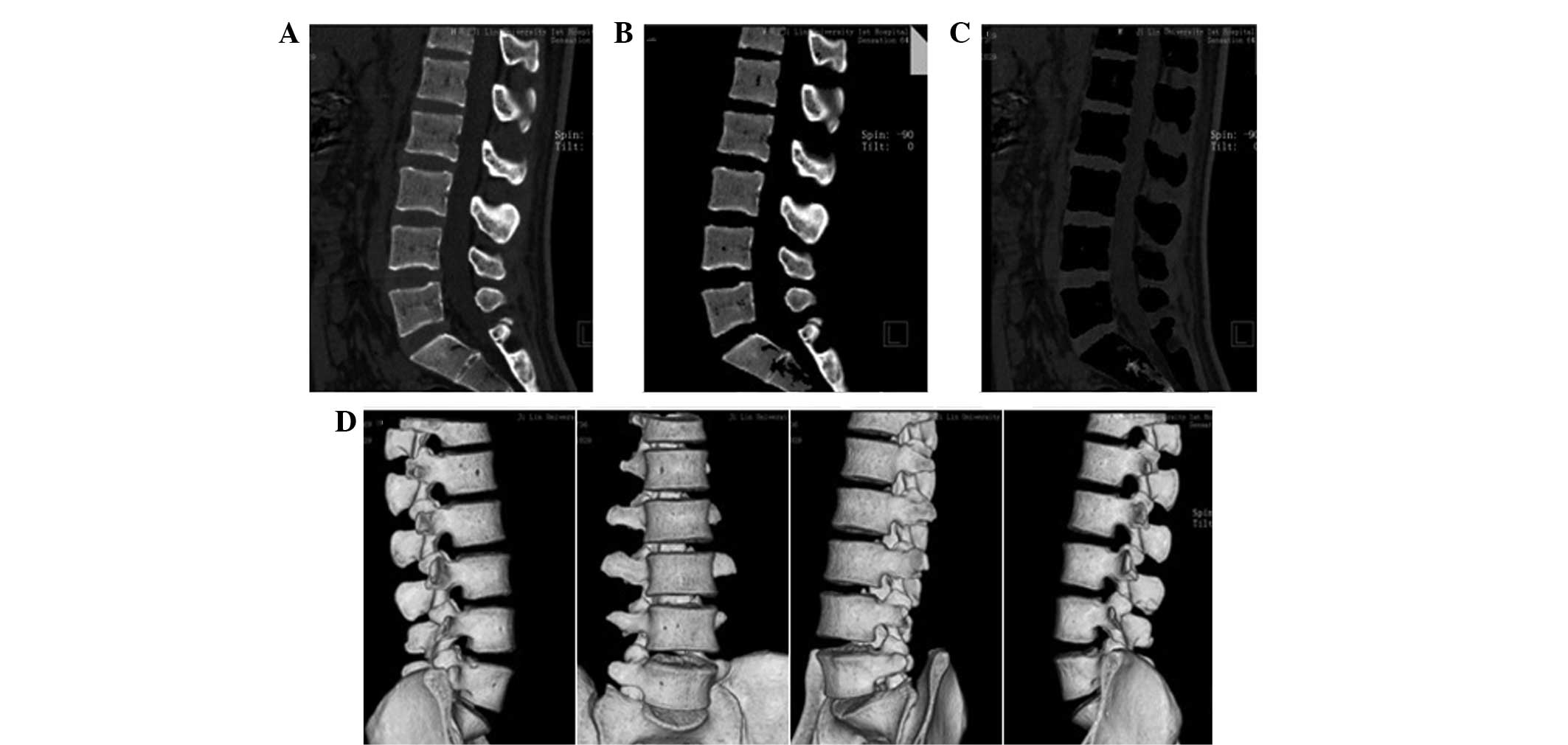
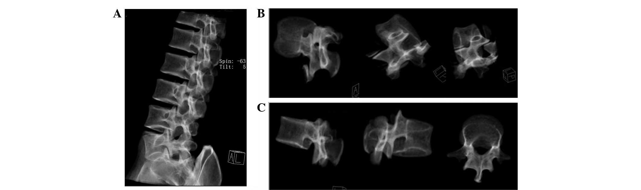
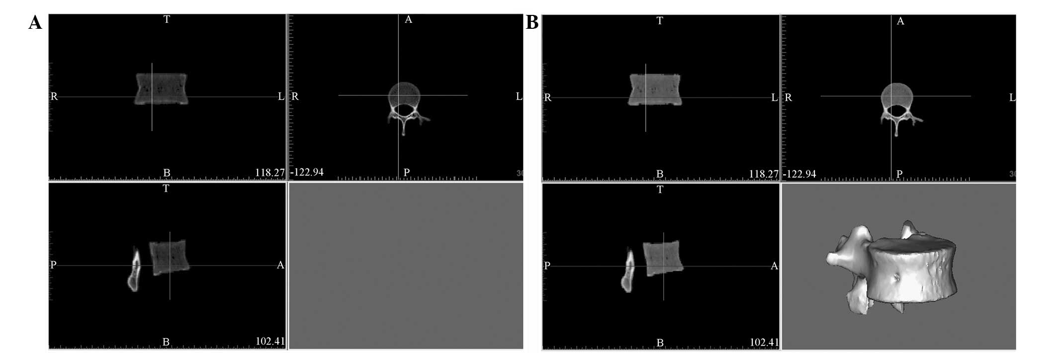
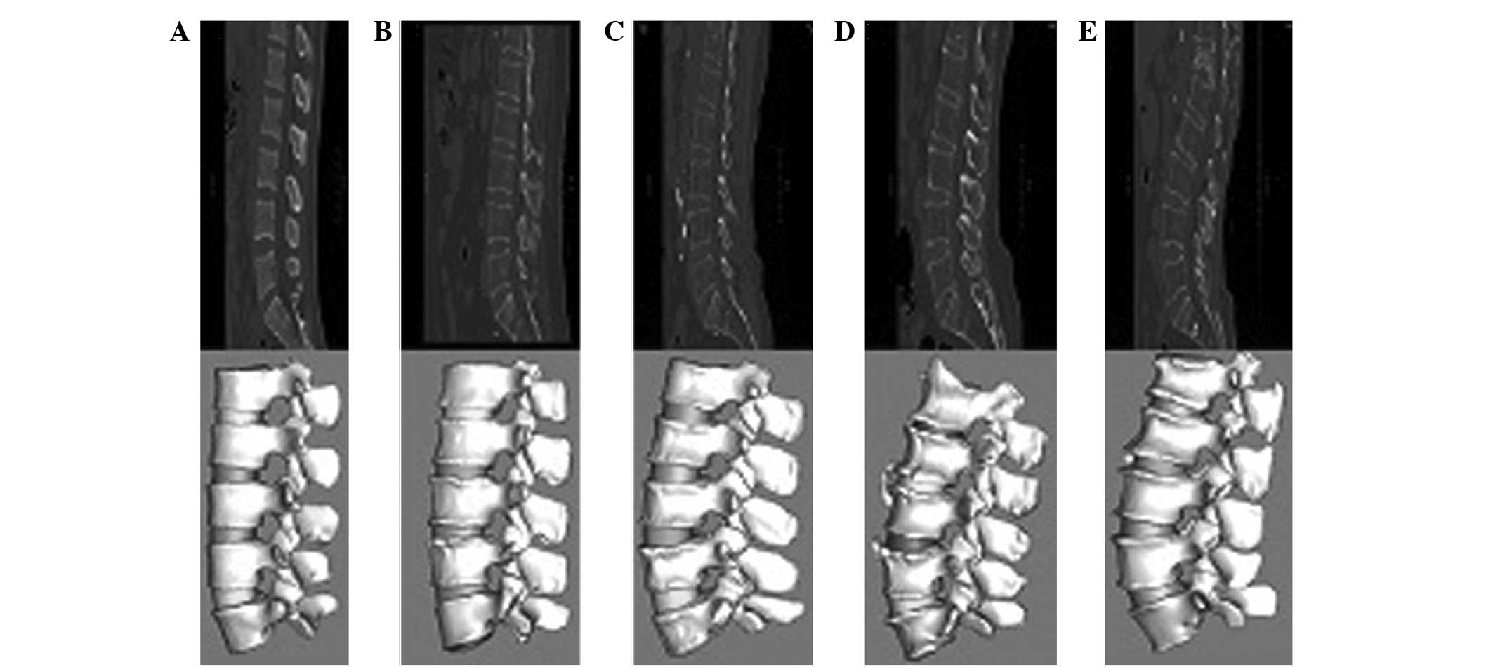
|
1
|
Turner MJ, Clough RW, Martin HC and Topp
LJ: Stiffness and deflection analysis of complex structures. J Aero
Sci. 23:805–823. 1956. View
Article : Google Scholar
|
|
2
|
Belytschko TB, Andriacchi TP, Schultz AB
and Galante JO: Analog studies of forces in the human spine:
computational techniques. J Biomech. 6:361–371. 1973. View Article : Google Scholar : PubMed/NCBI
|
|
3
|
Wang JL, Parnianpour M, Shirazi-Adl A and
Engin AE: Rate effect on sharing of passive lumbar motion segment
under load-controlled sagittal flexion: viscoelastic finite element
analysis. Theor Appl Fract Mec. 32:119–128. 1999. View Article : Google Scholar
|
|
4
|
El-Rich M, Arnoux PJ, Wagnac E, Brunet C
and Aubin CE: Finite element investigation of the loading rate
effect on the spinal load-sharing changes under impact conditions.
J Biomech. 42:1252–1262. 2009. View Article : Google Scholar : PubMed/NCBI
|
|
5
|
Hakim NS and King AI: A three dimensional
finite element dynamic response analysis of a vertebra with
experimental verification. J Biomech. 12:277–285. 1979. View Article : Google Scholar : PubMed/NCBI
|
|
6
|
Schmidt H, Heuer F, Simon U, et al:
Application of a new calibration method for a three-dimensional
finite element model of a human lumbar annulus fibrosus. Clin
Biomech (Bristol, Avon). 21:337–344. 2006. View Article : Google Scholar : PubMed/NCBI
|
|
7
|
Renner SM, Natarajan RN, Patwardhan AG, et
al: Novel model to analyze the effect of a large compressive
follower pre-load on range of motions in a lumbar spine. J Biomech.
40:1326–1332. 2007. View Article : Google Scholar : PubMed/NCBI
|
|
8
|
Rohlmann A, Bauer L, Zander T, Bergmann G
and Wilke HJ: Determination of trunk muscle forces for flexion and
extension by using a validated finite element model of the lumbar
spine and measured in vivo data. J Biomech. 39:981–989. 2006.
View Article : Google Scholar : PubMed/NCBI
|
|
9
|
Zander T, Rohlmann A, Burra NK and
Bergmann G: Effect of a posterior dynamic implant adjacent to a
rigid spinal fixator. Clin Biomech (Bristol, Avon). 21:767–774.
2006. View Article : Google Scholar : PubMed/NCBI
|
|
10
|
Zeng XL, Peng L and Bai J:
Three-dimensional finite element modeling and analysis of human
L3–L4 lumbar segment based on CT data. Beijing Biomed Eng.
26:266–269. 2007.(In Chinese).
|
|
11
|
Jiang HB: Static and dynamic mechanics
analysis on artificial hip joints with different interface designs
by the finite element method. J Bionic Eng. 4:123–131. 2007.
View Article : Google Scholar
|
|
12
|
Chevalier Y, Charlebois M, Pahra D, et al:
A patient-specific finite element methodology to predict damage
accumulation in vertebral bodies under axial compression, sagittal
flexion and combined loads. Comput Methods Biomech Biomed Engin.
11:477–487. 2008. View Article : Google Scholar
|
|
13
|
Kim TY, Kang KT, Yoon do H, et al: Effects
of lumbar arthrodesis on adjacent segments: differences between
surgical techniques. Spine (Phila Pa 1976). 37:1456–1462. 2012.
View Article : Google Scholar : PubMed/NCBI
|
|
14
|
Gong H, Wu W, Fang J, Dong X, Zhao M and
Guo T: Effects of materials of cementless femoral stem on the
functional adaptation of bone. J Bionic Eng. 9:66–74. 2012.
View Article : Google Scholar
|
|
15
|
Gu WY, Mao XG, Foster RJ, Weidenbaum M,
Mow VC and Rawlins BA: The anisotropic hydraulic permeability of
human lumbar anulus fibrosus. Influence of age, degeneration,
direction, and water content. Spine (Phila Pa 1976). 24:2449–2455.
1999. View Article : Google Scholar : PubMed/NCBI
|
|
16
|
Nadzadi ME, Pedersen DR, Callaghan JJ and
Brown TD: Effects of acetabular component orientation on
dislocation propensity for small-head-size total hip arthroplasty.
Clin Biomech (Bristol, Avon). 17:32–40. 2002. View Article : Google Scholar : PubMed/NCBI
|
|
17
|
Lu S, Xu YQ, Zhang YZ, et al: A novel
computer-assisted drill guide template for lumbar pedicle screw
placement: a cadaveric and clinical study. Int J Med Robot.
5:184–191. 2009. View
Article : Google Scholar : PubMed/NCBI
|
|
18
|
Xiao Z, Wang L, Gong H, et al:
Establishment and verification of a non-linear finite element model
for human L4–L5 lumbar segment. Biomedical Engineering and
Informatics (BMEI), 2010 3rd International Conference on IEEE.
3:1171–1175. 2010.
|
|
19
|
Francis A, Nareliya R and Kumar V:
Three-dimensional finite element analysis of human femur: A
comparative study, proceedings of all India seminar on biomedical
engineering 2012 (AISOBE 2012). Springer India. 37–48. 2013.
|
|
20
|
Sun W, Starly B, Darling A and Gomez C:
Computer-aided tissue engineering: application to biomimetic
modelling and design of tissue scaffolds. Biotechnol Appl Biochem.
39(Pt 1): 49–58. 2004. View Article : Google Scholar : PubMed/NCBI
|
|
21
|
Kuo CS, Hu HT, Lin RM, et al:
Biomechanical analysis of the lumbar spine on facet joint force and
intradiscal pressure-a finite element study. BMC Musculoskelet
Disord. 11:1512010. View Article : Google Scholar : PubMed/NCBI
|
|
22
|
Xiao Z, Wang L, Gong H and Zhu D:
Biomechanical evaluation of three surgical scenarios of posterior
lumbar interbody fusion by finite element analysis. Biomed Eng
Online. 11:312012. View Article : Google Scholar : PubMed/NCBI
|
|
23
|
Harrysson OL, Hosni YA and Nayfeh JF:
Custom-designed orthopedic implants evaluated using finite element
analysis of patient-specific computed tomography data:
femoral-component case study. BMC Musculoskelet Disord. 8:912007.
View Article : Google Scholar
|
|
24
|
Niemeyer F, Wilke HJ and Schmidt H:
Geometry strongly influences the response of numerical models of
the lumbar spine - a probabilistic finite element analysis. J
Biomech. 45:1414–1423. 2012. View Article : Google Scholar : PubMed/NCBI
|