Introduction
In recent years, the incidence of colon cancer has
shown a gradual upward trend in China, becoming the third most
common tumor type endangering human health (1,2).
Liver metastasis from colon cancer is extremely common and is the
main reason for clinical treatment failure, which has subsequently
impacted long-term survival and prognosis (3–5). As
a result, the prevention and treatment of colon cancer with liver
metastasis is a clinically important topic that requires further
investigation. At present, surgical resection is the preferred
method of treatment for colon cancer with liver metastasis, while
other conventional treatment methods include chemotherapy,
radiation, radio frequency therapy, freezing, microwaving or using
a laser, percutaneous ethanol injection and radioactive seed
implantation. However, gene therapy and immunotherapy techniques
performed in recent years have become highly promising strategies
for colon cancer treatment (6,7). The
establishment of animal models of colon cancer with radical
resection of liver metastasis has improved the investigations into
the mechanisms underlying liver metastasis in human colon cancer;
thus, improving the development of novel, effective liver
metastasis prevention and treatment programs. Previously, an SW480
nude mouse subcutaneous transplanted tumor model was established
and the wild type (WT) p53 gene was combined with thymidine
kinase/ganciclovir (TK/GCV) and cytosine deaminase/5-fluorocytosine
(CD/5-FC) systems, which were shown to significantly inhibit the
growth of subcutaneous transplanted tumors and prolong the survival
of mice (8). However, whether this
method can affect liver metastasis in colon cancer remains unclear.
On the basis of the aforementioned evidence, the present study
successfully established an SW480 nude mouse liver metastasis model
and further observed the role of combined gene therapy in the
prevention and treatment of liver metastatic tumors. The aim of the
present study was to provide a novel approach to clinically prevent
colon cancer radical resection of liver metastasis and treat liver
metastatic tumors of colon cancer.
Materials and methods
Collection and preparation of the cell
suspension
A human colon cancer cell line, SW480, was purchased
from Shanghai Institute of Biological Cells (Shanghai, China).
These cells have point mutations in the eighth and ninth exons of
the p53 gene. SW480/p53 and SW480/TK-CD were built in an in
vitro experiment and were shown to stably transfect p53 and TK
and CD double suicide genes, respectively (8). Human colon cancer SW480 cells,
SW480/p53 cells and SW480/TK-CD cells were cultured and passaged in
RPMI-1640 medium (Hyclone, Logan, UT, USA) containing newborn calf
serum (Hyclone) and incubated at 37°C constant temperature to
obtain a sufficient number of cells. Next, the well-grown cells of
~80% confluence (Hyclone, Logan, UT, USA) were collected to
establish a cell suspension at a concentration of 5×10⁷,
which were then inoculated to the spleen on an ice bath.
Grouping
A total of 32 BALB/c female nude mice (age, 4–6
weeks; weight, 14–20 g) were purchased from the Experimental Animal
Center of Shandong University (Jinan, China) and reared under
specific-pathogen free conditions. The study was conducted in
strict accordance with the recommendations in the Guide for the
Care and Use of Laboratory Animals of the National Institutes of
Health, and the animal use protocol was reviewed and approved by
the Institutional Animal Care and Use Committee of the Affiliated
Hospital of Shandong Academy of Medical Science (Jinan, China). A
total of 32 nude mice were randomly divided into four groups (n=8
per group). Group 1 mice received splenic injections of SW480 cells
(control group), while group 2 mice were injected with SW480/p53
cells in the spleen. Group 3 mice were administered splenic
injections of SW480/TK-CD cells, and GCV (Roche, Basel,
Switzerland) and 5-FC were injected into the abdominal cavity.
Finally, group 4 mice received splenic injections of SW480/p53
cells mixed in equal proportion with SW480/TK-CD cells, as well as
GCV and 5-FC injections (Sigma, San Francisco, CA, USA) in the
abdominal cavity (9).
Establishment of a liver metastasis
animal model with colon cancer
An injection method was used to establish the model,
which firstly involved weighing the nude mice and injecting 45
mg/kg sodium pentobarbital (1%) into the abdominal cavity for
anesthesia. Following forced entry, the skin in the surgical field
was disinfected and a 0.5–1.0-cm back left oblique incision was
made (below the junction of the left armpit bottom line and costal
margin) into the abdomen to expose the spleen, which was then
raised out of the abdominal cavity gently. The SW480, SW480/p53 and
SW480/TK-CD colon cancer cells were then slowing injected,
according to the group, into the spleen of the nude mice using a
fifth mode of a needle. Each nude mouse was injected with 0.2 ml
cell suspension (1×10⁷/per) for at least 3 min,
following which the spleen capsule was found to swell and grow
white. Following the injection, it was a requirement that the
needle eye was oppressed with a 75% alcohol swab for 2 min to stop
the bleeding and kill the cancer cells, which may exosmose in order
to prevent metastasis in the abdominal cavity. Next, the spleen was
placed in the original position and the abdomen was closed. The
principles of aseptic surgery were followed during the whole
process.
Suicide gene prodrug therapy
One day after the spleen inoculation, normal saline
(the same volume as suicide gene prodrugs) was injected into the
abdominal cavity of the mice in groups 1 and 2, while the suicide
gene prodrugs, GCV and 5-FC, were injected into the abdominal
cavity of the mice in groups 3 and 4 for ten consecutive days; the
injection doses were 100 mg/(kg/d) GCV and 500 mg/(kg/d) 5-FC.
Efficacy observations
Nude mice in each group were autopsied six weeks
following surgery to observe whether metastasis of the liver and
other areas had occurred. The livers were removed and fixed with
10% formalin solution for one night. Next, the number of metastatic
tumors (metastatic nodules on the liver surface that were visually
counted), liver metastasis rate, conventional pathology, electron
microscopy and other indicators were monitored.
Statistical analysis
SPSS 16.0 (SPSS, Inc., Chicago, IL, USA) software
was used for statistical analysis. The χ2-test and
Student’s t-test were used to compare the liver metastasis rate and
average number of liver metastatic tumors, respectively. P<0.05
was considered to indicate a statistically significant
difference.
Results
Metastasis cases of the liver and other
areas in each group
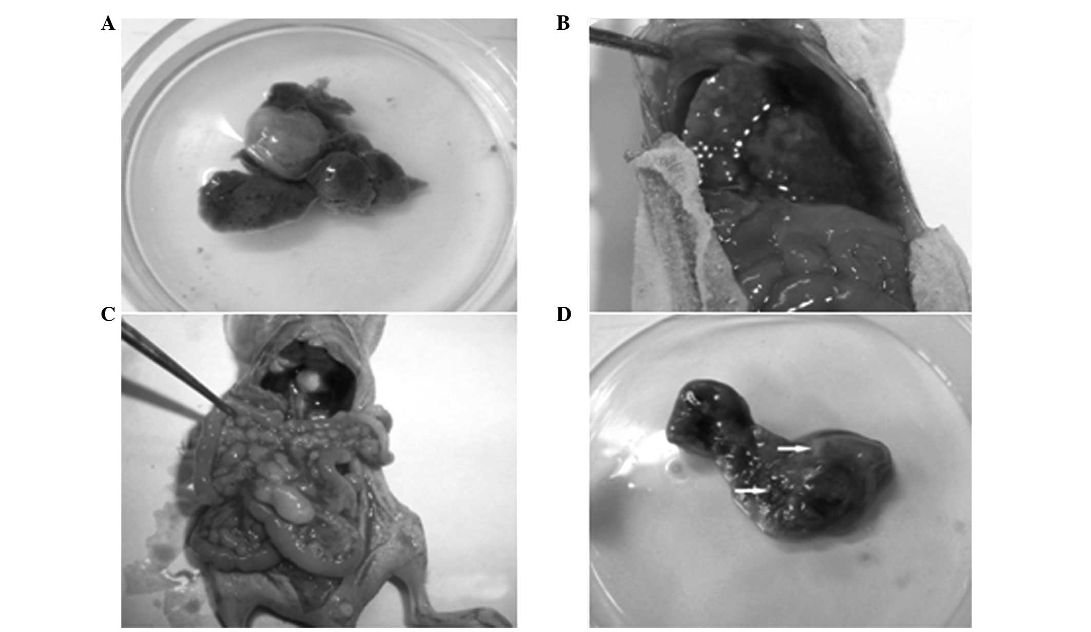
Grey nodules of varying numbers were present on the
liver surface of nude mice with liver metastasis; as shown in
Fig. 1, a large isolating nodule
with a maximum diameter of 3 cm (Fig.
1A) and multiple miliary nodules (Fig. 1B) were observed. The liver volume
decreased and the texture was brittle and hard, with ulceration
observed in areas with large nodules. A normal liver is usually
bright red with a soft texture.
The liver metastasis rate and metastatic tumor
numbers of each group are shown in Table I. Comparisons between each
treatment group and the control for the average number of liver
metastatic tumors were statistically significant (P<0.01). With
regard to the liver metastasis rate, no statistically significant
difference was observed when comparing groups 2 and 1 (P>0.05).
However, when comparing groups 3 and 4 with group 1, statistically
significant differences were observed (P=0.013 and P=0.001,
respectively), indicating that combined gene therapy significantly
reduced the number of liver metastatic tumors from colon cancer and
reduced the incidence of liver metastasis.
 | Table IEffect of combined gene therapy on the
average number of liver metastatic tumors and the liver metastasis
rate. |
Table I
Effect of combined gene therapy on the
average number of liver metastatic tumors and the liver metastasis
rate.
| Group (n=8) | Liver metastatic
tumors, n | Liver metastasis
rate, % |
|---|
| 1 | 12.13±6.20 | 100 |
| 2 | 3.13±2.23 | 87.5 |
| 3 | 0.50±0.76 | 37.5 |
| 4 | 0.13±0.35 | 12.5 |
With regard to metastasis to other organs, one or
more cases were observed in the lymph nodes, diaphragm and
abdominal cavity in the nude mice of the control group (Fig. 1C); however, metastasis was not
observed in the heart, lung, brain or kidney. Metastasis was not
present in other organs of the nude mice in the treatment groups.
The mice with liver metastasis all had in situ implanted
tumor formation in the spleen (Fig.
1D; all metastasis cases were confirmed by pathological
examination from the same adenocarcinoma).
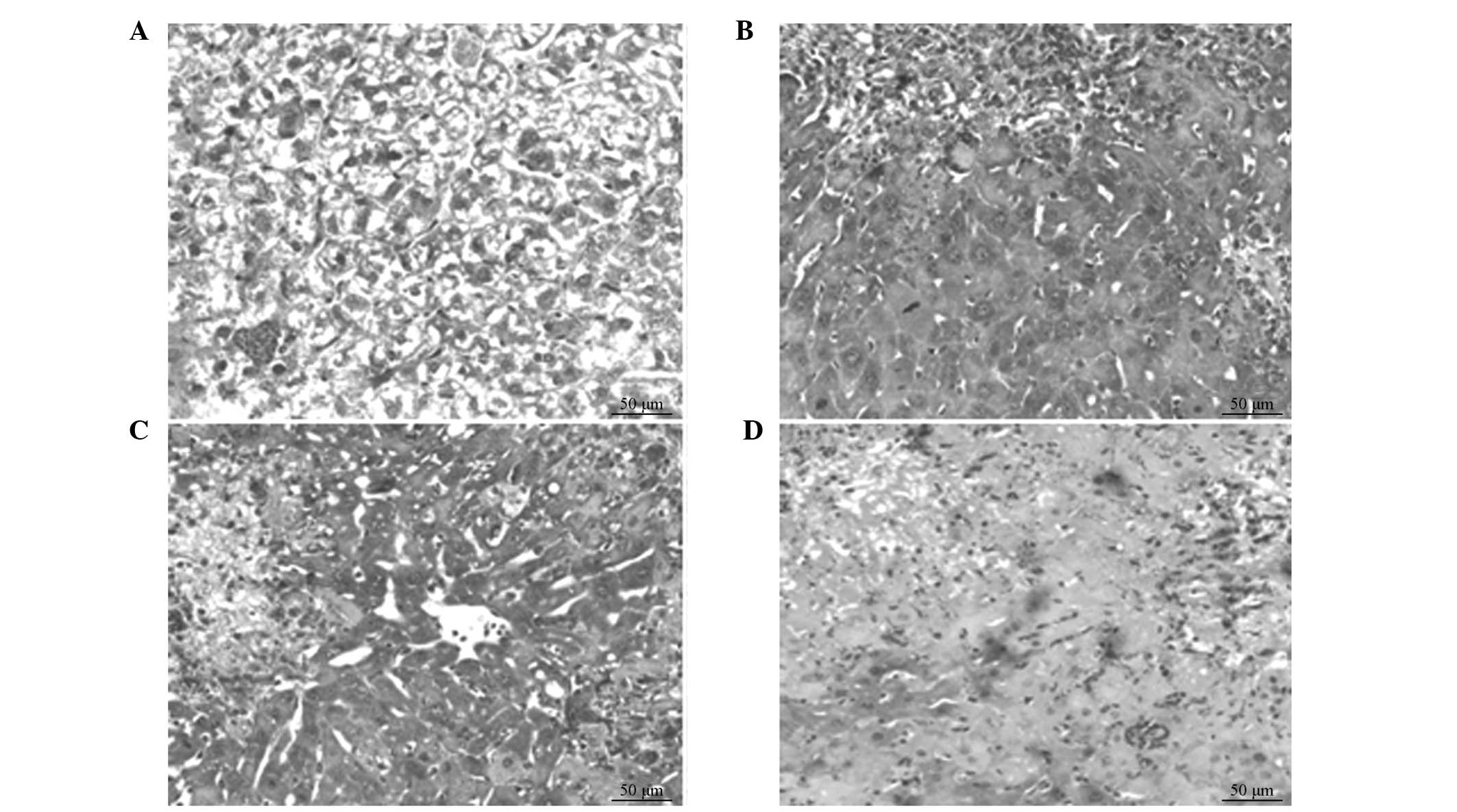
Light microscopy observations
Tumor cell growth in the control group was active
and a lobular structure remained distinguishable. The tumor cells
were significantly heteromorphic, with a round nucleus that was
placed in the middle of the cell. The nuclei were large and deeply
stained, and nucleus division increased while the amount of
cytoplasm was lower. Fibrosis and inflammatory cell infiltration
were not observed, with apoptosis present only in small areas
(Fig. 2A). Necrotic tissue was
present in the treatment groups and apoptotic cells were observed,
with cell shrinkage and cytoplasm and nuclear condensation.
Apoptotic bodies were observed following nuclear fragmentation and
dissolution. In groups 2 and 3, the tumor tissues of the nude mice
exhibited a small area of tumor necrosis (Fig. 2B and C); however, necrosis was
observed in the majority of the regions in group 4. Tumor cell
structures were unclear and inflammatory cells were observed in the
necrotic tissues (Fig. 2D).
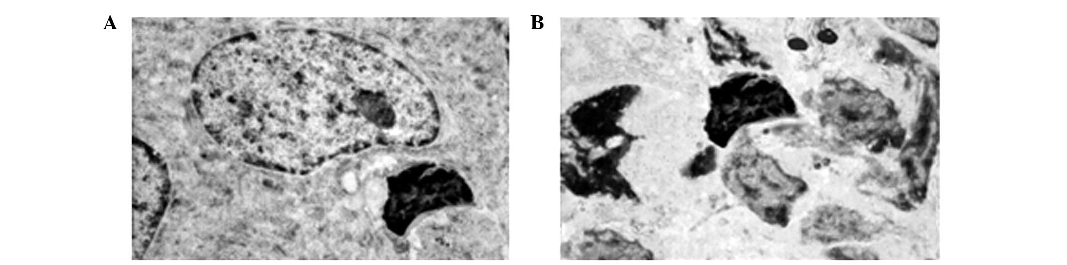
Electron microscopy observations
As demonstrated in Fig.
3A, tumor cells in the control group were irregular with a
large volume and large nucleus. Nucleoli were prominent, nucleus
division was easy to observe and there were a high number of
organelles in the cytoplasm. A large number of tumor cells in the
treatment groups were soma-condensed, with cytoplasmic
concentration, ribosomes and mitochondria aggregation, nuclear
condensation and nuclear fragmentation. The complete pericellular
membrane of the apoptotic bodies was also observed (Fig. 3B).
Discussion
Spleen planting and cecal wall planting methods are
most commonly used to establish an experimental liver metastasis
model with colon cancer. Other routes are possible, including
implantation within the portal vein, ileocolic vein and liver,
however, these are rarely used due to operational difficulties
(10,11). When using the cecal wall planting
method, the range of tumor metastases is wide, with a relatively
low incidence and slow development of liver metastasis. With regard
to the spleen planting method, the incidence of liver metastasis
may be as high as 100%, extrahepatic metastasis rarely occurs and
the development of liver metastasis is rapid (12,13).
The establishment of this model aims to study liver metastasis of
colon cancer; thus, the spleen injection route was selected in the
present study. Although concurrent spleen in situ tumors
were present, the spleen immune function and the inherent host
antitumor immunity were retained, and were more consistent with the
complex environment of tumor occurrence and development in the
human body.
The ability to maintain the original biological
characteristics of tumor cells is key to animal model
establishment. The present study simulated the process of cancer
cells backflowing into the portal vein and liver, which resulted in
hematogenous dissemination and liver metastasis following the
resection of colon cancer; thus, an SW480 nude mouse liver
metastasis model was successfully constructed. This model did not
affect the invasion and metastasis biological characteristics of
human colon cancer cells. Pathological sections in the control
group demonstrated complete consistency with regard to the
histological structure of the liver metastatic tumors with human
colon adenocarcinoma.
In the present study, a number of elements of the
model establishment were noted for future reference. Firstly, the
biological characteristics of the original tumor cells in
vitro cultivation and preparation of suspensions should be
maintained. Following enzyme digestion and treatment, certain
components in the surface of tumor cells changed, which may affect
the growth and metastasis potential of tumor cells. Therefore, the
enzyme digestion time should be well controlled, the cells should
be gently pipetted and the centrifugation speed should not exceed
1,200 g. Secondly, an incision, 0.5–1.0 cm in length, should be cut
in the middle back to minimize the trauma and reveal the spleen
under easy and secure surgery. Thirdly, during the injection, the
under part of the spleen should be pulled from the incision and the
needle should be inserted horizontally along the spleen axis with a
depth of ≥0.5 cm in order to prevent the spleen from being easily
punctured and suspensions from overflowing from the injection site.
Fourthly, the number of injection cells should be large enough to
reach 1×10⁷ in each mouse, as the implanted tumor cells
must escape attack from numerous natural killer cells and activated
phagocytic cells, which secrete tumor necrosis factors, in order to
cause vascular invasion and liver metastasis. Finally, the
injection time should be at least 3 min in order to avoid splenic
capsule rupture due to excessive tension.
Currently, combined gene therapy is emerging as a
popular tumor therapeutic strategy. p53 gene mutations are the most
common genetic alterations in colon cancer (14), and tumor cells with p53 mutations
can compete with the killing effect of suicide gene transformation
prodrugs on tumor cells; thus, affecting the therapeutic effect of
the suicide gene (15–18). In a number of suicide gene systems,
studies on the TK and CD genes have been highly detailed, with
definite effects observed. The TK and CD double suicide gene
alliance has been demonstrated to be more efficient, more
broad-spectrum, more secure, less simple to produce drug resistance
and able to reduce the doses of prodrugs (18–20).
The most important feature of suicide gene therapy is the bystander
effect, which overcomes the disadvantages of low gene transduction
and significantly expands the killing effect of suicide genes.
Numerous studies have demonstrated that double suicide gene
systems, as compared with a single suicide gene, may produce a more
powerful bystander effect (21–23).
In addition, the bystander effect may expand the therapeutic effect
of the WTp53 gene in tumors into adjacent cells and consequently
produce an enhanced tumor suppressive effect (24,25).
The application of gene therapy firstly demands the
transfected gene to exhibit certain expression efficiency. In
addition, the spliced gene should be limited among the target cells
in order to maximize the killing of tumor cells without damaging
normal cells. Therefore, improving the targeted gene transfer
techniques and the efficiency of gene targeting transfection is key
to current gene therapy. In the present study, prebuilt SW480/p53
and SW480/TK-CD cells containing the target genes were transplanted
into the spleen to produce liver metastasis, and the problem of
gene targeting transduction was successfully resolved (25). Following the transplantation of
cancer cells into the spleen, the prodrugs, GCV and 5-FC, were
timely administered on the portal vein system into the abdominal
cavity. This administration route allowed the abdominal cavity,
portal vein and liver to be subjected to a constant, high
concentration of the drugs. The selected route and timings of the
aforementioned genes and prodrugs were the most efficient
combination to produce a more direct, targeted and specific gene
therapy effect. The introduction of the WTp53 gene not only
incorporates its normal tumor suppressor function to compete with
the mutated p53, but also enhances the performance of the double
suicide genes and demonstrates a more powerful bystander effect.
The results demonstrated that the combination of the WTp53 gene
with the TK/GCV and CD/5-FC systems exhibited a synergistic effect.
In addition, compared with single gene application, this therapy
markedly reduced the incidence of colon cancer liver metastasis and
the number of liver metastatic tumors. Therefore, the present study
has provided a novel approach for the prevention of liver
metastasis following radical resection of colon cancer and for the
treatment of colon cancer liver metastatic tumors.
The role of combined gene therapy is not a simple
composition, but an organic combination, the complementary and
synergistic effects of which may markedly improve the tumoricidal
effect (26). However, in
vivo animal experiments differ from humans. In reality, it is
not possible to transfect the target genes into tumor cells and
then transform the tumor cells containing the target genes into
tumors, as has been performed in the present study. Thus, the
target genes are unable to be transfected into each tumor cell.
Therefore, future studies are required to design efficiently
targeted gene delivery vectors that are able to achieve safe and
controllable gene expression with more gene combinations.
Acknowledgements
The study was supported by a grant from the Shandong
Academy of Medical Science Foundation, Shandong, China (no.
200944).
References
|
1
|
Kruse J, von Bernstorff W, Evert K, et al:
Macrophages promote tumour growth and liver metastasis in an
orthotopic syngeneic mouse model of colon cancer. Int J Colorectal
Dis. 28:1337–1349. 2013. View Article : Google Scholar : PubMed/NCBI
|
|
2
|
Schiesser M, Chen JW, Maddern GJ and
Padbury RT: Perioperative morbidity affects long-term survival in
patients following liver resection for colorectal metastases. J
Gastrointest Surg. 12:1054–1060. 2008. View Article : Google Scholar : PubMed/NCBI
|
|
3
|
Huang X, Zou Y, Lian L, et al: Changes of
T cells and cytokines TGF-β1 and IL-10 in mice during liver
metastasis of colon carcinoma: implications for liver anti-tumor
immunity. J Gastrointestinal Surg. 17:1283–1291. 2013.
|
|
4
|
Min BS, Kim NK, Jeong HC and Chung HC:
High levels of serum VEGF and TIMP-1 are correlated with colon
cancer liver metastasis and intrahepatic recurrence after liver
resection. Oncology Lett. 4:123–130. 2012.PubMed/NCBI
|
|
5
|
Ceauşu RA, Cîmpean AM, Gaje P, Gurzu S,
Jung I and Raica M: CD105/Ki67 double immunostaining expression in
liver metastasis from colon carcinoma. Rom J Morphol Embryol.
52:613–616. 2011.PubMed/NCBI
|
|
6
|
Yoshida D, Ikeda Y, Waki K, Shirabe K,
Kakeji Y, Tsujitani S and Maehara Y: Different incidence of
synchronous liver metastasis between proximal and distal colon
cancer. Surg Today. 42:426–430. 2012. View Article : Google Scholar : PubMed/NCBI
|
|
7
|
Rees M, Tekkis PP, Welsh FK, O’Rourke T
and John TG: Evaluation of long-term survival after hepatic
resection for metastatic colorectal cancer: A multifactorial model
of 929 patients. Ann Surg. 247:125–135. 2008. View Article : Google Scholar : PubMed/NCBI
|
|
8
|
Zhang SX, Zhang SL and Lan XP: The
establishment of a subcutaneously transplanted tumor model with
human colon cancer cell line SW480 in nude mice. Military Medical
Journal of Southeast China. 14:108–110. 2012.(In Chinese).
|
|
9
|
Niu HX, He QS, Leng YD and Liu YZ: The
establishment of the model of hepatic metastasis with human colon
cancer cell line SW480 in nude mice. Chin J Curr Adv Gen Surg.
10:540–542. 2007.(In Chinese).
|
|
10
|
Metildi CA, Kaushal S, Snyder CS, Hoffman
RM and Bouvet M: Fluorescence-guided surgery of human colon cancer
increases complete resection resulting in cures in an orthotopic
nude mouse model. J Surg Res. 179:87–93. 2012. View Article : Google Scholar
|
|
11
|
Schoffelen R, van der Graaf WT, Sharkey
RM, et al: Quantitative immuno-SPECT monitoring of pretargeted
radioimmunotherapy with a bispecific antibody in an intraperitoneal
nude mouse model of human colon cancer. J Nucl Med. 53:1926–1932.
2012. View Article : Google Scholar
|
|
12
|
Rada T, Carvalho PP, Santos TC, Castro AG,
Reis RL and Gomes ME: Chondrogenic potential of two hASCs
subpopulations loaded onto gellan gum hydrogel evaluated in a nude
mice model. Curr Stem Cell Res Ther. 8:357–364. 2013. View Article : Google Scholar : PubMed/NCBI
|
|
13
|
Yang L, Zhou J, Ma Q, et al: Knockdown of
PPARδ gene promotes the growth of colon cancer and reduces the
sensitivity to bevacizumab in nude mice model. PloS One.
8:e607152013.
|
|
14
|
Okal A, Mossalam M, Matissek KJ, Dixon AS,
Moos PJ and Lim CS: A chimeric p53 evades mutant p53 transdominant
inhibition in cancer cells. Mol Pharm. 10:3922–3933.
2013.PubMed/NCBI
|
|
15
|
Khoury MP and Bourdon JC: The isoform of
the p53 protein. Cold Spring Harb Perspect Biol. 2:a0009272010.
View Article : Google Scholar : PubMed/NCBI
|
|
16
|
Ladelfa MF, Peche LY, Toledo MF, Laiseca
JE, Schneider C and Martín M: Tumor-specific MAGE proteins as
regulators of p53 function. Cancer Lett. 325:11–17. 2012.
View Article : Google Scholar : PubMed/NCBI
|
|
17
|
Menendez D, Nguyen TA, Freudenberg JM,
Mathew VJ, Anderson CW, Jothi R and Resnick MA: Diverse stresses
dramatically alter genome-wide p53 binding and transactivation
landscape in human cancer cells. Nucleic Acids Res. 41:7286–7301.
2013. View Article : Google Scholar : PubMed/NCBI
|
|
18
|
Liu X, Wilcken R, Joerger AC, Chuckowree
IS, Amin J, Spencer J and Fersht AR: Small molecule induced
reactivation of mutant p53 in cancer cells. Nucleic Acids Res.
41:6034–6044. 2013. View Article : Google Scholar : PubMed/NCBI
|
|
19
|
Dong XY, Wang WQ, Zhao Y, et al:
Antibody-directed double suicide gene therapy targeting of
MUC1-positive leukemia cells in vitro and in vivo. Curr Gene Ther.
13:346–357. 2013. View Article : Google Scholar : PubMed/NCBI
|
|
20
|
Chen Y, Wang G, Kong D, et al:
Double-targeted and double-enhanced suicide gene therapy mediated
by generation 5 polyamidoamine dendrimers for prostate cancer. Mol
Carcinog. 52:237–246. 2011. View
Article : Google Scholar : PubMed/NCBI
|
|
21
|
Li LQ, Shen F, Xu XY, Zhang H, Yang XF and
Liu WG: Gene therapy with HSV1-sr39TK/GCV exhibits a stronger
therapeutic efficacy than HSV1-TK/GCV in rat C6 glioma cells.
ScientificWorldJournal. 2013:9513432013.PubMed/NCBI
|
|
22
|
Lu Z, Zhang TY, Han MM, et al: Antitumor
activity of the recombinant rClone30-CD/5-FC system. Yao Xue Xue
Bao. 48:261–268. 2013.(In Chinese).
|
|
23
|
Chai LP, Wang ZF, Liang WY, Chen L, Chen
D, Wang AX and Zhang ZQ: In vitro and in vivo effect of 5-FC
combined gene therapy with TNF-α and CD suicide gene on human
laryngeal carcinoma cell line Hep-2. PloS One. 8:e611362013.
|
|
24
|
Torimura T, Ueno T, Taniguchi E, et al:
Interaction of endothelial progenitor cells expressing cytosine
deaminase in tumor tissues and 5-fluorocytosine administration
suppresses growth of 5-fluorouracil-sensitive liver cancer in mice.
Cancer Sci. 103:542–548. 2011. View Article : Google Scholar
|
|
25
|
Niu HX, Du T, Xu ZF, Zhang XK and Wang RG:
Role of wild type p53 and double suicide genes in interventional
therapy of liver cancer in rabbits. Acta Cir Bras. 27:522–528.
2012. View Article : Google Scholar : PubMed/NCBI
|
|
26
|
Pasquale EB: Eph receptors and ephrins in
cancer: bidirectional signaling and beyond. Nat Rev Cancer.
10:165–180. 2010. View
Article : Google Scholar : PubMed/NCBI
|