Introduction
Despite rapid advances in the understanding of
ovarian carcinogenesis, ovarian cancer remains the most common
cause of mortality from gynecological malignancies and the fifth
most common cause of cancer mortalities in females in the United
States (1,2). Since mouse models of ovarian cancer
can mimic the clinical processes of human ovarian cancer, several
models have been developed during the past decades to promote in
vivo research on human ovarian cancer (3–6).
However, a major limitation of these models is the lack of a human
microenvironment and an inaccurate replication of the interactions
between ovarian cancer cells and the human ovarian microenvironment
(7,8), which is now known to play a crucial
role in the regulation of tumor growth and metastasis (9,10).
To precisely mimic the clinical processes of human
cancer, an increasing number of humanized factors have been
gradually added to these mouse models. The development of
‘humanized’ mice is now an important research tool for the in
vivo study of human biological processes (11). Since the first study reported that
severe combined immunodeficient (SCID) mice were able to be
successfully engrafted with human tissues, normal and neoplastic
human tissues have been successfully engrafted into SCID mice
(12). The implantation of human
tissue xenografts in immunodeficient mice has provided insight into
the biology of human cancer, autoimmunity and infectious diseases
(11). Proia and Kuperwasser
(13) reproducibly established
functionally normal breast tissue in mice by implanting reduction
mammoplasty tissue samples in an orthotopic xenograft model. The
endogenous mouse epithelium was cleared, and comixed human
epithelial and stromal cells were implanted to construct a
humanized mouse model. However, whether this model may be used to
understand normal human breast development or tumorigenesis remains
unknown. Bankert et al (14) described a humanized mouse model of
ovarian cancer that recapitulated the solid tumor progression,
ascites formation and metastasis observed in patients. In this
model, the tumor and tumor stroma were successfully engrafted into
the peritoneal cavity of SCID mice. This model may be used to
determine how fibroblasts and lymphocytes within the tumor
microenvironment contribute to tumor growth and metastasis.
However, this humanized mouse model focused solely upon the
engraftment of ovarian cancer tissues, not cancer cells grown in a
human microenvironment.
In previous studies, orthotopic implantation of
human breast tissues in mice was demonstrated to result in a novel
mouse model with a human mammary microenvironment (15), while the implantation of human
gastric tissues in mice resulted in a novel mouse model with a
human gastric microenvironment (16). To the best of our knowledge, there
have been no studies investigating the implantation of normal human
ovarian tissue in a SCID mouse model. Therefore, the aim of the
present study was to develop a novel protocol for the establishment
of human ovarian stroma within a mouse model subcutaneously. It was
hypothesized that the resulting stroma may serve as a useful
preclinical tool that may allow the progression of human ovarian
cancer to be investigated in a humanized ovarian
microenvironment.
Materials and methods
Animals and materials
A total of 28 female SCID mice (age, 5–6 weeks;
C.B-17IcrCrl-scid-bgBR) were purchased from the Model Animal
Research Center of Nanjing University (MARC, Nanjing, China). The
mice were housed under specific pathogen-free,
temperature-controlled conditions. Their cages, bedding and
drinking water were autoclaved and changed regularly. Food was
sterilized by irradiation. The mice were maintained in a daily
cycle of alternating 12-h periods of light and darkness. All
experimental procedures were conducted according to the Guide for
the Care and Use of Laboratory Animals and were approved by the
Animal Care and Use Committee of Nanjing Medical University
(Nanjing, China).
Normal, human, noncancerous ovarian tissues were
obtained from patients that had undergone a total hysterectomy with
prophylactic oophorectomy at the First Affiliated Hospital of
Nanjing Medical University. The use of human samples in the study
was approved by the Committee for Ethical Review of Research at
Nanjing Medical University, according to the ethical guidelines of
the Declaration of Helsinki and each patient provided informed
consent to participate in the study.
Implantation of human ovarian tissues in
mice
Under sterile conditions, normal human ovarian
tissues were sliced into small sections (~4×4×4 mm in size). Three
small sections of normal human ovarian tissue were randomly
selected for subsequent histological examination; the other pieces
were stored in phosphate-buffered saline at 4°C until implantation
into the mice.
Prior to implantation, the mice were anesthetized
with an intraperitoneal injection of 1% pentobarbital sodium (10
μl/g body weight; Sigma-Aldrich, Steinheim, Germany). The surgical
procedure was performed as previously described, with certain
modifications (9,10). In brief, 5–6 mm incisions were made
using a scalpel on the skin of the left mid-dorsal flank, through
which four or five small sections of human ovarian tissue were
subcutaneously implanted. The procedure was finished within 6 h
following the prophylactic oophorectomy. All the mice received
gentamicin in the drinking water (800,000 U/l) for up to one week
following implantation. A total of 14 SCID mice were implanted with
xenografts from one individual patient, while an additional 14 SCID
mice were implanted with xenografts from two patients in this phase
of the study where the xenografts come from three human
patients.
Gross observation and specimen
collection
Following implantation, the ovarian xenografts were
subjected to weekly gross examinations. Mice were sacrificed at
one, two or four weeks following surgery, and the human xenografts,
including the mouse tissue surrounding the implanted human ovarian
tissues, were harvested for histological assessment and
immunohistochemical analysis. The harvested specimens were fixed in
10% formalin for examination.
Histological and immunohistochemical
examination
Specimens were dehydrated and embedded in paraffin.
Sections (4-μm thick) were stained with hematoxylin and eosin and
examined under a microscope. For immunohistochemical staining, the
specimens were deparaffinized and rehydrated using xylene and
graded alcohol. Sections were mounted on slides, subjected to
antigen retrieval and then incubated with rabbit monoclonal
antibodies against human estrogen receptor (ER), progesterone
receptor (PR) and CD34 (Maxim Biotech, San Francisco, CA, USA), as
well as human cytokeratin (CK)-7, CK-20 and α-smooth muscle actin
(CK-7, CK-20, α-SMA; LabVision, Kalamazoo, MI, USA).
Immunocomplexes were visualized using the diaminobenzidine method
and sections were counterstained with hematoxylin. Negative
controls were serial sections processed without the primary
antibody. These reagents exhibited no cross-reactivity between the
relevant species (mouse and human). All histological examinations
were performed by one experienced pathologist.
Results
Acceptance of xenografts in SCID
mice
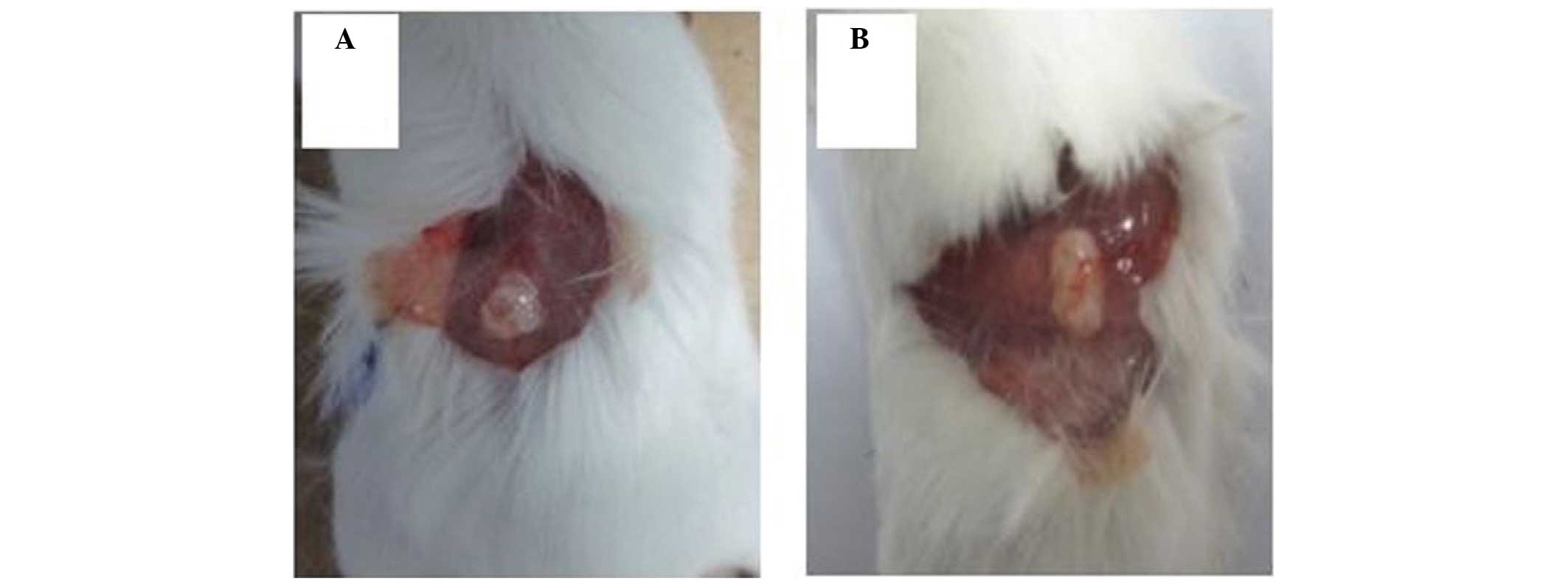
From one week following the implantation, the
ovarian xenografts were subjected to weekly gross examinations. The
embedded human ovarian tissues survived in the SCID mouse hosts for
up to six weeks. No infection or tissue rejection reactions were
observed at the time of tissue harvest, and all the implants became
vascularized and survived well on the sheath of mouse muscles in
the subcutaneous tissue (Fig. 1A and
B).
Histological examination
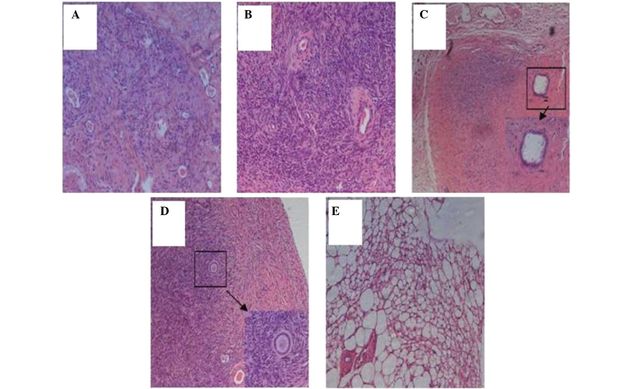
Histological evaluation revealed that the morphology
of the human ovarian tissues embedded in the SCID mice was similar
to the morphology of the original tissues prior to implantation.
The viability of the cells within the xenografts was confirmed by
the intact nature of the cell membranes, normal granulation of the
cytoplasm and normal size and staining characteristics of the
nucleus (Fig. 2A and B). In a
number of the engrafted ovarian tissues, which were derived from a
perimenopausal female, the follicular architecture of the original
tissues was retained (Fig. 2C and
D). However, ovarian tissues embedded in the nude mice in the
preliminary experiments were unable to survive, and histological
evaluation indicated that the specific morphology of the ovarian
tissues was not retained in the implanted tissues (Fig. 2E).
Immunohistochemical analysis
Immunohistochemical analysis demonstrated that the
harvested specimens were positive for ER and partially positive for
PR, indicating that the inoculated ovarian tissues had remained
estrogen- and progesterone-dependent, similar to the original
tissues (Fig. 3A and B). Positive
staining was also observed for anti-human CK-7, indicating that the
majority of the ovarian epithelium in the original and transplanted
tissues was derived from the human ovarian tissues (Fig. 3C and D). Positive staining for
anti-human CD34 indicated that the implanted ovarian tissues were
well vascularized and remained viable (Fig. 3E). In addition, α-SMA, which is
often expressed in cancer-associated fibroblasts, was only detected
in the vascular pericytes and not in the ovarian epithelium or
ovarian stroma, indicating that the transplanted tissues were
normal ovarian tissues similar to the original tissue (Fig. 3F).
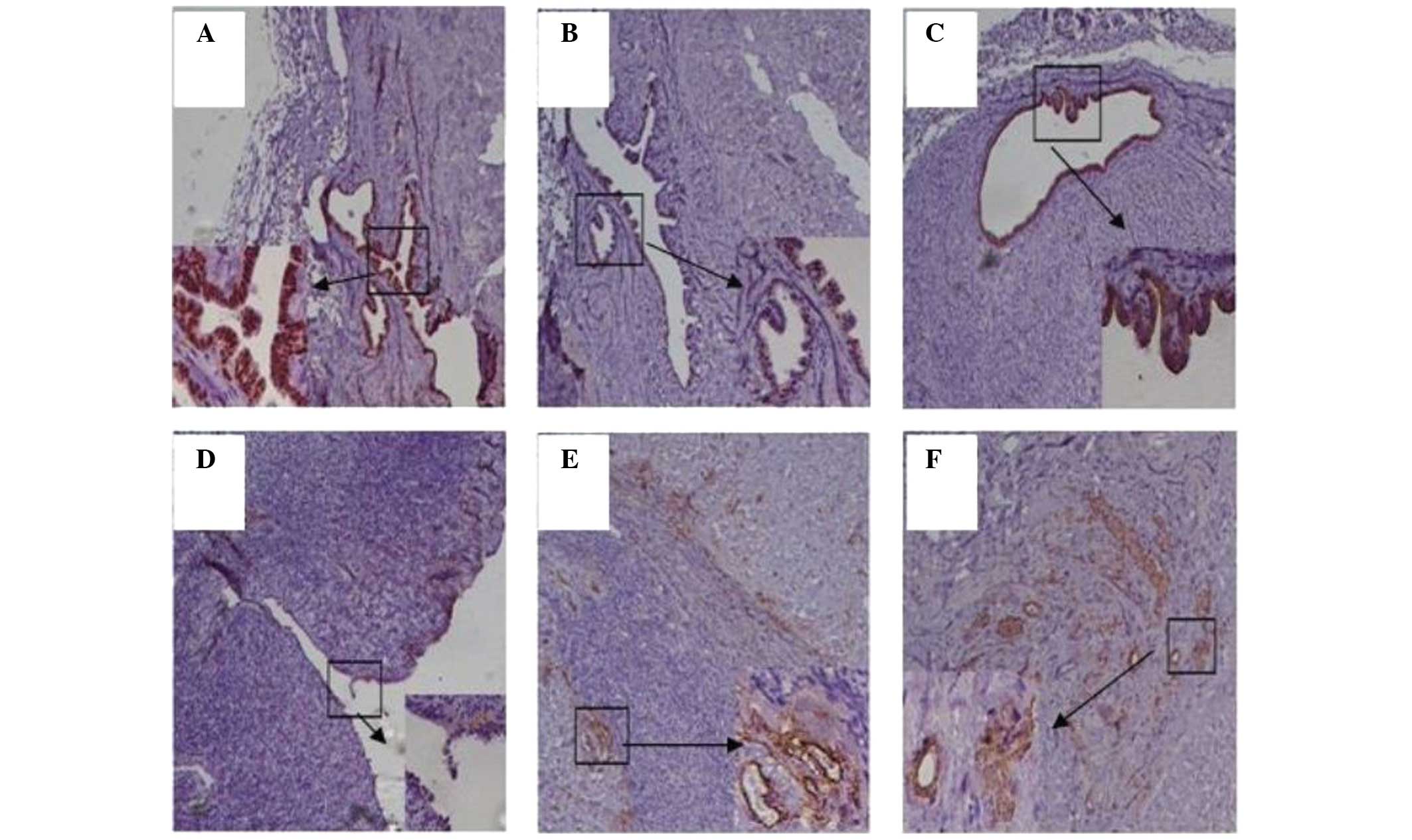 | Figure 3Immunohistochemical analysis of human
ovarian tissues implanted in severe combined immunodeficient mice.
(A) Positive staining for the estrogen receptor in implanted human
ovarian tissue (magnification, ×100; inset magnification, ×400).
(B) Partially positive staining for the progesterone receptor in
implanted human ovarian tissue (magnification, ×100; inset
magnification, ×400). (C) Positive staining for cytokeratin (CK)-7
in implanted human ovarian tissue, indicating that the normal
ovarian architecture was retained (magnification, ×100; inset
magnification, ×400). (D) Positive staining for CK-7 in normal
ovarian tissue prior to transplantation (magnification, ×100; inset
magnification, ×400). (E) Positive staining for CD34 in the
implanted human ovarian tissue (magnification, ×100; inset
magnification, ×400). (F) Positive staining for α-smooth muscle
actin in the implanted human ovarian tissue (magnification, ×100;
inset magnification, ×400). |
Discussion
Complex biological processes often require in
vivo analysis, and a number of important research advances have
been made using mice as a model for the study of various biological
systems. However, mice are not the same as humans, and in
vivo studies of human biology are severely limited by ethical
and technical constraints. There is a growing need for animal
models, which can be used to construct in vivo studies of
human cells, tissues and organs without putting individuals at
risk. Humanized mice, or mouse-human chimeras, have been developed
to overcome these constraints and are now an important research
tool for the in vivo study of human cells and tissues
(11). Humanized mice are defined
as immunodeficient mice engrafted with hematopoietic cells or
tissues, or mice that transgenically express human genes (11). The development of mice that are
‘humanized’ by the engraftment of human tissues provides an
opportunity for the in vivo study of human biological
processes, which may otherwise not be possible. In the present
study, a novel protocol was developed for the establishment of
normal human ovarian stroma within the subcutaneous tissues of
mice. It was hypothesized that the implanted stroma would
accurately mimic the human ovarian microenvironment, and result in
a humanized mouse model that may potentially serve as a useful
preclinical tool for investigating the progression of human ovarian
cancer.
A key factor in the success of this engraftment and
the long-term survival of human xenografts is the use of SCID mice.
Failure to develop mature T and B lymphocytes and the lack of an
immune system makes SCID mice an ideal model for cell and
tissue-transfer experiments. As a classical model, the subcutaneous
mouse model has been widely used for a number of years. In the
present study, normal, human ovarian tissues were subcutaneously
implanted into SCID mice with the aim of developing a novel human
ovarian tissue-derived mouse model.
Numerous studies have used mice with xenografts
implanted under the renal capsule (17). However, in the present study, human
ovarian tissue was embedded subcutaneously in the SCID mice. The
advantages of subcutaneous implantation include a plentiful blood
supply at the implantation site and minimal surgical damage to the
tissues. Between day 7 and 14 following implantation,
neovascularization was observed in the implanted tissue. This model
has the potential to improve the understanding of the crosstalk
between tissue stroma and the epithelium, as well as the factors
involved in tumor initiation and progression. The humanized ovarian
microenvironment established in this mouse model offers a basis to
study the proliferation of human ovarian cancer cells in a human
ovarian milieu.
A normal, human tissue microenvironment contains
three-dimensional intercellular interactions between the stroma
cells, epithelial cells and cytokines. These are essential for
maintaining epithelial polarity and modulating the growth
inhibition of normal cells (18,19).
However, when cancer cells emerge in normal tissue, they produce a
range of stroma-modulating factors to activate the stromal cells in
their microenvironment. The activated stromal cells then secrete
matrix metalloproteinases and cytokines to promote tumor growth,
stimulate angiogenesis, increase inflammatory responses and induce
cell differentiation (20–22). Therefore, the normal tissue
microenvironment is disrupted and transforms into the tumorous
tissue microenvironment.
In the present study, the epithelial cells in the
xenografts were shown to be derived from normal ovarian tissues, as
these cells were positive for CK-7, which is diffusely distributed
throughout the cytoplasm of ovarian cells. Furthermore,
human-derived vessels within the implants remained viable for a
period of four weeks following implantation. Positive staining for
the ER and partially positive staining for the PR indicated that
the inoculated ovarian tissues continued to be estrogen- and
progesterone-dependent. α-SMA was found to be expressed only in the
vascular pericytes and not in the epithelium or stromal
fibroblasts, indicating that the subcutaneous xenografts of human
ovarian tissues in the SCID mice consisted of cells derived from
normal tissues. These observations indicated that the implanted
human ovarian tissues survived well in the SCID mice.
The results of the present study indicate that the
humanized SCID mouse ovarian transplant model has the potential to
serve as a useful preclinical tool, which may be used to build a
humanized mouse model of ovarian cancer to investigate the efficacy
of chemotherapeutic strategies. The absence of an immune system is
one of the major limitations inherent in this type of model.
However, SCID mice are likely to continue to be critical in
establishing xenograft models that recapitulate the tumor growth
and spread observed in patients.
In conclusion, the present study demonstrated that
human ovarian tissue successfully survives in a SCID mouse host and
retains the properties of the original normal ovarian tissues. In
addition, the experimental protocol used in the current study may
be used to establish a humanized mouse model of ovarian cancer for
preclinical research.
Acknowledgements
The study was supported by grants from the Priority
Academic Program Development of Jiangsu Higher Education
Institutions (no. JX10231081) and the Natural Science Foundation of
Jiangsu Province (no. BK2012878).
Abbreviations:
|
SCID
|
severe combined immunodeficient
|
|
ER
|
estrogen receptor
|
|
PR
|
progesterone receptor
|
|
CK
|
cytokeratin
|
|
α-SMA
|
α-smooth muscle actin
|
References
|
1
|
Siegel R, Naishadham D and Jemal A: Cancer
statistics. CA Cancer J Clin. 63:11–30. 2013.
|
|
2
|
Jemal A, Bray F, Center MM, Ferlay J, Ward
E and Forman D: Global cancer statistics. CA Cancer J Clin.
61:69–90. 2011. View Article : Google Scholar
|
|
3
|
Quinn BA, Xiao F, Bickel L, Martin L, Hua
X, et al: Development of a syngeneic mouse model of epithelial
ovarian cancer. J Ovarian Res. 3:242010. View Article : Google Scholar : PubMed/NCBI
|
|
4
|
Mullany LK and Richards JS: Minireview:
animal models and mechanisms of ovarian cancer development.
Endocrinology. 153:1585–1592. 2012. View Article : Google Scholar : PubMed/NCBI
|
|
5
|
Zhang J, Chen X, Shi G, Xie X, Liu H, et
al: Establishment of a new representative model of human ovarian
cancer in mice. J Ovarian Res. 6:92013. View Article : Google Scholar : PubMed/NCBI
|
|
6
|
Sloan Stakleff KD, Rouse AG, Ryan AP,
Haller NA and Von Gruenigen VE: A novel early-stage orthotopic
model for ovarian cancer in the Fischer 344 rat. Int J Gynecol
Cancer. 15:246–254. 2005.PubMed/NCBI
|
|
7
|
Fong MY and Kakar SS: Ovarian cancer mouse
models, a summary of current models and their limitations. J
Ovarian Res. 2:122009. View Article : Google Scholar : PubMed/NCBI
|
|
8
|
Garson K, Gamwell LF, Pitre EM and
Vanderhyden BC: Technical challenges and limitations of current
mouse models of ovarian cancer. J Ovarian Res. 5:392012. View Article : Google Scholar : PubMed/NCBI
|
|
9
|
Allen M and Louise Jones J: Jekyll and
Hyde: the role of the microenvironment on the progression of
cancer. J Pathol. 223:162–176. 2011. View Article : Google Scholar : PubMed/NCBI
|
|
10
|
Balkwill FR, Capasso M and Hagemann T: The
tumor microenvironment at a glance. J Cell Sci. 125:5591–5596.
2012. View Article : Google Scholar : PubMed/NCBI
|
|
11
|
Shultz LD, Ishikawa F and Greiner DL:
Humanized mice in translational biomedical research. Nat Rev
Immunol. 7:118–130. 2007. View
Article : Google Scholar : PubMed/NCBI
|
|
12
|
Bankert RB, Egilmez NK and Hess SD:
Human-SCID mouse chimeric models for the evaluation of anti-cancer
therapies. Trends Immunol. 22:386–393. 2001. View Article : Google Scholar : PubMed/NCBI
|
|
13
|
Proia DA and Kuperwasser C: Reconstruction
of human mammary tissues in a mouse model. Nat Protoc. 1:206–214.
2006. View Article : Google Scholar : PubMed/NCBI
|
|
14
|
Bankert RB, Balu-Iyer SV, Odunsi K, Shultz
LD, Kelleher RJ Jr, et al: Humanized mouse model of ovarian cancer
recapitulates patient solid tumor progression, ascites formation,
and metastasis. PLoS One. 6:e244202011. View Article : Google Scholar : PubMed/NCBI
|
|
15
|
Wang J, Xia TS, Liu XA, Ding Q, Du Q, et
al: A novel orthotopic and metastatic mouse model of breast cancer
in human mammary microenvironment. Breast Cancer Res Treat.
120:337–344. 2010. View Article : Google Scholar : PubMed/NCBI
|
|
16
|
Zheng MJ, Wang J, Chen YW, Xu L, Xue DD,
et al: A novel mouse model of gastric cancer with human gastric
microenvironment. Cancer Lett. 325:108–115. 2012. View Article : Google Scholar : PubMed/NCBI
|
|
17
|
Priolo C, Agostini M, Vena N, Ligon AH,
Fiorentino M, et al: Establishment and genomic characterization of
mouse xenografts of human primary prostate tumors. Am J Pathol.
176:1901–1913. 2010. View Article : Google Scholar : PubMed/NCBI
|
|
18
|
Weinberg RA: Coevolution in the tumor
microenvironment. Nat Genet. 40:494–495. 2008. View Article : Google Scholar : PubMed/NCBI
|
|
19
|
Pietras K and Ostman A: Hallmarks of
cancer: interactions with the tumor stroma. Exp Cell Res.
316:1324–1331. 2010. View Article : Google Scholar : PubMed/NCBI
|
|
20
|
Mueller MM and Fusenig NE: Friends or foes
- bipolar effects of the tumour stroma in cancer. Nat Rev Cancer.
4:839–849. 2004. View
Article : Google Scholar : PubMed/NCBI
|
|
21
|
Mantovani A, Allavena P, Sica A and
Balkwill F: Cancer-related inflammation. Nature. 454:436–444. 2008.
View Article : Google Scholar
|
|
22
|
Weis SM and Cheresh DA: Tumor
angiogenesis: molecular pathways and therapeutic targets. Nat Med.
17:1359–1370. 2011. View
Article : Google Scholar : PubMed/NCBI
|